. 2020 Oct;146(10):2559-2574.
doi: 10.1007/s00432-020-03317-4. Epub 2020 Jul 17.

Potential of platinum-resensitization by Wnt signaling modulators as treatment approach for epithelial ovarian cancer

Affiliations

Potential of platinum-resensitization by Wnt signaling modulators as treatment approach for epithelial ovarian cancer

Till Kaltofen et al. J Cancer Res Clin Oncol. 2020 Oct.

Abstract

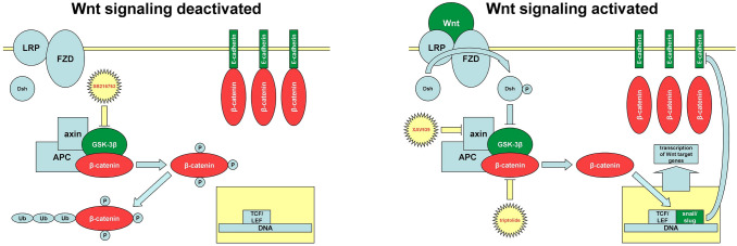
Purpose: Canonical Wnt/ β-catenin pathway is one mechanism being activated in platinum-resistant epithelial ovarian cancer (EOC). Detecting potential targets for Wnt pathway modulation as a putative future therapeutic approach was the aim of this study.

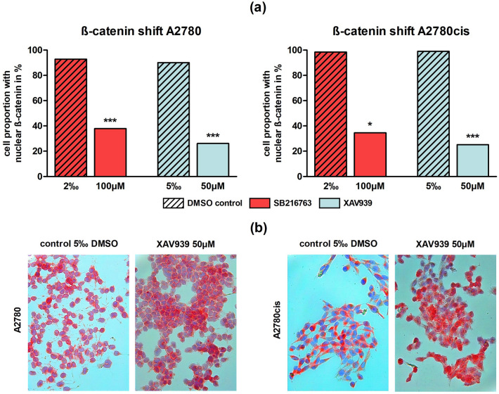
Methods: Biological effects of different Wnt modulators (SB216763, XAV939 and triptolide) on the EOC cell lines A2780 and its platinum-resistant clone A2780cis were investigated via multiple functional tests. Immunohistochemistry (IHC) was carried out to compare the expression levels of Wnt marker proteins (β-catenin, snail/ slug, E-cadherin) in patient specimens and to correlate them with lifetime data.

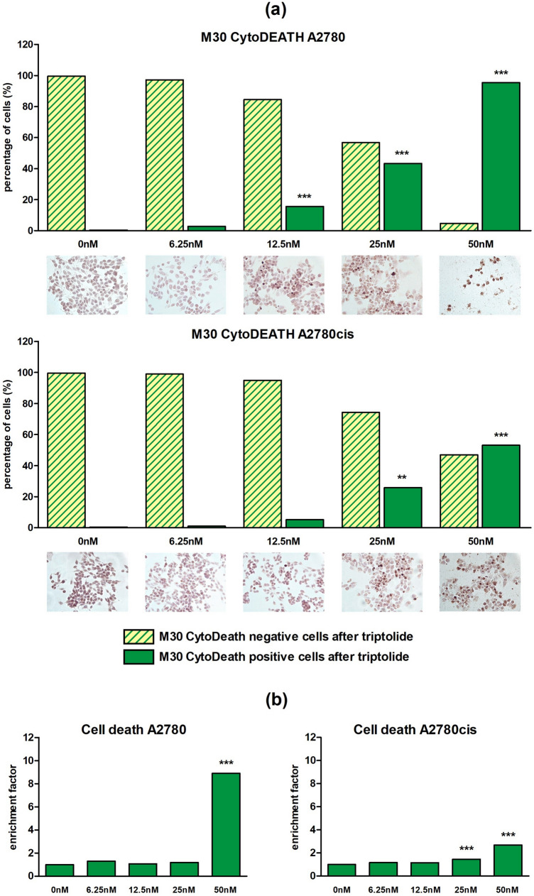
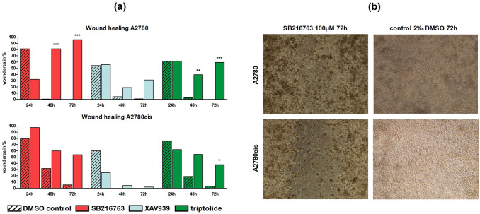
Results: We could show that activated Wnt signaling of the platinum-resistant EOC cell line A2780cis can be reversed by Wnt manipulators through SB216763 or XAV939. All Wnt manipulators tested consecutively decreased cell proliferation and cell viability. Apoptosis of A2780 and A2780cis was enhanced by triptolide in a dose-dependent manner, whereas cell migration was inhibited by SB216763 and triptolide. IHC analyses elucidated significantly different expression patterns for Wnt markers in the serous subtype. Herein, higher plasmatic snail/ slug expression is associated with improved progression-free (PFS) and overall survival (OS).

Conclusion: According to the described effects on EOC biology, all three Wnt manipulators seem to have the potential to augment the impact of a platinum-based chemotherapy in EOC. This is promising as a dominance of this pathway was confirmed in serous histology.

Keywords: E-cadherin; Ovarian cancer; Platinum-resistance; Snail/ slug; Wnt; β-catenin.


Conflict of interest statement

Sven Mahner reports grants and personal fees from AstraZeneca, Clovis, Medac, MSD, PharmaMar, Roche, Sensor Kinesis, Tesaro and Teva outside the submitted work. Fabian Trillsch has received grants and personal fees from AstraZeneca, Clovis, Medac, PharmaMar, Roche and Tesaro outside the submitted work. The other authors declare no conflict of interest.

Figures

Fig. 1
Fig. 1
Canonical Wnt signaling pathway: In absence of Wnt ligand (left), the destruction complex, consisting of GSK-3β, adenomatous polyposis coli (APC) and axin, hyperphosphorylates β-catenin, which marks it for ubiquitination (Ub) and proteasomal degradation. Binding of Wnt ligand (right) to a frizzled (FZD)/lipoprotein receptor-related protein (LRP) receptor complex leads to the phosphorylation of dishevelled (Dsh) inactivating GSK-3β and preventing the phosphorylation of β-catenin. Non-phosphorylated β-catenin shifts into the nucleus and forms a complex with T-cell factor (TCF)/lymphoid enhancer factor (LEF) activating transcription of Wnt target genes. Furthermore, the expression of zinc-finger transcription factors snail and slug is upregulated. They bind to E-boxes in E-cadherins promoter region and prevent its transcription. Suppresion of E-cadherin offers more available cytoplasmic β-catenin and induces a self-driven positive feedback loop. In conclusion, by losing E-cadherin as an adhesion molecule cells undergo EMT (Arend et al. ; Gasior et al. ; Gatcliffe et al. 2008). The three inhibitors (SB216763, XAV939 triptolide) used in this study, are shown in their typical domain
Fig. 2
Fig. 2
ß-catenin shift after treatment with different Wnt signaling manipulators in A2780 and A2780cis: a In A2780 and A2780cis the proportion of cells with nuclear localization of ß-catenin was significantly decreased through the addition of 100 µM SB216763 or 50 µM XAV939 compared to controls (n = 3 per column, mean, *p < 0.05/ ***p < 0.001 by Sidak's multiple comparisons test). b Representative images of A2780 and A2780cis after treatment with 50 µM XAV939 compared to controls with 5‰ DMSO. ß-catenin is red-colored. While in controls ß-catenin is mainly localized in the nucleus, addition of the Wnt inhibitor XAV939 leads to a shift towards the cell membrane
Fig. 3
Fig. 3
BrdU cell proliferation and MTT cell viability assay for different Wnt signaling manipulators (100 µM SB216763, 50 µM XAV939, 50 nM triptolide) in A2780 and A2780cis after 72 h: a In the BrdU assay only triptolide displayed a significant impairment of proliferation in A2780 and A2780cis, whereas SB216763 revealed a significant reduction of cell proliferation in A2780cis and XAV939 showed no influence (n = 9 per column, mean, *p < 0.05/ ***p < 0.001 by Dunnett's multiple comparisons test, controls with 0‰ DMSO were set 100%). b In the MTT assay all three inhibitors in platinum-sensitive and -resistant cells reduced metabolic activity with different levels of significance (n = 9 per column, mean, **p < 0.01/ ***p < 0.001 by Dunnett's multiple comparisons test, controls with 0‰ DMSO were set 100%)
Fig. 4
Fig. 4
CytoDEATH apoptosis assay after 48 h and Cell death detection ELISA after 24 h for different triptolide concentrations in A2780 and A2780cis: a Apoptosis of cell lines A2780 and A2780cis was enhanced by triptolide in a dose-dependent manner. The percental rate of apoptic cells (M30 CytoDEATH positive) significantly increased in both cell lines with rising concentrations. Meanwhile, M30 CytoDEATH negative cells rapidly decreased (n = 3 per column, mean, **p < 0.01/ ***p < 0.001 by Dunnett's multiple comparisons test). According to documented concentrations representative microphotographs of EOC cells with an apoptic cell proportion in intense brown after DAB treatment are added. b Incubation for 24 h with different triptolide concentrations revealed a dose-dependent rise of absorbance up to a maximum at 50 nM, which confirms enhancement of apoptosis by triptolide (n = 6 per column, mean, ***p < 0.001 by Dunnett's multiple comparisons test, controls without DMSO were set 1)
Fig. 5
Fig. 5
Wound healing assay for different Wnt signaling manipulators in A2780 and A2780cis: a Cell migration in A2780 and A2780cis was significantly reduced through the addition of 100 µM SB216763 or 50 nM triptolide after 48 and 72 h compared to controls. Treatment with XAV939 did not affect migration ability at all (n = 3 per column, mean, *p < 0.05/ **p < 0.01/ ***p < 0.001 by Sidak's multiple comparisons test, wound area after 0 h was set 100%). b Representative images of the wound area in A2780 and A2780cis 72 h after treatment with 100 µM SB216763 compared to controls with 2‰ DMSO. While controls showed a confluent monolayer of cells, in SB216763 treated cells a remaining wound area is visible
Fig. 6
Fig. 6
Comparison of IHC scores of Wnt signaling markers dependent on FIGO stages: Analyzing expression of Wnt signaling markers dependent on FIGO stages (n = 153) revealed significant differences for β-catenin and E-cadherin in two different locations (membrane and plasma) (mean ± SEM, *p < 0.05/ **p < 0.01/ ***p < 0.001 by Mann–Whitney-U test)
Fig. 7
Fig. 7
PFS depending on plasmatic snail/ slug IHC scores in serous EOC and representative microphotographies: a The Kaplan–Meier curves demonstrate PFS and OS as a function of plasmatic snail/ slug expression in serous OC cases. Green lines (IHC score > 7, PFS = 33.6 ± 4.8 months, OS = 50.4 ± 13.2 months) show a significant benefit in PFS and a trend in OS compared to the red line (IHC score ≤ 7, PFS = 15.6 ± 2.4 months, OS = 27.6 ± 3.6 months) (median ± SEM, p = 0.001/ p = 0.058 by chi-square statistic of the Log-Rank test [Mantel-Cox]). b Plasmatic snail/ slug accumulation in serous subtype is marked in brown after DAB treatment. While the left microphotography shows a higher expression (IHC score > 7), on the right photography a lower expression (IHC score ≤ 7) is shown

References

    1. Anglesio MS, Wiegand KC, Melnyk N, Chow C, Salamanca C, Prentice LM, Senz J, Yang W, Spillman MA, Cochrane DR, Shumansky K, Shah SP, Kalloger SE, Huntsman DG (2013) Type-specific cell line models for type-specific ovarian cancer research. PLoS ONE 8:e72162. 10.1371/journal.pone.0072162 - PMC - PubMed
    1. Arend RC, Londoño-Joshi AI, Straughn JM Jr, Buchsbaum DJ (2013) The Wnt/β-catenin pathway in ovarian cancer: A review. Gynecol Oncol 131:772–779. 10.1016/j.ygyno.2013.09.034 - PubMed
    1. Baldwin LA, Hoff JT, Lefringhouse J, Zhang M, Jia C, Liu Z, Erfani S, Jin H, Xu M, She Q-B, van Nagell JR, Wang C, Chen L, Plattner R, Kaetzel DM, Luo J, Lu M, West D, Liu C, Ueland FR, Drapkin R, Zhou BP, Yang XH (2014) CD151-α3β1 integrin complexes suppress ovarian tumor growth by repressing slug-mediated EMT and canonical Wnt signaling. Oncotarget 5:12203–12217. 10.18632/oncotarget.2622 - PMC - PubMed
    1. Barghout SH, Zepeda N, Xu Z, Steed H, Lee C-H, Fu Y (2015) Elevated β-catenin activity contributes to carboplatin resistance in A2780cp ovarian cancer cells. Biochem Biophys Res Commun 468:173–178. 10.1016/j.bbrc.2015.10.138 - PubMed
    1. Berx G, van Roy F (2009) Involvement of members of the cadherin superfamily in cancer. Cold Spring Harb Perspect Biol 1:a003129. 10.1101/cshperspect.a003129 - PMC - PubMed

MeSH terms