. 2020:2161:209-228.
doi: 10.1007/978-1-0716-0680-3_15.

Characterization of R-Loop Structures Using Single-Molecule R-Loop Footprinting and Sequencing

Affiliations

Characterization of R-Loop Structures Using Single-Molecule R-Loop Footprinting and Sequencing

Maika Malig et al. Methods Mol Biol. 2020.

Abstract

R-loops are three-stranded structures that form during transcription when the nascent RNA hybridizes with the template DNA resulting in a DNA:RNA hybrid and a looped-out single-stranded DNA (ssDNA) strand. These structures are important for normal cellular processes and aberrant R-loop formation has been implicated in a number of pathological outcomes, including certain cancers and neurodegenerative diseases. Mapping R-loops has primarily been performed using DRIP (DNA:RNA immunoprecipitation) based methods that are dependent on the anti-DNA:RNA hybrid S9.6 antibody and short-read sequencing. While DRIP-based methods are robust and report R-loop formation genome-wide, they only do so at the population average level; interrogating R-loop formation at the single molecule level is not feasible with such approaches. Here we present single molecule R-loop footprinting (SMRF-seq), a method that relies on the chemical reactivity of the displaced ssDNA strand to non-denaturing sodium bisulfite and single molecule long-read sequencing as a readout, to characterize R-loops. SMRF-seq can be used independently of S9.6 to generate high resolution, strand-specific, maps of individual R-loops at ultra-deep coverage on kilobases-length DNA fragments.

Keywords: DNA:RNA hybrid; Non-denaturing bisulfite conversion; R-loop; SMRT sequencing; Transcription.


Figures
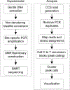
Fig. 1
Fig. 1
Overall workflow for both experimental procedures and computational analysis
Fig. 2
Fig. 2
Example gradient PCR with 2990 bp product. Agarose gel stained with 1× GelRed. Run for 20 min at 150 V
Fig. 3
Fig. 3
Example BioAnalyzer trace of a SMRTbell library. Pooled amplicons range from 2938 to 4379 bp
Fig. 4
Fig. 4
Example index file. Columns are as follows: chr, start, end, gene, 0, strand. Tab delimited and no header format
Fig. 5
Fig. 5
Example footprinted region with PNG output after footPeak_graph.pl run. (a) SNRPN70 footprinted region (hg19) and DRIPc-seq data with (+) and (−) strands shown in red and blue, respectively. (b) Example output showing random 200 peaks reads on the non-template strand. (c) Example output showing random 100 reads for template strand. For panels (b) and (c), each horizontal line corresponds to one read. Red tick lines indicate converted Cs within a called peak, yellow indicates non-converted Cs, green indicates converted Cs not called a peak; gray is missing/ambiguous sequence
Fig. 6
Fig. 6
Genome Browser snapshot of same SNRPN70 terminal region in previous figure. Converted cytosines within called peaks are annotated as red tick marks on the positive strand (blue tick marks for footprints called in the negative strand). Each horizontal line is a single molecule read mapping to the region
Fig. 7
Fig. 7
Example target region in SNRPN70. Red bar indicates possible target region to amplify. Blue arrow indicates forward and reverse primer sites flanking peaks of DRIPc-seq signal (below)

References

    1. Ginno PA, Lott PL, Christensen HC, Korf I, Chedin F (2012) R-loop formation is a distinctive characteristic of unmethylated human CpG island promoters. Mol Cell 45(6):814–825. 10.1016/j.molcel.2012.01.017 - DOI - PMC - PubMed
    1. Sanz LA, Hartono SR, Lim YW, Steyaert S, Rajpurkar A, Ginno Pa , Xu X, Chedin F (2016) Prevalent, dynamic, and conserved R-loop structures associate with specific epigenomic signatures in mammals. Mol Cell 63 :167–178. 10.1016/j.molcel.2016.05.032 - DOI - PMC - PubMed
    1. Wahba L, Costantino L, Tan FJ, Zimmer A, Koshland D (2016) S1-DRIP-seq identifies high expression and polyA tracts as major contributors to R-loop formation. Genes Dev 30 :1327–1338. 10.1101/gad.280834.116 - DOI - PMC - PubMed
    1. El Hage A, Webb S, Kerr A, Tollervey D (2014) Genome-wide distribution of RNA-DNA hybrids identifies RNase H targets in tRNA genes, retrotransposons and mitochondria. PLoS Genet 10(10):e1004716. 10.1371/journal.pgen.1004716 - DOI - PMC - PubMed
    1. Hartono SR, Malapert A, Legros P, Bernard P, Chedin F, Vanoosthuyse V (2018) The affinity of the S9.6 antibody for double-stranded RNAs impacts the accurate mapping of R-loops in fission yeast. J Mol Biol 430 (3):272–284. 10.1016/j.jmb.2017.12.016 - DOI - PMC - PubMed

Publication types