Review
. 2020 Sep:126:105804.
doi: 10.1016/j.biocel.2020.105804. Epub 2020 Jul 15.

Cardiac optical mapping - State-of-the-art and future challenges

Affiliations
Review

Cardiac optical mapping - State-of-the-art and future challenges

Christopher O'Shea et al. Int J Biochem Cell Biol. 2020 Sep.

Abstract

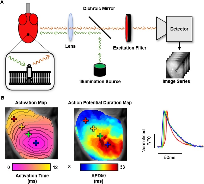
Cardiac optical mapping utilises fluorescent dyes to directly image the electrical function of the heart at a high spatio-temporal resolution which far exceeds electrode techniques. It has therefore become an invaluable tool in cardiac electrophysiological research to map the propagation of heterogeneous electrical signals across the myocardium. In this review, we introduce the principles behind cardiac optical mapping and discuss some of the challenges and state of the art in the field. Key advancements discussed include newly developed fluorescent indicators, tools for the analysis of complex datasets, panoramic imaging systems and technical and computational approaches to realise optical mapping in freely beating hearts.

Keywords: Action potential; Arrhythmia; Calcium transient; Cardiac optical mapping; Electrophysiology; Fluorescent imaging.


Figures

Fig. 1
Fig. 1
Cardiac optical mapping setup, data and analysis. A) Schematic representation of a typical optical mapping setup for imaging a potentiometric dye loaded cardiac preparation (left). Inset shows the fluorescent indicator embedded within the cellular membrane. The potentiometric dye is excited by photons (green arrows) from an illumination source. This causes the release of fluorescent photons (red arrows) whose spectral properties depend on the transmembrane voltage. Fluorescent photons are filtered from the illumination photons and directed to a high-density imaging detector to produce a time series dataset. B) Example of analyses possible from optical mapping datasets, including activation and signal morphology mapping (left panel). Right panel shows examples of the raw signals produced at each pixel of an optical mapping dataset, in this case optical action potentials from mouse ventricles. Y axis denotes normalised fractional fluorescence change (F/F0) from baseline fluorescence level (F0). (For interpretation of the references to colour in this figure legend, the reader is referred to the web version of this article).
Fig. 2
Fig. 2
Challenges in cardiac optical mapping. A) Example of processing techniques that are often applied on all pixels in an optical mapping dataset to enhance the signal to noise ratio of the raw data (left) to allow effective parameter quantification (right). Note: The exact methods and sequence of processing methods used will vary depending on experimental setup, model and analysis software used. B) Schematic representation of a typical imaged area (green) from a mouse whole heart using a single camera setup. The red area shows the area of the epicardial ventricular surface that is not imaged. C) Example signals from a mouse atrium where motion artefacts are present. In the area with prolonged APD (red), contraction has not been successfully uncoupled, distorting the measured optical signal. (For interpretation of the references to colour in this figure legend, the reader is referred to the web version of this article).

References

    1. Bachtel A.D., Gray R.A., Stohlman J.M., Bourgeois E.B., Pollard A.E., Rogers J.M. A novel approach to dual excitation ratiometric optical mapping of cardiac action potentials with Di-4-ANEPPS using pulsed LED excitation. IEEE Trans. Biomed. Eng. 2011;58:2120–2126. doi: 10.1038/jid.2014.371. - DOI - PMC - PubMed
    1. Benjamin E.J., Muntner P., Alonso A., Bittencourt M.S., Callaway C.W., Carson A.P. 2019. Heart Disease and Stroke Statistics-2019 Update: A Report From the American Heart Association. - DOI - PubMed
    1. Brack K.E., Narang R., Winter J., Andr G. The mechanical uncoupler blebbistatin is associated with significant electrophysiological effects in the isolated rabbit heart. Exp. Physiol. 2013;5:1009–1027. doi: 10.1113/expphysiol.2012.069369. - DOI - PMC - PubMed
    1. Bray M.A., Lin S.F., Wikswo J.P. Three-dimensional surface reconstruction and flourescent visualization of cardiac activation. IEEE Trans. Biomed. Eng. 2000;47:1382–1391. doi: 10.1109/10.871412. - DOI - PubMed
    1. Broyles C., Robinson P., Daniels M. Fluorescent, bioluminescent, and optogenetic approaches to study excitable physiology in the single cardiomyocyte. Cells. 2018;7:51. doi: 10.3390/cells7060051. - DOI - PMC - PubMed

Publication types

LinkOut - more resources