

Target-shaped combined halo and reversed-halo sign, an atypical chest CT finding in COVID-19
- PMID: 32682246
- PMCID: PMC7329690
- DOI: 10.1016/j.clinimag.2020.06.038
Target-shaped combined halo and reversed-halo sign, an atypical chest CT finding in COVID-19
Abstract
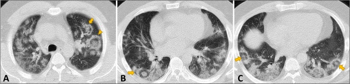
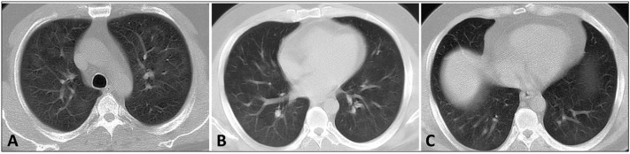
Typical chest CT findings in COVID-19 have been described as bilateral peripheral ground glass opacities, with or without consolidation. Halo sign and reversed halo sign have been reported as atypical imaging findings in this disease. However, to the best of our knowledge, combined presence of these signs has never been reported before. Herein, we present a COVID-19 patient with numerous atypical target-shaped, combined halo and reversed halo pulmonary lesions, in the absence of any other underlying disease.
Keywords: COVID-19; Lung diseases; Severe acute respiratory syndrome coronavirus 2; Tomography, X-ray computed.
Copyright © 2020 Elsevier Inc. All rights reserved.
Figures
Similar articles
-
Interpretation of CT signs of 2019 novel coronavirus (COVID-19) pneumonia.Eur Radiol. 2020 Oct;30(10):5455-5462. doi: 10.1007/s00330-020-06915-5. Epub 2020 May 4. Eur Radiol. 2020. PMID: 32367422 Free PMC article.
-
Radiology of COVID-19 - Imaging the pulmonary damage.J Pak Med Assoc. 2020 May;70(Suppl 3)(5):S60-S63. doi: 10.5455/JPMA.21. J Pak Med Assoc. 2020. PMID: 32515387
-
Halo, Reversed Halo, or Both? Atypical Computed Tomography Manifestations of Coronavirus Disease (COVID-19) Pneumonia: The "Double Halo Sign".Korean J Radiol. 2020 Oct;21(10):1161-1164. doi: 10.3348/kjr.2020.0687. Epub 2020 Jul 17. Korean J Radiol. 2020. PMID: 32729264 Free PMC article.
-
Chest CT features of coronavirus disease 2019 (COVID-19) pneumonia: key points for radiologists.Radiol Med. 2020 Jul;125(7):636-646. doi: 10.1007/s11547-020-01237-4. Epub 2020 Jun 4. Radiol Med. 2020. PMID: 32500509 Free PMC article. Review.
-
One year of COVID-19 pandemic: what we Radiologists have learned about imaging.Rofo. 2022 Feb;194(2):141-151. doi: 10.1055/a-1522-3155. Epub 2021 Oct 14. Rofo. 2022. PMID: 34649291 Review. English, German.
Cited by
-
The "Target Sign" in a 46-Year-Old Patient with COVID-19 Pneumonia.Case Rep Radiol. 2021 Oct 29;2021:9956927. doi: 10.1155/2021/9956927. eCollection 2021. Case Rep Radiol. 2021. PMID: 34721918 Free PMC article.
-
"Pulmonary target sign" as a diagnostic feature in chest computed tomography of COVID-19.World J Radiol. 2021 Jul 28;13(7):233-242. doi: 10.4329/wjr.v13.i7.233. World J Radiol. 2021. PMID: 34367510 Free PMC article.
-
Simultaneous presence of the "bullseye" and "reversed halo" sign at CT of COVID-19 pneumonia: A case report.Radiol Case Rep. 2021 Sep;16(9):2442-2446. doi: 10.1016/j.radcr.2021.05.074. Epub 2021 Jun 3. Radiol Case Rep. 2021. PMID: 34099964 Free PMC article.
-
Differences and Similarities between the Double Halo Sign, the Chest CT Target Sign and the Reversed Halo Sign in Patients with COVID-19 Pneumonia.Korean J Radiol. 2021 Apr;22(4):672-676. doi: 10.3348/kjr.2020.1150. Epub 2021 Feb 2. Korean J Radiol. 2021. PMID: 33660464 Free PMC article. No abstract available.
-
COVID-19 Pneumonia Presenting with Multiple Nodules Mimicking Metastases: An Atypical Case.J Belg Soc Radiol. 2022 Aug 11;106(1):72. doi: 10.5334/jbsr.2681. eCollection 2022. J Belg Soc Radiol. 2022. PMID: 36042788 Free PMC article.
References
-
- World Health Organization Coronavirus disease (COVID-19) situation report. 2020. https://www.who.int/emergencies/diseases/novel-coronavirus-2019/situatio...
-
- Salehi S., Abedi A., Balakrishnan S., Gholamrezanezhad A. Coronavirus disease 2019 (COVID-19): a systematic review of imaging findings in 919 patients. AJR Am J Roentgenol. 2020:1–7. - PubMed
MeSH terms
LinkOut - more resources
Full Text Sources
Other Literature Sources
Medical
