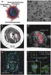
Employment of targeted nanoparticles for imaging of cellular processes in cardiovascular disease
- PMID: 32682272
- PMCID: PMC7744313
- DOI: 10.1016/j.copbio.2020.06.003
Employment of targeted nanoparticles for imaging of cellular processes in cardiovascular disease
Abstract
Cardiovascular disease (CVD) is a leading cause of global mortality, accounting for pathologies that are primarily of atherosclerotic origin and driven by specific cell populations. A need exists for effective, non-invasive methods to assess the risk of potentially fatal major adverse cardiovascular events (MACE) before occurrence and to monitor post-interventional outcomes such as tissue regeneration. Molecular imaging has widespread applications in CVD diagnostic assessment, through modalities including magnetic resonance imaging (MRI), positron emission tomography (PET), and acoustic imaging methods. However, current gold-standard small molecule contrast agents are not cell-specific, relying on non-specific uptake to facilitate imaging of biologic processes. Nanomaterials can be engineered for targeted delivery to specific cell populations, and several nanomaterial systems have been developed for pre-clinical molecular imaging. Here, we review recent advances in nanoparticle-mediated approaches for imaging of cellular processes in cardiovascular disease, focusing on efforts to detect inflammation, assess lipid accumulation, and monitor tissue regeneration.
Copyright © 2020 Elsevier Ltd. All rights reserved.
Conflict of interest statement
Declaration of interests
The authors declare that they have no known competing financial interests or personal relationships that could have appeared to influence the work reported in this paper.
Figures


References
-
- Mensah GA; Roth GA; Fuster V, The Global Burden of Cardiovascular Diseases and Risk Factors. Journal of the American College of Cardiology 2019, 74 (20), 2529. - PubMed
-
- Miao B; Hernandez Adrian V; Alberts Mark J; Mangiafico N; Roman Yuani M; Coleman Craig I, Incidence and Predictors of Major Adverse Cardiovascular Events in Patients With Established Atherosclerotic Disease or Multiple Risk Factors. Journal of the American Heart Association 2020, 9 (2), e014402. - PMC - PubMed
Publication types
MeSH terms
Grants and funding
LinkOut - more resources
Full Text Sources

