Review
. 2021;17(1):10-16.
doi: 10.2174/1573403X16666200719022142.

The Wenckebach Phenomenon

Affiliations
Review

The Wenckebach Phenomenon

Simon P Hansom et al. Curr Cardiol Rev. 2021.

Abstract

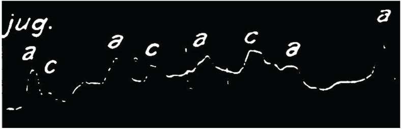
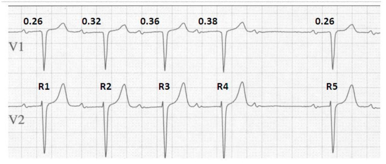
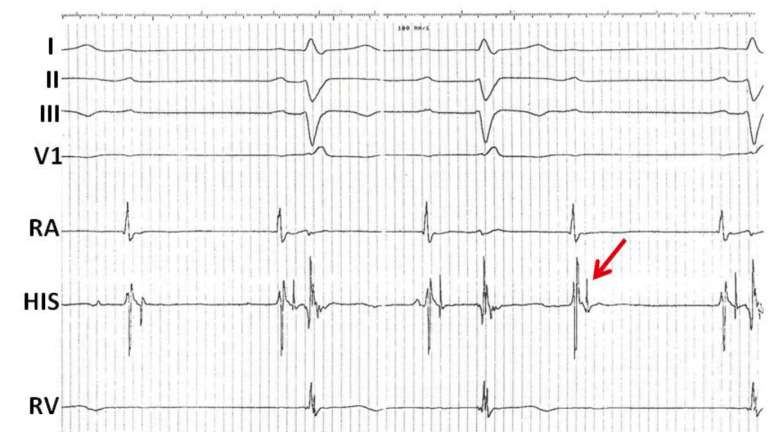
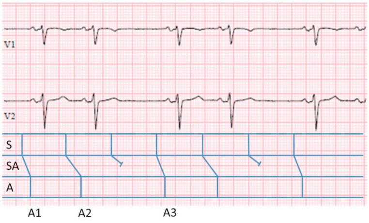
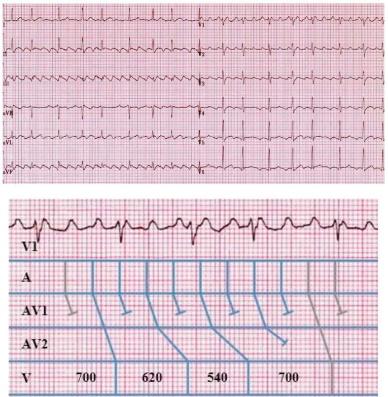
Medicine has many great pioneers, and in 1899, one such pioneer - Karel Frederik Wenckebach made a discovery which, even to this day, remains one of the fundamental concepts within electrophysiology. Since the Wenckebach Phenomenon was first described, the field of electrophysiology has developed at a rapid pace, allowing us to observe this behaviour, and its complexities, in many new ways. In a similar way, this chapter will illustrate Wenckebach behaviour across a spectrum of modalities from the 12 lead ECG, through to the intra-cardiac recordings from both electrophysiological studies and implantable cardiac devices. In doing so, we continue to shed light on the phenomenon first identified through Wenckebach's meticulous attention to detail some 120 years ago.

Keywords: AV node; ECG; Mobitz type 1; Wenckebach; atrioventricular block; electrophysiology.


Figures

Fig. (1)
Fig. (1)
– Smoked drum kymograph tracing of a JVP waveform. Copy of the original smoked drum kymograph recording of the JVP waveform by Wenckebach. Close inspection demonstrates a progressive lengthening in the interval between the a and c waves resulting in an absent c wave followed by a relative pause before the cycle repeats. Reproduced and modified from original article with permission.1
Fig. (2)
Fig. (2)
Typical Wenckebach behaviour. An ECG demonstrating the 4 key elements of typical Wenckebach behaviour. The duration of the ventricular pause can be calculated by deducting the difference in the longest and the shortest PR intervals from 2x the PP interval i.e. 2(PP) - (PR4-PR1) = 2(780) - (380-260) = 1560-120 = 1440ms. The change in RR interval is determined by the difference between the 2 PR intervals forming the resulting RR interval that is being assessed, such that; R1 - R2 = PP + PR2 - PR1 = PP + 0.32 - 0.26 = PP + 0.06. Repeating this approach demonstrates that with each successive prolongation in PR interval, the relative RR intervals decrease by R2-R3 = 0.04 ms and R3-R4 = 0.02 ms respectively.
Fig. (3)
Fig. (3)
Infra-nodal Wenckebach conduction. Intracardiac electrograms demonstrating infra-nodal Wenckebach. Shown are surface leads I, II, III, and V1 as well as intracardiac electrograms from the right atrium (RA), AV node His region (HIS) and the right ventricle (RV). There is a regular atrial rhythm. The first P wave is not conducted; however the second and third P waves are followed by a wide QRS. Each atrial electrogram is followed by a His electrogram in a 1:1 relationship with a stable AH interval. However, the conducted beats demonstrate progressive prolongation in the HV interval until the third beat in the sequence is no longer conducted and is blocked below the His (arrow). This appearance is in keeping with infra-nodal Wenckebach.
Fig. (4)
Fig. (4)
3:2 S-A Wenckebach. ECG leads V1 and V2 demonstrating SA nodal Wenckebach with an associated ladder diagram. The classical depiction of the A, AV and V have been replaced by S, SA, A. To calculate the unseen input within the SA node we apply the same (n+1)/n formula. The total cycle length, equal to the interval from A1 to A3 = 1880 ms. During this time there have been a total of 2 outputs and therefore 3 inputs from within the SA node. Therefore, the overall ‘unseen’ input cycle length is 1880/3= 626ms.
Fig. (5)
Fig. (5)
a) Complex Wenckebach with 8:3 conduction. b). Enlarged copy of lead V1 with associated ladder diagram. (a):12 lead ECG demonstrating typical atrial flutter. Note the underlying repetitive appearance of the QRS complexes with grouped beating and overall 8:3 Wenckebach block. (b) demonstrates an enlarged copy of surface lead V1 with the repeating sequence and associated ladder diagram demonstrating assumed multi-level block resulting in repeating 8:3 conduction. AVN1 and AVN2 denote 2 levels within the AV node (AVN). Note that at the first AVN level there is 2:1 block. The second AVN level demonstrates typical 4:3 Wenckebach periodicity.
Fig. (6)
Fig. (6)
Wenckebach conduction demonstrated from the ventricular output of an implantable cardioverter defibrillator. A ventricular interval plot obtained from an implantable cardioverter defibrillator and designated initially by the device as VT/VF. Note the appearance of grouped beating and the classical reduction in the cycle length between successive ventricular beats in the sequence, therefore initially demonstrating ‘typical’ 3:2, 4:3 and 5:4 Wenckebach block. It is not until the final sequences with 6:5 and 8:7 block that the typical behaviour is lost. This Wenckebach behaviour shows that the rhythm is being driven by a regular atrial input and that the ventricular rhythm is actually due to atrial flutter with variable (Wenckebach) conduction.

References

    1. Upshaw C.B., Jr, Silverman M.E. The Wenckebach phenomenon: a salute and comment on the centennial of its original description. Ann. Intern. Med. 1999;130(1):58–63. doi: 10.7326/0003-4819-130-1-199901050-00011. - DOI - PubMed
    1. Pérez-Riera A.R., Femenía F., McIntyre W.F., Baranchuk A. Karel Frederick Wenckebach (1864-1940): a giant of medicine. Cardiol. J. 2011;18(3):337–339. - PubMed
    1. Wenckebach K.F. On the analysis of irregular pulses. Z. Klin. Med. 1899;37:475–488.
    1. Decherd G.M., Ruskin A. The mechanism of the Wenckebach type of AV block. Br. Heart J. 1946;8(1):6–16. doi: 10.1136/hrt.8.1.6. - DOI - PMC - PubMed
    1. Barold S.S. Lingering misconceptions about type I second-degree atrioventricular block. Am. J. Cardiol. 2001;88(9):1018–1020. doi: 10.1016/S0002-9149(01)01980-4. - DOI - PubMed

MeSH terms