

Histopathology and ultrastructural findings of fatal COVID-19 infections in Washington State: a case series
- PMID: 32682491
- PMCID: PMC7365650
- DOI: 10.1016/S0140-6736(20)31305-2
Histopathology and ultrastructural findings of fatal COVID-19 infections in Washington State: a case series
Erratum in
-
Department of Error.Lancet. 2020 Aug 1;396(10247):312. doi: 10.1016/S0140-6736(20)31646-9. Lancet. 2020. PMID: 32738953 Free PMC article. No abstract available.
Abstract
Background: Severe acute respiratory syndrome coronavirus 2 (SARS-CoV-2) is the cause of an ongoing pandemic, with increasing deaths worldwide. To date, documentation of the histopathological features in fatal cases of the disease caused by SARS-CoV-2 (COVID-19) has been scarce due to sparse autopsy performance and incomplete organ sampling. We aimed to provide a clinicopathological report of severe COVID-19 cases by documenting histopathological changes and evidence of SARS-CoV-2 tissue tropism.
Methods: In this case series, patients with a positive antemortem or post-mortem SARS-CoV-2 result were considered eligible for enrolment. Post-mortem examinations were done on 14 people who died with COVID-19 at the King County Medical Examiner's Office (Seattle, WA, USA) and Snohomish County Medical Examiner's Office (Everett, WA, USA) in negative-pressure isolation suites during February and March, 2020. Clinical and laboratory data were reviewed. Tissue examination was done by light microscopy, immunohistochemistry, electron microscopy, and quantitative RT-PCR.
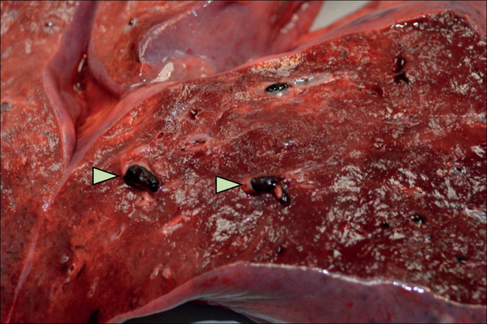
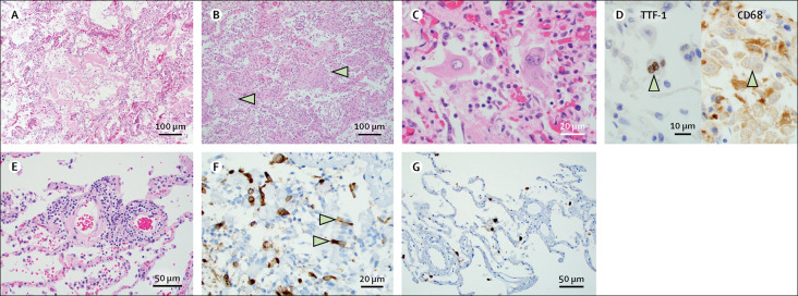
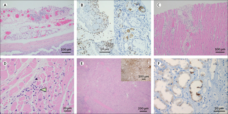
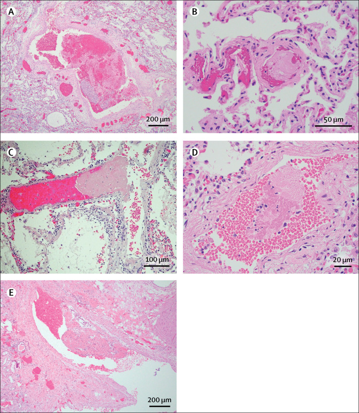
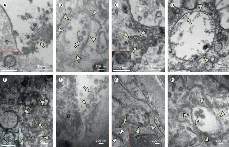
Findings: The median age of our cohort was 73·5 years (range 42-84; IQR 67·5-77·25). All patients had clinically significant comorbidities, the most common being hypertension, chronic kidney disease, obstructive sleep apnoea, and metabolic disease including diabetes and obesity. The major pulmonary finding was diffuse alveolar damage in the acute or organising phases, with five patients showing focal pulmonary microthrombi. Coronavirus-like particles were detected in the respiratory system, kidney, and gastrointestinal tract. Lymphocytic myocarditis was observed in one patient with viral RNA detected in the tissue.
Interpretation: The primary pathology observed in our cohort was diffuse alveolar damage, with virus located in the pneumocytes and tracheal epithelium. Microthrombi, where observed, were scarce and endotheliitis was not identified. Although other non-pulmonary organs showed susceptibility to infection, their contribution to the pathogenesis of SARS-CoV-2 infection requires further examination.
Funding: None.
Copyright © 2020 Elsevier Ltd. All rights reserved.
Figures
Comment in
-
Why misinterpretation of electron micrographs in SARS-CoV-2-infected tissue goes viral.Lancet. 2020 Oct 31;396(10260):e64-e65. doi: 10.1016/S0140-6736(20)32079-1. Epub 2020 Oct 5. Lancet. 2020. PMID: 33031763 Free PMC article. No abstract available.
References
-
- Wu Z, McGoogan JM. Characteristics of and important lessons from the coronavirus disease 2019 (COVID-19) outbreak in China: summary of a report of 72 314 cases from the Chinese Center for Disease Control and Prevention. JAMA. 2020;323:1239–1242. - PubMed
MeSH terms
LinkOut - more resources
Full Text Sources
Research Materials
Miscellaneous
