

Brain abnormalities in COVID-19 acute/subacute phase: A rapid systematic review
- PMID: 32682993
- PMCID: PMC7366124
- DOI: 10.1016/j.bbi.2020.07.014
Brain abnormalities in COVID-19 acute/subacute phase: A rapid systematic review
Abstract
Objective: This systematic review aimed to synthesize early data on typology and topography of brain abnormalities in adults with COVID-19 in acute/subacute phase.
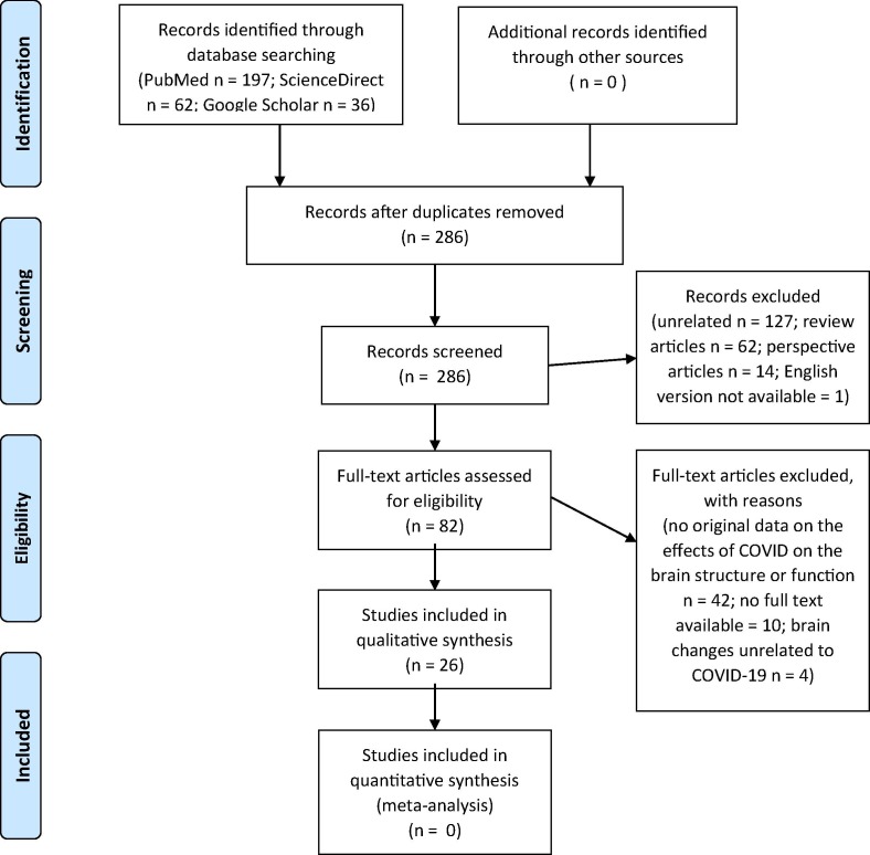
Methods: We performed systematic literature search via PubMed, Google Scholar and ScienceDirect on articles published between January 1 and July 05, 2020, using the following strategy and key words: ((covid[Title/Abstract]) OR (sars-cov-2[Title/Abstract]) OR (coronavirus[Title/Abstract])) AND (brain[Title/Abstract]). A total of 286 non-duplicate matches were screened for original contributions reporting brain imaging data related to SARS-Cov-2 presentation in adults.
Results: The selection criteria were met by 26 articles (including 21 case reports, and 5 cohort studies). The data analysis in a total of 361 patients revealed that brain abnormalities were noted in 124/361 (34%) reviewed cases. Neurologic symptoms were the primary reason for referral for neuroimaging across the studies. Modalities included CT (-angiogram, -perfusion, -venogram), EEG, MRI (-angiogram, functional), and PET. The most frequently reported brain abnormalities were brain white matter (WM) hyperintensities on MRI 66/124 (53% affected cases) and hypodensities on CT (additional 23% affected cases), followed by microhemorrhages, hemorrhages and infarcts, while other types were found in <5% affected cases. WM abnormalities were most frequently noted in bilateral anterior and posterior cerebral WM (50% affected cases).
Conclusion: About a third of acute/subacute COVID-19 patients referred for neuroimaging show brain abnormalities suggestive of COVID-19-related etiology. The predominant neuroimaging features were diffuse cerebral WM hypodensities / hyperintensities attributable to leukoencephalopathy, leukoaraiosis or rarefield WM.
Keywords: CNS; COVID-19; Infection; Leukoaraiosis; Leukoencephalopathy; Microangiopathy; Neurologic; SARS-Cov-2.
Copyright © 2020 Elsevier Inc. All rights reserved.
Figures
References
-
- Coronavirus disease (COVID-19) Pandemic. Geneva: World Health Organization. 2020. https://covid19.who.int/. Accessed July 10, 2020.
-
- Hutton B., Salanti G., Caldwell D.M., et al. The PRISMA extension statement for reporting of systematic reviews incorporating network meta-analyses of health care interventions: checklist and explanations. Ann. Intern. Med. 2015;162:777–784. - PubMed
-
- Moher D., Liberati A., Tetzlaff J., Altman D.G. Preferred reporting items for systematic reviews and meta-analyses: the PRISMA statement. Ann. Intern. Med. 2009;151:264–269. - PubMed
Publication types
MeSH terms
LinkOut - more resources
Full Text Sources
Other Literature Sources
Medical
Miscellaneous
