. 2020 Aug 10;38(36):5803-5813.
doi: 10.1016/j.vaccine.2020.06.049. Epub 2020 Jul 17.

A listeriolysin O subunit vaccine is protective against Listeria monocytogenes

Affiliations

A listeriolysin O subunit vaccine is protective against Listeria monocytogenes

Christopher C Phelps et al. Vaccine. .

Abstract

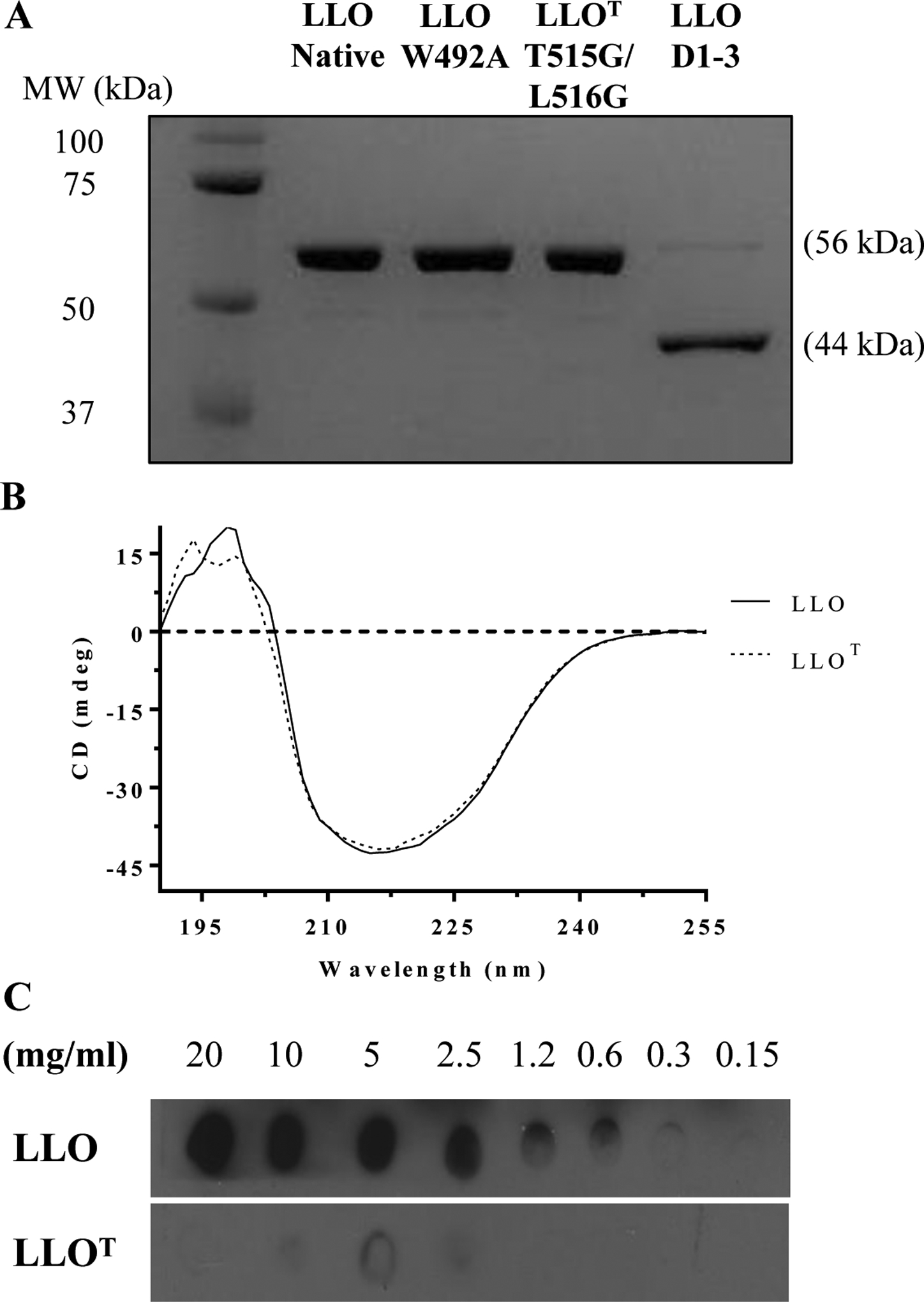
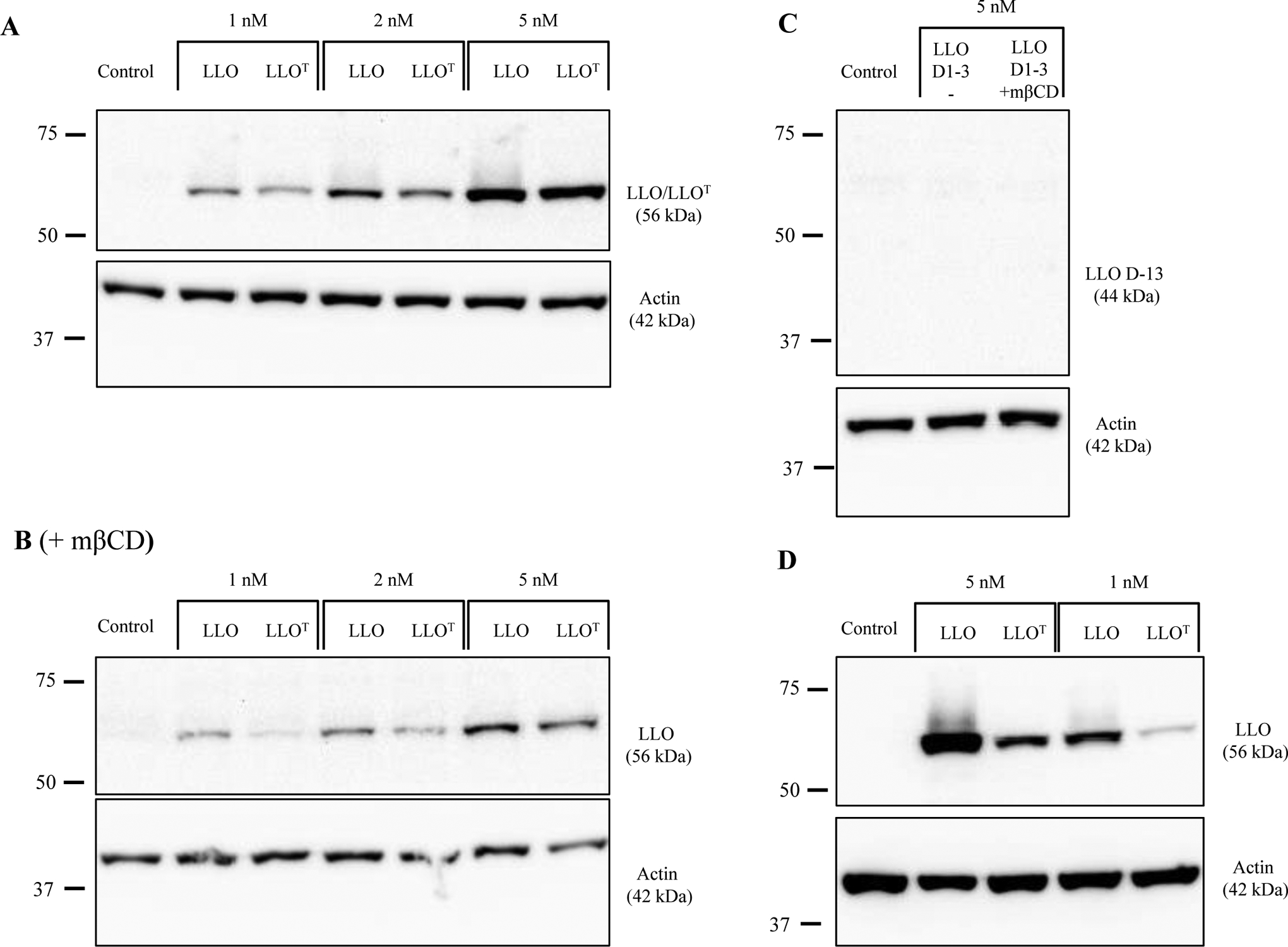
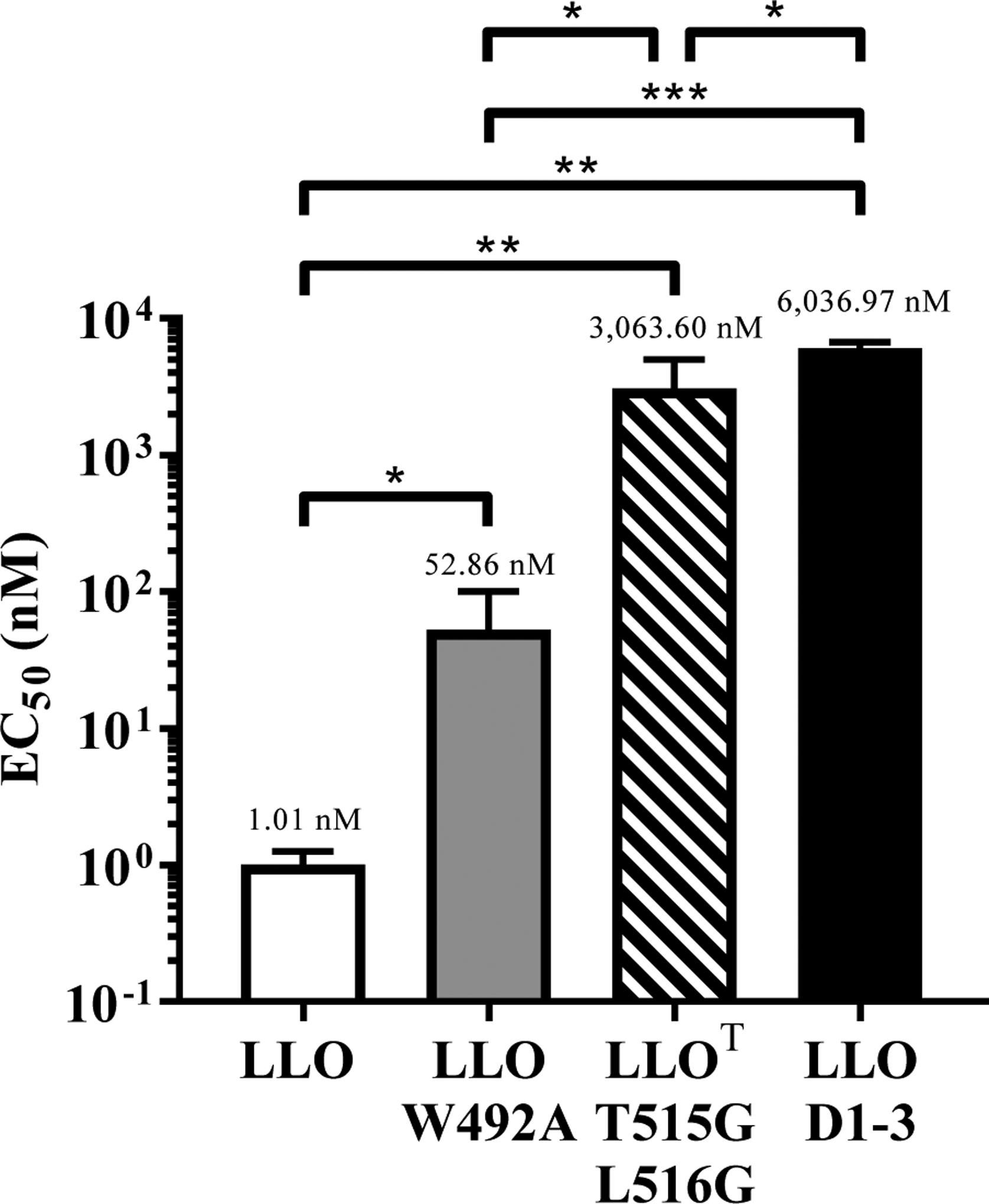
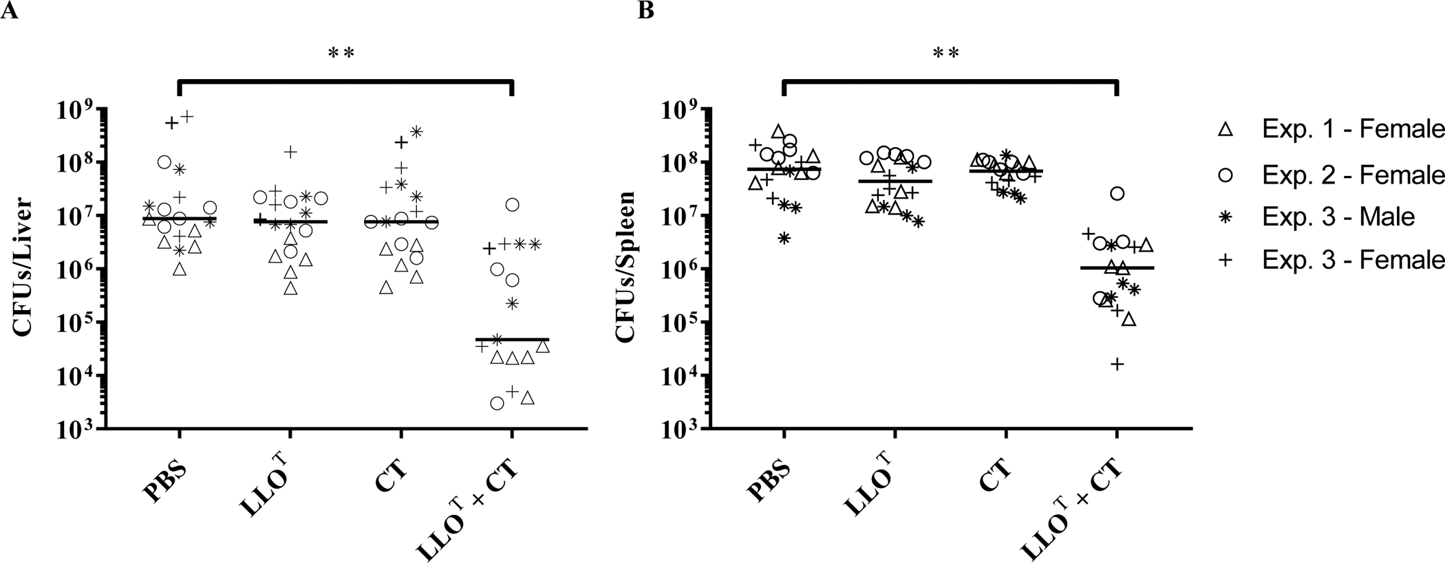
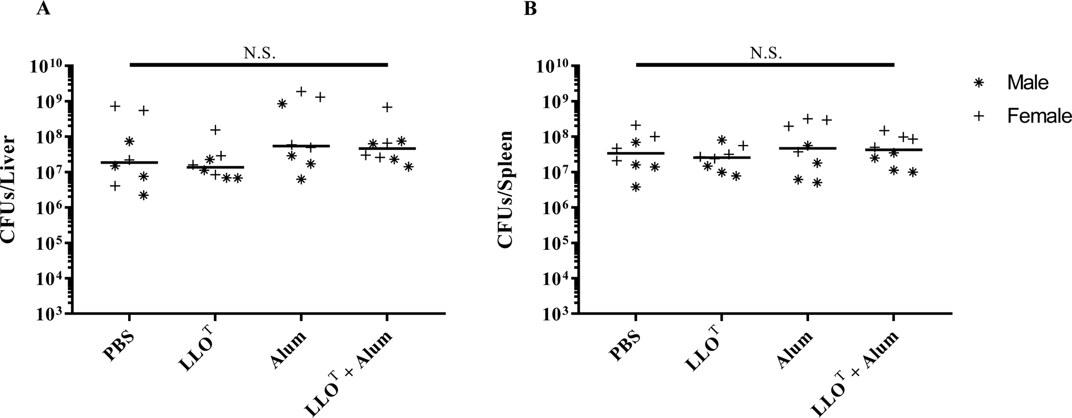
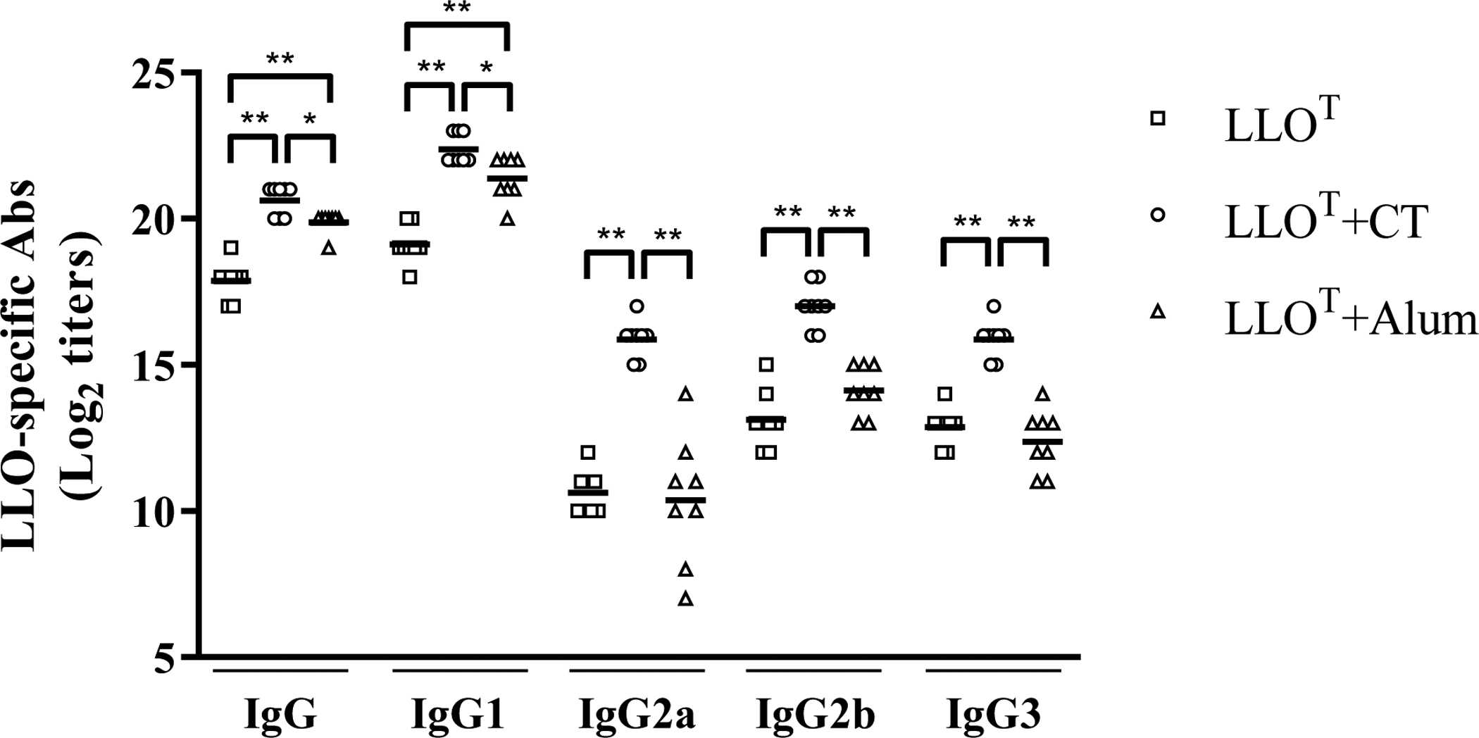
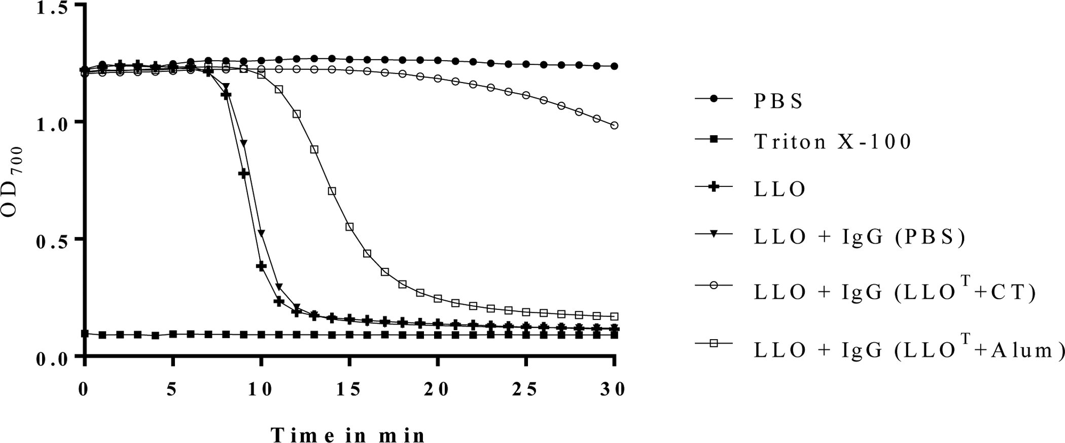
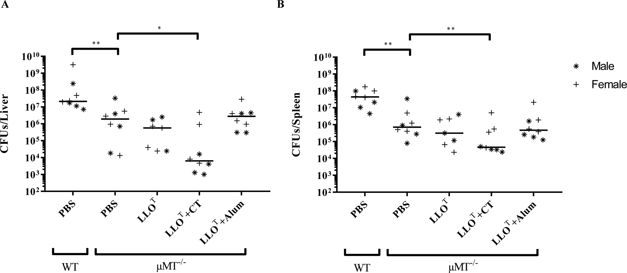
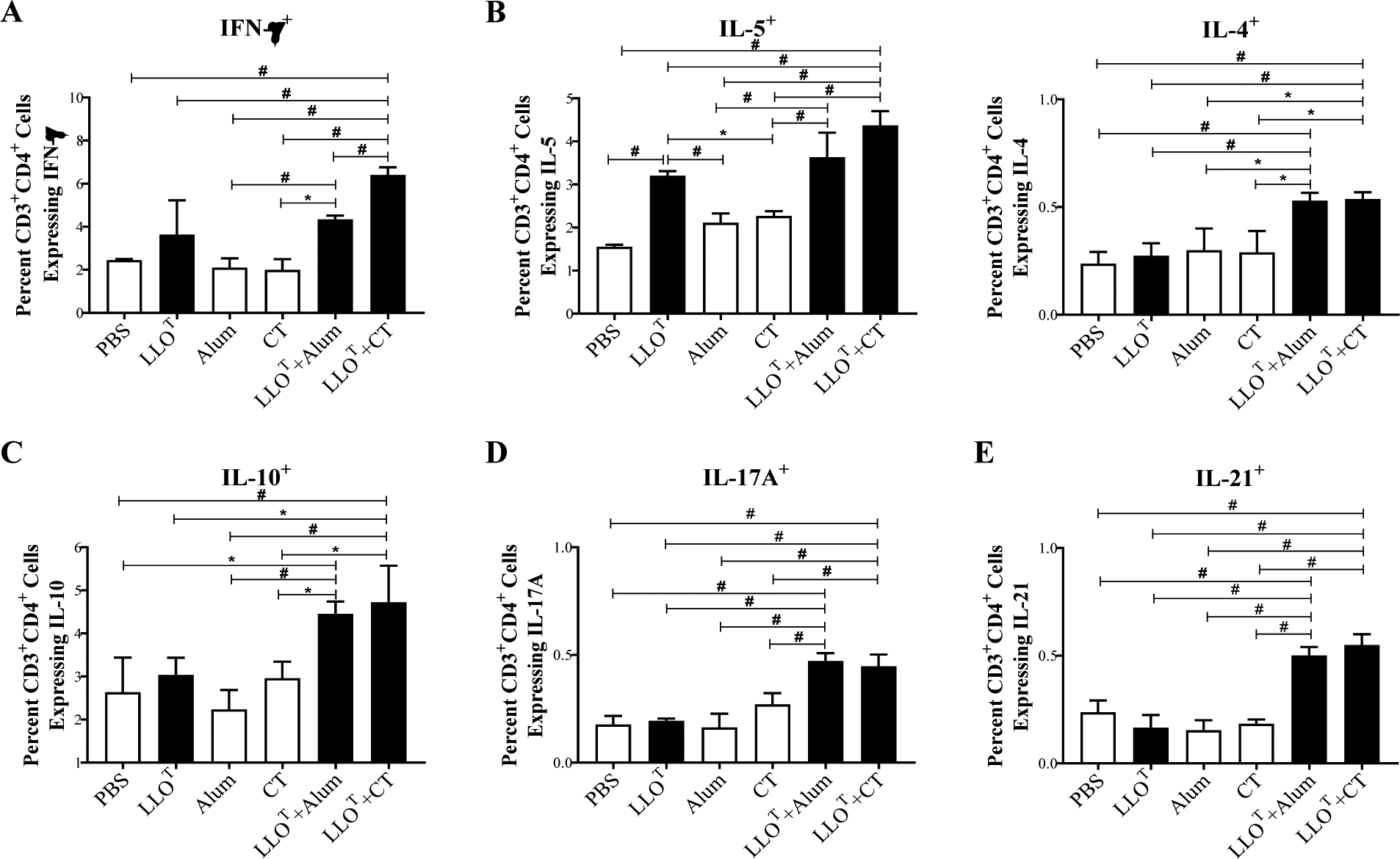
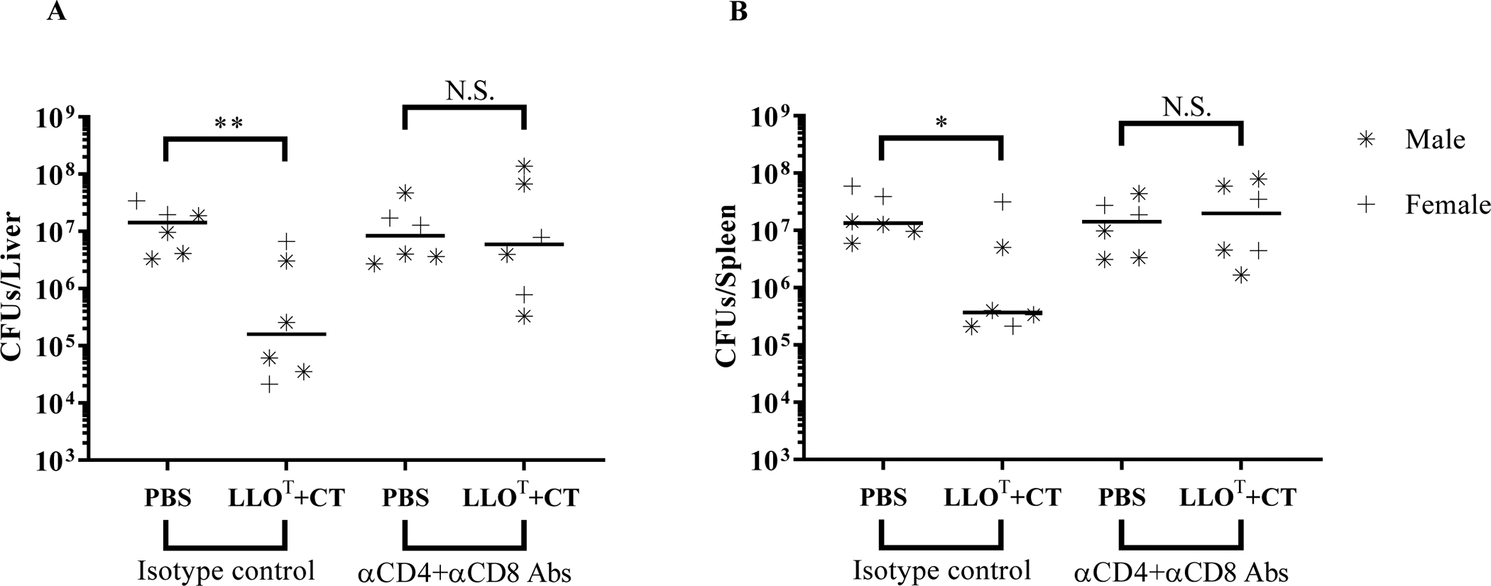
Listeria monocytogenes is a facultative intracellular pathogen responsible for the life-threatening disease listeriosis. The pore-forming toxin listeriolysin O (LLO) is a critical virulence factor that plays a major role in the L. monocytogenes intracellular lifecycle and is indispensable for pathogenesis. LLO is also a dominant antigen for T cells involved in sterilizing immunity and it was proposed that LLO acts as a T cell adjuvant. In this work, we generated a novel full-length LLO toxoid (LLOT) in which the cholesterol-recognition motif, a threonine-leucine pair located at the tip of the LLO C-terminal domain, was substituted with two glycine residues. We showed that LLOT lost its ability to bind cholesterol and to form pores. Importantly, LLOT retained binding to the surface of epithelial cells and macrophages, suggesting that it could efficiently be captured by antigen-presenting cells. We then determined if LLOT can be used as an antigen and adjuvant to protect mice from L. monocytogenes infection. Mice were immunized with LLOT alone or together with cholera toxin or Alum as adjuvants. We found that mice immunized with LLOT alone or in combination with the Th2-inducing adjuvant Alum were not protected against L. monocytogenes. On the other hand, mice immunized with LLOT along with the experimental adjuvant cholera toxin, were protected against L. monocytogenes, as evidenced by a significant decrease in bacterial burden in the liver and spleen three days post-infection. This immunization regimen elicited mixed Th1, Th2, and Th17 responses, as well as the generation of LLO-neutralizing antibodies. Further, we identified T cells as being required for immunization-induced reductions in bacterial burden, whereas B cells were dispensable in our model of non-pregnant young mice. Overall, this work establishes that LLOT is a promising vaccine antigen for the induction of protective immunity against L. monocytogenes by subunit vaccines containing Th1-driving adjuvants.

Keywords: Cancer immunotherapy; Cholesterol-dependent cytolysin; Listeria monocytogenes; Listeriolyin O; Vaccine.


Conflict of interest statement

Declaration of Competing Interest The authors declare that they have no known competing financial interests or personal relationships that could have appeared to influence the work reported in this paper.

Figures

Figure 1.
Figure 1.. LLOT does not bind to cholesterol.
(A) Recombinant LLO, LLOT, LLO W492A, and LLO D1–3 (1 μg) were subjected to SDS-PAGE and stained with Coomassie blue. (B) Representative CD spectra of LLO and LLOT (0.5 mg/ml). (C) LLO and LLOT were incubated on a PVDF membrane pre-coated with a serial dilution of an ethanol-cholesterol solution. Pre-coated membranes were then incubated with LLO or LLOT, and binding to cholesterol was visualized by immunoblot analysis using anti-LLO antibodies.
Figure 2.
Figure 2.. LLOT binds to host cells.
Control HeLa cells (A), and HeLa cells pre-treated with 5 mM methly-β-cyclodextrin (mβCD) (B), were incubated 10 min at 4°C with LLO or LLOT. (C) HeLa cells pre-treated, or not, with 5 mM mβCD were incubated 10 min at 4°C with LLO D1–3. (D) THP-1 cells were incubated 10 min at 4°C with LLO or LLOT. (A, B, C, D) After incubation at 4°C, cells were washed and lysed at 4°C, and cell lysates were analyzed by immunoblotting using anti-LLO and anti-actin primary antibodies. Representative immunoblots were selected from at least 3 independent experiments.
Figure 3:
Figure 3:. LLOT is non-hemolytic.
Hemolytic activity of LLO, LLO W492A, LLOT, and LLO D1–3 was measured from three independent experiments, each performed in duplicate, as EC50, i.e. the toxin/toxoid concentration required to cause 50% hemolysis. Average EC50 is shown above each column. P values were calculated using a two-tailed Student’s t-test (* = P, <0.05; ** = P <0.01; *** = P <0.001).
Figure 4.
Figure 4.. Immunization with LLOT plus cholera toxin protects mice against infection by L. monocytogenes.
Mice were immunized at weekly intervals, for 3 consecutive weeks, by intraperitoneal injection of PBS (negative control), cholera toxin adjuvant (CT, 1 μg), LLOT (20 μg), or LLOT (20 μg) plus cholera toxin (1 μg) (LLOT + CT). On day 28, mice were intravenously inoculated with 2 × 10⁴ L. monocytogenes and sacrificed after 72 h to collect organs and enumerate bacterial colony forming units (CFUs) in the liver (A) and spleen (B). Results are expressed as CFUs/organ and medians are presented. Data are from 3 independent experiments with 5 females per condition in experiment 1, 5 females per condition in experiment 2, and 4 males and 4 females per condition in experiment 3. Statistical significance was calculated using a two-sided Mann-Whitney test, ** P < 0.01.
Figure 5.
Figure 5.. Immunization with LLOT plus Alum does not protect mice against infection by L. monocytogenes.
Mice were immunized at weekly intervals, for 3 consecutive weeks, by intraperitoneal injection of PBS (negative control), LLOT (20 μg), Alum (40 μg), or LLOT (20 μg) plus Alum (40 μg). At day 28, mice were intravenously inoculated with 2 × 10⁴ L. monocytogenes and sacrificed after 72 h to collect organs and enumerate bacterial colony forming units (CFUs) in the liver (A) and spleen (B). Data are from one experiment, including 4 males plus 4 females for each experimental condition. Results are expressed as CFUs per organ and medians are presented. Statistical analysis was made using a two-sided Mann-Whitney test, N.S. = Not statistically significant.
Figure 6.
Figure 6.. LLOT-specific IgG production in mice immunized with LLOT and adjuvants.
The titers of LLOT-specific IgG, IgG1 and IgG2a, IgG2b, and IgG3 were determined by ELISA in serially diluted (1:2) sera from mice immunized with LLOT, LLOT+CT (cholera toxin), or LLOT + Alum. LLO-specific IgGs in sera from mice inoculated with adjuvants alone were similar to naïve mice (data not shown). Antibody titers were determined as the last dilution of sera with an absorbance > 0.1 above that of control sera from naïve mice. Antibody titers are expressed as Log2 values for each mouse with the mean presented. Statistical significance was calculated using a one-way ANOVA, * P < 0.05, ** P < 0.01. N = titers from 8 mice for each group.
Figure 7.
Figure 7.. Immunization with LLOT plus cholera toxin generates LLO-neutralizing antibodies.
IgGs (15 μg/ml) were purified from pooled sera isolated from 8 mice per immunized group: PBS, LLOT + CT adjuvant, or LLOT + Alum adjuvant, and tested for their ability to neutralize 5 nM LLO mixed with human erythrocytes. Erythrocyte lysis of was measured at OD700 at 60 sec intervals for 30 min. As hemolysis negative and positive controls, erythrocytes were incubated with PBS or Triton X-100, respectively. Data are representative of 4 independent experiments.
Figure 8.
Figure 8.. Immunization with LLOT plus cholera toxin is protective in μMT−/− mice that lack mature B cells.
WT and μMT−/− mice (4 males and 4 females per experimental condition, with the exception of the LLOT condition that included 4 females and 3 males, were immunized at weekly intervals for 3 consecutive weeks by intraperitoneal injection of PBS (negative control), LLOT (20 μg), LLOT (20 μg) plus CT adjuvant (1 μg), or LLOT (20 μg) plus Alum adjuvant (40 μg). At day 28, mice were intravenously inoculated with 2 × 10⁴ L. monocytogenes and sacrificed after 72 h to enumerate bacterial colony forming units (CFUs) in the liver (A) and spleen (B). Results are expressed as CFUs per organ and medians are presented. Statistical significance was calculated using a two-sided Mann-Whitney test, * P <0.05, ** P < 0.01.
Figure 9.
Figure 9.. Analysis of T cell responses in the different immunized groups.
Splenocytes from the different immunized groups were isolated at day 38 after initial immunization and cultured 5 days in the presence of 5 μg/ml LLOT. Cultured splenocytes were then stimulated with PMA and ionomycin for 1 h and subsequently treated with Golgistop for 5 h. Splenocytes were then labeled with fluorescently tagged antibodies against CD3, CD4, IFN-γ, IL-5, IL-4, IL-10, IL-17a, and IL-21 and analyzed by flow cytometry. The frequencies of LLOT-specific Th1 (CD3+CD4+IFN-γ+) (A); Th2 (CD3+CD4+IL-5+, CD3+CD4+IL-4+, and CD3+CD4+IL-10+) (B and C); Th17 (CD3+CD4+IL-17A+) (D); and Tfh (CD3+CD4+IL-21+) (E) were expressed as the average percentage of positive cells for indicated cytokines ± standard deviation among the CD3/CD4 double positive cells. Statistical differences were determined by one-way ANOVA and significant differences were considered at: * p ≤ 0.05 and # p ≤ 0.005.
Figure 10.
Figure 10.. T cells are required for the protective immunity of the LLOT+CT immunization group.
Mice were immunized at weekly intervals for 3 consecutive weeks by intraperitoneal injection of PBS (negative control), or LLOT (20 μg) plus CT (1 μg). Mice received 300 μg of CD4 plus CD8 depleting antibodies or 300 μg of corresponding isotype controls on day 26 via intraperitoneal injection (600 μg total). On day 28, mice were intravenously inoculated with 2 × 10⁴ L. monocytogenes. Mice were given a second 100 μg dose of depleting antibodies or isotype control antibodies 24 h post-infection (200 μg total). Mice were sacrificed after 72 h of infection to collect organs and enumerate bacterial colony forming units (CFUs) in the liver (A) and spleen (B). Data are from 1 experiment with 4 males and 2 females per experimental condition. Results are expressed as CFUs per organ and medians are presented. Statistical significance was calculated using a two-sided Mann-Whitney test, N.S. = Not statistically significant, * P < 0.05 and ** P < 0.01.

References

    1. Vazquez-Boland JA, Dominguez-Bernal G, Gonzalez-Zorn B, Kreft J & Goebel W Pathogenicity islands and virulence evolution in Listeria. Microbes Infect 3, 571–584 (2001). - PubMed
    1. Teberg AJ, Yonekura ML, Salminen C & Pavlova Z Clinical manifestations of epidemic neonatal listeriosis. Pediatr Infect Dis J 6, 817–820 (1987). - PubMed
    1. Mylonakis E, Paliou M, Hohmann EL, Calderwood SB & Wing EJ Listeriosis during pregnancy: a case series and review of 222 cases. Medicine (Baltimore) 81, 260–269 (2002). - PubMed
    1. Robbins JR & Bakardjiev AI Pathogens and the placental fortress. Curr Opin Microbiol 15, 36–43, doi:10.1016/j.mib.2011.11.006 (2012). - DOI - PMC - PubMed
    1. Berche P Bacteremia is required for invasion of the murine central nervous system by Listeria monocytogenes. Microb Pathog 18, 323–336, doi:10.1006/mpat.1995.0029 (1995). - DOI - PubMed

Publication types