

Review
doi: 10.1016/j.jcot.2020.05.040.
Epub 2020 Jun 8.
When a volar locking plate is not the right choice in fractures of the distal radius: Case based technical considerations
Affiliations
- PMID: 32684691
- PMCID: PMC7355094
- DOI: 10.1016/j.jcot.2020.05.040
Item in Clipboard
Review
When a volar locking plate is not the right choice in fractures of the distal radius: Case based technical considerations
J Clin Orthop Trauma.
2020 Jul-Aug.
Erratum in
-
Erratum regarding previously published articles.J Clin Orthop Trauma. 2020 Nov-Dec;11(6):1169-1171. doi: 10.1016/j.jcot.2020.09.032. Epub 2020 Sep 26. J Clin Orthop Trauma. 2020. PMID: 33013141 Free PMC article.
-
Erratum regarding previously published articles.J Clin Orthop Trauma. 2021 Aug 5;21:101560. doi: 10.1016/j.jcot.2021.101560. eCollection 2021 Oct. J Clin Orthop Trauma. 2021. PMID: 34414073 Free PMC article.
Abstract
A volar approach is commonly used for fixation of distal radius fractures with a volar locking plate. There are certain fracture patterns for which volar locked plating is not suitable. This case based review outlines such case examples.
Keywords: Distal radius fracture; Dorsal approach; Dorsal rim; Fragment specific fixation; Radiocarpal fracture dislocation; Scaphoid impression.
© 2020 Delhi Orthopedic Association. All rights reserved.
Figures

PA (1a) and Lateral (1b) radiographs show a radiocarpal fracture dislocation with a radial styloid fragment and dorsal rim fracture fragments.

Coronal (2a) and Sagittal CT (2b) show a radial styloid fragment with horizontal fracture line. Dorsal rim fracture fragment with impaction. Seen dorsal to that are small fragments representing capsular avulsion.

Radial styloid and dorsal rim fracture fragments are clearly visualized. Periosteum and capsule has been stripped from the injury itself. Hand is to the right and forearm to the left in this image.

Hand is to the right and forearm to the left. Fixation was started by obtaining provisional fixation of radial styloid fragment with a Kirschner wire. Dorsal rim fragment was dis-impacted, reduced and then held with a carefully contoured fragment specific dorsal plate.
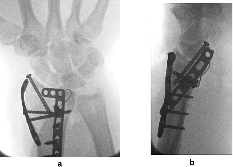
PA (5a) and Lateral (5b) views show fragment specific fixation. Radial column fixation has been achieved using a cannulated screw and a radial column plate. Dorsal rim fragment fixation has been achieved with a buttress plate and supported with cancellous bone chips after dis-impaction. Volar lip fragments have been restored by a tension band construct.

Lateral and PA radiographs after fixation of a radiocarpal fracture dislocation. Bottom views show radiographs after Kirschner wire removal and healing. Two suture anchors were used volarly to obtain fixation of volar radiocarpal ligaments. 1.6mm Kirschner wires were used dorsally to obtain fixation of dorsal rim fragments. Headed cannulated screw (3.0 mm) was used for fixation of the radial column.
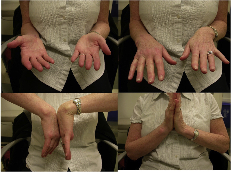
Excellent forearm rotation and slightly diminished wrist flexion-extension arc obtained at 6 months for a patient who sustained radiocarpal fracture dislocation.

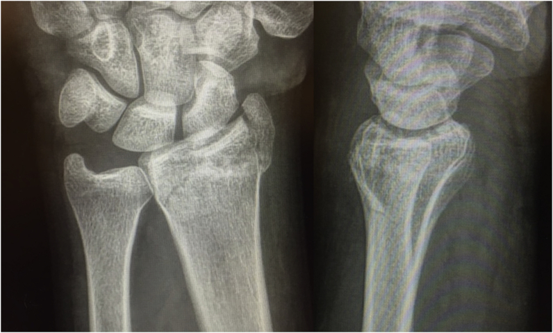
Dorsal shear fracture of distal radius with dorsal subluxation of carpus.

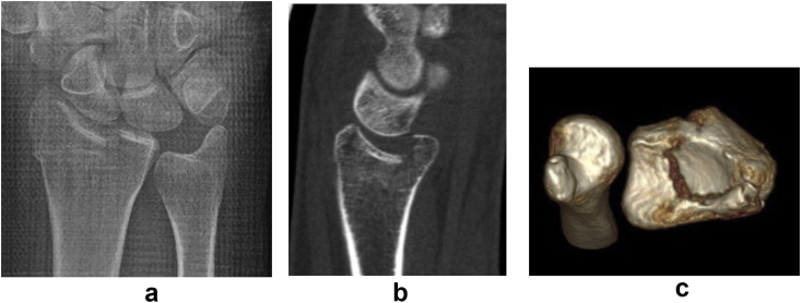
Sagittal, Coronal and 3D CT showing a dorsal shear fracture pattern with dorsal subluxation of carpus. Volar cortex is mostly intact.

Fragment specific fixation using a buttress plate for the dorsal shear component and a cannulated screw for the styloid fragment.

Excellent functional outcome obtained at 9 month follow up.
PA and Lateral radiographs show articular incongruity and comminution of the radial column.

Sagittal and 3D CT show a scaphoid impression fracture with articular depression and dorsal rim fragments.

Well contoured dorsal plate to achieve fixation of the dorsal rim to the shaft and buttresses dorsal rim to the articular fragment after it has been elevated to restore the joint surface. Additional Kirschner wire fixation of radial column comminution is also demonstrated.
PA radiograph (15a) showing a scaphoid impression fracture with joint incongruity. Sagittal CT and 3D formatting (15b,c) show articular depression with fracture of the dorsal rim and comminution of radial styloid.

PA view (16a) showing restoration of joint congruity. Dorsal plate contoured to radius metaphysis achieving fixation of the dorsal rim to shaft. Locking screws are placed through the plate distally to function as rafting screws for the depressed articular fragment (16b).

PA view (17a) shows an isolated volar ulnar corner fracture. Lateral view (17b) shows increase in teardrop angle. Sagittal (17c) and axial (17d) CTs show isolated lunate facet fracture.

Extended carpal tunnel approach is suitable to get exposure of this fracture fragment (18a). Dissection performed with a bump behind the wrist so that tension on flexors and median nerve is reduced (18b). Fixation achieved with a buttress plate and a cannulated screw with washer through the fragment

PA and Lateral radiographs showing restoration of the teardrop angle and radiocarpal alignment.
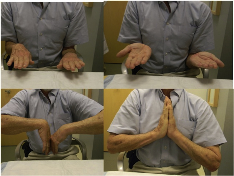
Patient obtained full forearm rotation and slightly restricted wrist flexion-extension arc at short term follow up.

PA and Lateral radiographs showing a tension band construct with a screw used as a post to obtain fixation of small lunate facet fragments. Also seen is fixation of radial styloid and scaphoid fractures.
References
-
- Scheck M. Long-term follow-up of treatment of comminuted fractures of the distal end of the radius by transfixation with Kirschner wires and cast. J Bone Joint Surg Am. 1962 Mar;44-A:337–351. - PubMed
-
- Mandziak D.G., Watts A.C., Bain G.I. Ligament contribution to patterns of articular fractures of the distal radius. J Hand Surg Am. 2011;36(10):1621–1625. - PubMed
Publication types
LinkOut - more resources
Full Text Sources
