. 2020 Jun 24:2020:4243953.
doi: 10.1155/2020/4243953. eCollection 2020.

Mast Cell Activation, Neuroinflammation, and Tight Junction Protein Derangement in Acute Traumatic Brain Injury

Affiliations

Mast Cell Activation, Neuroinflammation, and Tight Junction Protein Derangement in Acute Traumatic Brain Injury

Duraisamy Kempuraj et al. Mediators Inflamm. .

Abstract

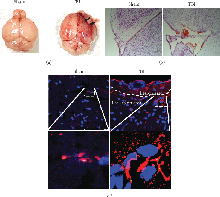
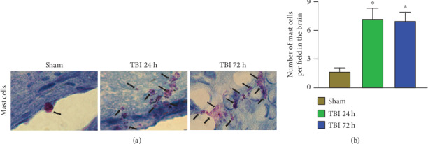
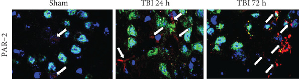
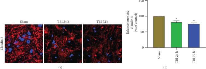
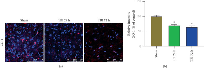
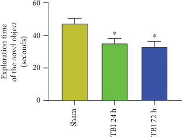
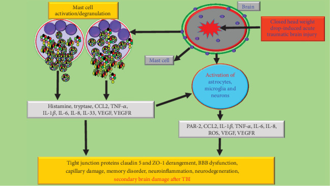
Traumatic brain injury (TBI) is one of the major health problems worldwide that causes death or permanent disability through primary and secondary damages in the brain. TBI causes primary brain damage and activates glial cells and immune and inflammatory cells, including mast cells in the brain associated with neuroinflammatory responses that cause secondary brain damage. Though the survival rate and the neurological deficiencies have shown significant improvement in many TBI patients with newer therapeutic options, the underlying pathophysiology of TBI-mediated neuroinflammation, neurodegeneration, and cognitive dysfunctions is understudied. In this study, we analyzed mast cells and neuroinflammation in weight drop-induced TBI. We analyzed mast cell activation by toluidine blue staining, serum chemokine C-C motif ligand 2 (CCL2) level by enzyme-linked immunosorbent assay (ELISA), and proteinase-activated receptor-2 (PAR-2), a mast cell and inflammation-associated protein, vascular endothelial growth factor receptor 2 (VEGFR2), and blood-brain barrier tight junction-associated claudin 5 and Zonula occludens-1 (ZO-1) protein expression in the brains of TBI mice. Mast cell activation and its numbers increased in the brains of 24 h and 72 h TBI when compared with sham control brains without TBI. Mouse brains after TBI show increased CCL2, PAR-2, and VEGFR2 expression and derangement of claudin 5 and ZO-1 expression as compared with sham control brains. TBI can cause mast cell activation, neuroinflammation, and derangement of tight junction proteins associated with increased BBB permeability. We suggest that inhibition of mast cell activation can suppress neuroimmune responses and glial cell activation-associated neuroinflammation and neurodegeneration in TBI.


Conflict of interest statement

The authors declare that they have no conflict of interest.

Figures

Figure 1
Figure 1
Closed-head weight drop-induced neuroinflammation. Representative images of (a) whole brains and 0.1% crystal violet staining indicate hemorrhage/inflammatory changes (b) in TBI brains as compared to sham control mouse brains (n = 6 mice/group). Representative microphotographs show increased GFAP immunoreactivity indicating an increased number of activated astrocytes in TBI mice (72 h) as compared to sham control mice (c). Image magnifications: (b) 200x and (c) 630x.
Figure 2
Figure 2
Acute TBI increases mast cell number and degranulation in the brain. Mast cell number and its degranulation were evaluated in the frozen sections (20 μm) cut from the brains after 24 h and 72 h of weight drop-induced TBI and sham control mouse brains without TBI (n = 6 mice/group). These brain sections were stained with 0.1% toluidine blue solution for mast cell detection. The number, as well as activation (purple, black arrows) of mast cells, was increased in 24 h and 72 h TBI brains as compared with sham control mouse brains without TBI (a). Note the presence of widespread extracellular granules in degranulated mast cells. Degranulated mast cells also appear irregular in shape. Control mast cells without degranulation did not show extracellular cytoplasmic granules. Photomicrograph original magnifications = 100x. Representative photomicrographs show increased number and activation of mast cells in 24 h and 72 h TBI brains as compared with sham control brains without TBI (a). Photomicrograph original magnifications = 100x. The total number of mast cells was increased in TBI brains as compared to sham control mouse brains (b; ∗p < 0.05, sham control vs. TBI).
Figure 3
Figure 3
Increased level of CCL2 in the brain and serum of acute TBI mice. CCL2 level was quantified by ELISA in the brain tissue lysate and serum of 24 and 72 h acute TBI mice and sham control mice without TBI (n = 3). Results show significantly increased level of CCL2 in the (a) brains and (b) sera of both 24 h and 72 h acute TBI as compared with sham control mice (∗p < 0.05, sham control vs. TBI).
Figure 4
Figure 4
Acute TBI increases PAR-2 expression in the brain. PAR-2 expression was analyzed in the frozen sections (20 μm) from the brains of 24 h and 72 h weight drop model of TBI and sham control mouse brains without TBI (n = 3 mice/group). We analyzed the expression of PAR-2 (red color) for inflammation and NeuN (green color) for neurons in these brain sections by triple immunofluorescence staining. Cellular nuclei were stained with DAPI (blue color). Representative photomicrographs show increased PAR-2 expression (red color, white arrows) in 24 h and 72 h acute TBI brains as compared with sham control mouse brains without TBI. Photomicrograph original magnifications = 630x.
Figure 5
Figure 5
Acute TBI increases VEGFR2 expression in the brain. VEGFR2 expression was analyzed in the frozen sections (20 μm) cut from the brains after 24 h and 72 h of weight drop model of TBI and sham control mouse brains without TBI (n = 3 mice/group) by immunofluorescence staining. Representative images and immunoreactivity intensity bar graphs show increased VEGFR2 expression (red color) in 24 h and 72 h acute TBI brains as compared with sham control brains without TBI (∗p < 0.05, sham control vs. TBI). The nuclei were stained with DAPI (blue color). Photomicrograph original magnifications = 630x.
Figure 6
Figure 6
Acute TBI affects claudin 5 expression in the brain. Claudin 5 expression was analyzed in the frozen sections (20 μm) of the brains after 24 h and 72 h of weight drop-induced TBI and sham control mouse brains without TBI (n = 3 mice/group) by immunofluorescent staining. Representative images and immunoreactivity intensity bar graphs show decreased/derangement of claudin 5 (red color, white arrows) expression in 24 h and 72 h acute TBI brains as compared with sham control mouse brains without TBI (∗p < 0.05, sham control vs. TBI). The nuclei were stained with DAPI (blue color). Photomicrograph original magnifications = 630x.
Figure 7
Figure 7
Acute TBI affects ZO-1 expression in the brain. ZO-1 expression was analyzed in the frozen sections (20 μm) of the brains after 24 h and 72 h of weight drop-induced TBI and sham control mouse brains without TBI (n = 3 mice/group). Representative images and immunoreactivity intensity bar graphs show the derangement of ZO-1 expression (red color) in 24 h and 72 h acute TBI brains as compared with sham control brains without TBI (∗p < 0.05, sham control vs. TBI). The cellular nuclei were stained with DAPI (blue color). Photomicrograph original magnifications = 400x.
Figure 8
Figure 8
NOR test was conducted to assess memory function in TBI mice. The mouse was exposed to two similar objects (A, A) to familiarize in an open field arena box apparatus for 5 min a day before TBI procedures (n = 6 mice/group). The time spent near the objects was recorded. Then, the mouse was exposed to one familiar object (A) and a novel object (B) for 5 min, and the time spent at each object was recorded after 24 h and 72 h of TBI procedure. Results show that sham control mice show increased time spent at the novel object than at the familiar object (∗p < 0.05). However, TBI mice show poor performance as they did not recognize the familiar object, and the time spent at the familiar object and novel object did not show any significant variation.
Figure 9
Figure 9
Schematic representation of mast cell activation in neuroinflammation, BBB disruption, and neuronal death in closed-head acute TBI brain. Neurotrauma/TBI can cause primary damage to neurovascular and gliovascular units in the brain with subsequent neuroimmune and neuroinflammatory responses. This leads to the activation of immune cells, including microglia, astrocytes, and mast cells in the brain. Mast cell activation causes degranulation and release of prestored and preactivated mediators such as histamine, proteases, and TNF-α followed by newly synthesized cytokines, chemokines, and neurotoxic mediators that can act on glial cells and neurons. Subsequently, peripheral immune as well as inflammatory cells can infiltrate the region of injury in the brain due to the BBB breach. CCL2 released from mast cells and brain cells induces the infiltration of inflammatory cells. Activated glial cells release neuroinflammatory mediators that further activate glial cells, neurons, and mast cells in a vicious fashion and induce PAR-2 and VEGFR2 expression and BBB breach with decreased tight junction protein such as claudin 5 and ZO-1 and its derangements. This continuous process can cause and upregulate neuroinflammation and neuronal death and secondary brain damage after TBI.

References

    1. Ma X., Aravind A., Pfister B. J., Chandra N., Haorah J. Animal models of traumatic brain injury and assessment of injury severity. Molecular Neurobiology. 2019;56(8):5332–5345. doi: 10.1007/s12035-018-1454-5. - DOI - PubMed
    1. Ladak A. A., Enam S. A., Ibrahim M. T. A review of the molecular mechanisms of traumatic brain injury. World Neurosurgery. 2019;131:126–132. doi: 10.1016/j.wneu.2019.07.039. - DOI - PubMed
    1. Khellaf A., Khan D. Z., Helmy A. Recent advances in traumatic brain injury. Journal of Neurology. 2019;266(11):2878–2889. doi: 10.1007/s00415-019-09541-4. - DOI - PMC - PubMed
    1. Zysk M., Clausen F., Aguilar X., Sehlin D., Syvanen S., Erlandsson A. Long-term effects of traumatic brain injury in a mouse model of Alzheimer’s disease. Journal of Alzheimer's Disease. 2019;72(1):161–180. doi: 10.3233/JAD-190572. - DOI - PMC - PubMed
    1. Kempuraj D., Ahmed M. E., Selvakumar G. P., et al. Brain injury-mediated neuroinflammatory response and Alzheimer’s disease. The Neuroscientist. 2020;26:134–155. doi: 10.1177/1073858419848293. - DOI - PMC - PubMed