Review
. 2020 Jun;16(2):200001.
doi: 10.1183/20734735.0001-2020.

Pulmonary alveolarproteinosis in children

Affiliations
Review

Pulmonary alveolarproteinosis in children

Andrew Bush et al. Breathe (Sheff). 2020 Jun.

Abstract

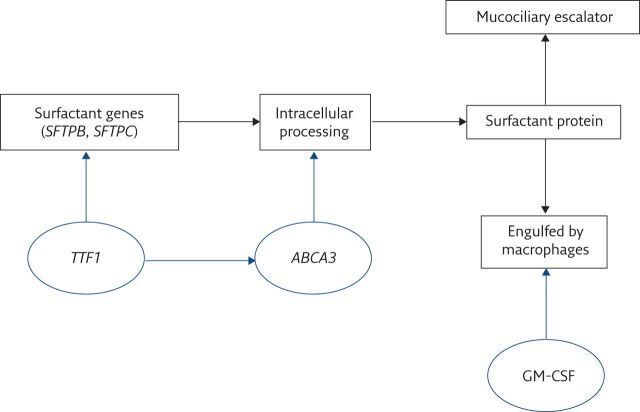
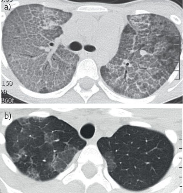
Pulmonary alveolar proteinosis (PAP) is an umbrella term for a wide spectrum of conditions that have a very characteristic appearance on computed tomography. There is outlining of the secondary pulmonary lobules on the background of ground-glass shadowing and pathologically, filling of the alveolar spaces with normal or abnormal surfactant. PAP is rare and the common causes in children are very different from those seen in adults; autoimmune PAP is rare and macrophage blockade not described in children. There are many genetic causes of PAP, the best known of which are mutations in the genes encoding surfactant protein (SP)-B, SP-C, thyroid transcription factor 1, ATP-binding cassette protein 3, and the granulocyte-macrophage colony-stimulating factor (GM-CSF) receptor α- and β- chains. PAP may also be a manifestation of rheumatological and metabolic disease, congenital immunodeficiency, and haematological malignancy. Precise diagnosis of the underlying cause is essential in planning treatment, as well as for genetic counselling. The evidence base for treatment is poor. Some forms of PAP respond well to whole-lung lavage, and autoimmune PAP, which is much commoner in adults, responds to inhaled or subcutaneous GM-CSF. Emerging therapies based on studies in murine models of PAP include stem-cell transplantation for GM-CSF receptor mutations.

Educational aims: To understand when to suspect that a child has pulmonary alveolar proteinosis (PAP) and how to confirm that this is the cause of the presentation.To show that PAP is an umbrella term for conditions characterised by alveolar filling by normal or abnormal surfactant, and that this term is the start, not the end, of the diagnostic journey.To review the developmental differences in the spectrum of conditions that may cause PAP, and specifically to understand the differences between causes in adults and children.To discuss when to treat PAP with whole-lung lavage and/or granulocyte-macrophage colony-stimulating factor, and review potential promising new therapies.


Conflict of interest statement

Conflict of interest: A. Bush has nothing to disclose. Conflict of interest: R. Pabary has nothing to disclose.

Figures

Figure 1
Figure 1
Pathways of surfactant processing. TTF1 is the transcription factor regulating SP-B and SP-C synthesis; ABCA3 is crucial for post-translational, intracellular surfactant processing; granulocyte–macrophage colony-stimulating factor (GM-CSF) regulates surfactant catabolism. The mucociliary escalator is not clinically important in surfactant clearance.
Figure 2
Figure 2
a) Typical computed tomography appearances of PAP in a teenage girl. There is septal thickening outlining the secondary pulmonary lobules on a ground-glass background, giving the typical cobblestoning appearance. The underlying diagnosis was autoimmune PAP. b) Same child after whole-lung lavage(Cliff Morgan, Royal Brompton Hospital, London, UK). There is extensive clearing of the alveolar filling.
Figure 3
Figure 3
Whole-lung lavage in a 2-year old girl with PAP due to a homozygous CSR2RB mutation. a) Chest radiograph prior to lavage. There is diffuse, bilateral nonspecific shadowing. The appearances are not specific for PAP. b) Three-dimensional model of trachea with two endotracheal tubes in situ. The model, printed from HRCT images, allows pre-procedure planning of ventilation, bronchial blocker and lavage strategy in patients in whom a double-lumen tube cannot be passed. c) Schematic diagram of the semiautomated circuit used for whole-lung lavage in smaller patients. d) Bronchoscopic view of the bronchial blocker in the left main bronchus, prior to instilling lavage fluid into the left lung. e) Whole-lung lavage. The bronchoscopist is carefully checking the position of the bronchial blocker, while a second operator is, in this case, using a syringe to perform the lavage. Note the creamy-coloured fluid in the aspirating syringe, typical of PAP. f) Serial aliquots of lavage fluid. Note that as the lavage has proceeded, the fluid becomes clearer. g) Chest radiograph following right sided lung lavage in the same child as in part a. There is substantial clearing of the changes in the right middle and lower lobes; the right upper lobe could not be lavaged because it was impossible to obtain a stable occlusion position in the right main bronchus and the bronchial blocker was placed in the right bronchus intermedius.

References

    1. Price A, Manson D, Cutz E, et al. . Pulmonary alveolar proteinosis associated with anti-GM-CSF antibodies in a child: successful treatment with inhaled GM-CSF. Pediatr Pulmonol 2006; 41: 367–370. doi:10.1002/ppul.20347 - DOI - PubMed
    1. Griese M, Bonella F, Cosyabel U, et al. . Quantitative lipidomics in pulmonary alveolar proteinosis. Am J Respir Crit Care Med 2019; 200: 881–887. doi:10.1164/rccm.201901-0086OC - DOI - PubMed
    1. Griese M. Pulmonary alveolar proteinosis: a comprehensive clinical perspective. Pediatrics 2017; 140: e20170610. doi:10.1542/peds.2017-0610 - DOI - PubMed
    1. Nishino M, Medoff BD, Mark EJ, et al. . Variant alveolar lipoproteinosis: a syndrome with distinct clinical and pathological features. Pathol Int 2011; 61: 509–517. doi:10.1111/j.1440-1827.2011.02710.x - DOI - PubMed
    1. Nathan N, Giraud V, Picard C, et al. . Germline SFTPA1 mutation in familial idiopathic interstitial pneumonia and lung cancer. Hum Mol Genet 2016; 25: 1457–1467. doi:10.1093/hmg/ddw014 - DOI - PubMed

LinkOut - more resources