

Case Reports
doi: 10.1155/2020/9260318.
eCollection 2020.
A Case of Gastric Intramural Hematoma Caused by Anisakis Infection
Affiliations
- PMID: 32685220
- PMCID: PMC7336193
- DOI: 10.1155/2020/9260318
Item in Clipboard
Case Reports
A Case of Gastric Intramural Hematoma Caused by Anisakis Infection
Case Rep Gastrointest Med.
.
Abstract
A sixty-year-old lady admitted complaining of epigastric pain and hematemesis. On admission, esophagogastroduodenoscopic examination revealed ruptured intramural hematoma on the antrum of stomach. Eight days later, follow-up EGD showed improving ruptured intramural hematoma and one anisakis larva. Therefore, the gastric intramural hematoma was considered to be caused by anisakis infection. She recovered after ten days of conservative treatment.
Copyright © 2020 Sang Jin Lee.
Conflict of interest statement
The authors declare that they have no conflicts of interest.
Figures
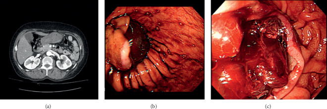
(a) Abdominal computed tomography scan showing about 8 × 3 cm nonenhancing mass (50∼60 Hounsfield unit) in the stomach antrum and lower body, suggesting submucosal hematoma. (b) Gastroscopy showing ruptured intramural hematoma on the anterior wall of antrum and low body. (c) Gastroscopy showing ruptured mucosa and hematoma in inside the mucosa wall.
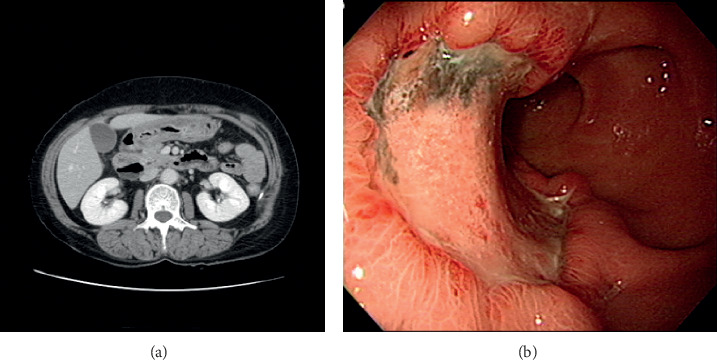
(a) Significant decreased size of hematoma in the stomach antrum and lower body compared with previous computed tomography. No visible active bleeding in stomach is observed. (b) Gastroscopy showing active ulcer on the anterior wall of the antrum and low body.
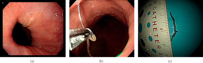
(a) Gastroscopy showing whitish thread-like worm on gastroesophageal junction. (b) Gastroscopic finding: the whitish thread-like worm is being removed by using a biopsy forcep. (c) Gross finding showing 2 cm whitish thread-like worm, identified as anisakis larva.
Similar articles
-
A Rare Case of Traumatic Colonic Intramural Hematoma in Saudi Arabia.Cureus. 2024 Jan 1;16(1):e51461. doi: 10.7759/cureus.51461. eCollection 2024 Jan. Cureus. 2024. PMID: 38169609 Free PMC article.
-
Anisakiasis: Report of 15 Gastric Cases Caused by Anisakis Type I Larvae and a Brief Review of Korean Anisakiasis Cases.Korean J Parasitol. 2015 Aug;53(4):465-70. doi: 10.3347/kjp.2015.53.4.465. Epub 2015 Aug 25. Korean J Parasitol. 2015. PMID: 26323845 Free PMC article.
-
Case Report: Gastric intramural hematoma with acute upper gastrointestinal bleeding in a child.Front Pediatr. 2025 May 19;13:1533324. doi: 10.3389/fped.2025.1533324. eCollection 2025. Front Pediatr. 2025. PMID: 40458449 Free PMC article.
-
A case of spontaneous intramural hematoma of the esophagus.Gastroenterol Jpn. 1993 Feb;28(1):81-7. doi: 10.1007/BF02775007. Gastroenterol Jpn. 1993. PMID: 8440426 Review.
-
Coumarin-induced intramural hematoma of the duodenum: case report and review of the literature.Scand J Gastroenterol. 2011 Mar;46(3):376-9. doi: 10.3109/00365521.2010.531484. Epub 2010 Nov 14. Scand J Gastroenterol. 2011. PMID: 21073371 Review.
Cited by
-
A Rare Case of Traumatic Colonic Intramural Hematoma in Saudi Arabia.Cureus. 2024 Jan 1;16(1):e51461. doi: 10.7759/cureus.51461. eCollection 2024 Jan. Cureus. 2024. PMID: 38169609 Free PMC article.
-
A Rare Case of Gastric Intramural Hematoma Secondary to Hemorrhagic Pancreatitis.Cureus. 2023 Sep 11;15(9):e45039. doi: 10.7759/cureus.45039. eCollection 2023 Sep. Cureus. 2023. PMID: 37701159 Free PMC article.
-
Spontaneous isolated gastric intramural hematoma combined with spontaneous superior mesenteric artery intermural hematoma: a rare case.BMC Geriatr. 2024 Apr 23;24(1):360. doi: 10.1186/s12877-024-04991-6. BMC Geriatr. 2024. PMID: 38654207 Free PMC article.
-
A Rare Case of Gastric Intramural Hematoma in Recurrent Leukemia.Cureus. 2022 Jan 18;14(1):e21385. doi: 10.7759/cureus.21385. eCollection 2022 Jan. Cureus. 2022. PMID: 35198296 Free PMC article.
References
-
- Lee W. H., Yoo S. S., Kim H. J., Kim T. H., Lee O. J. Endoscopic and clinical characteristics of gastrointestinal parasite infections. Clin Endosc. 2007;35(5):304–312.
Publication types
LinkOut - more resources
Full Text Sources
