. 2020 Jun 17:2020:4701563.
doi: 10.1155/2020/4701563. eCollection 2020.

Pathophysiological Changes in Female Rats with Estrous Cycle Disorder Induced by Long-Term Heat Stress

Affiliations

Pathophysiological Changes in Female Rats with Estrous Cycle Disorder Induced by Long-Term Heat Stress

GaiHong An et al. Biomed Res Int. .

Abstract

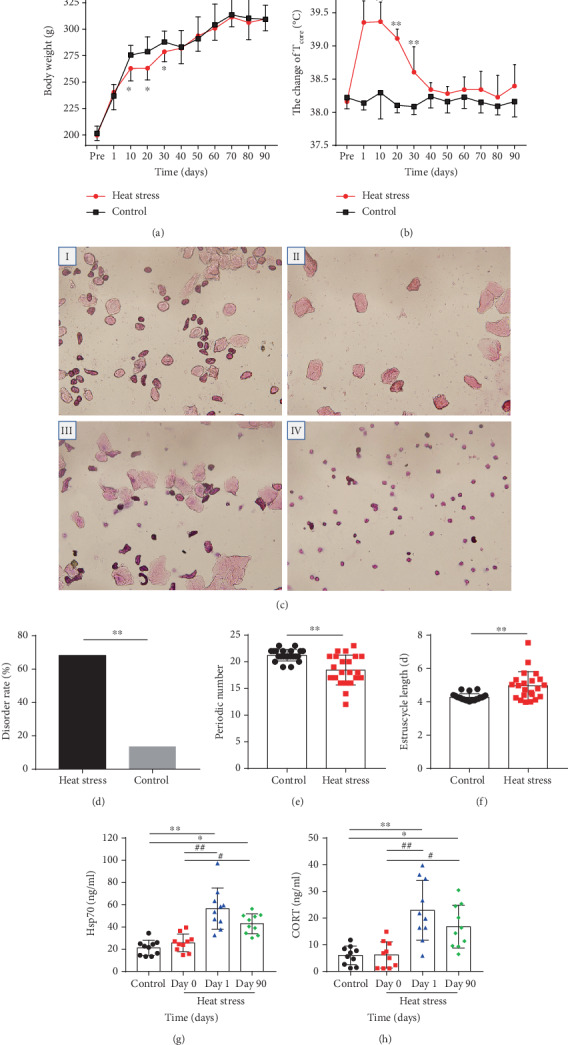
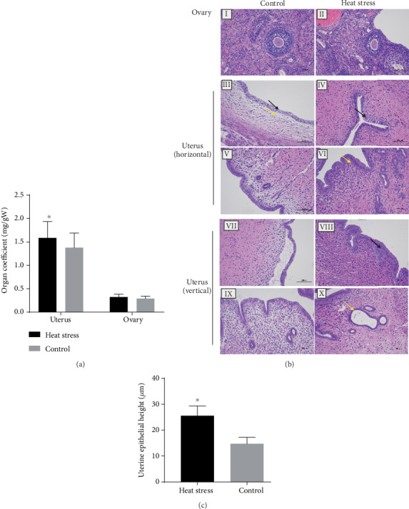
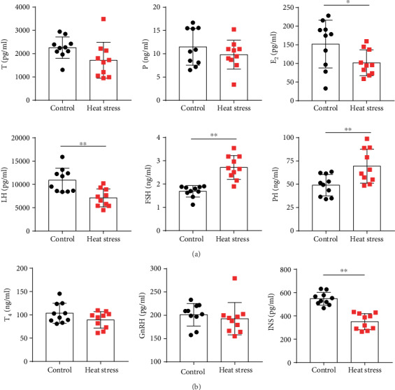
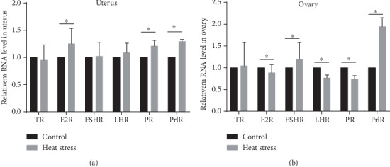
High-temperature exposure is detrimental to women's reproductive health; however, the impact caused by long-term high temperature is not comprehensive, and a stable model of estrous cycle disorder induced by a high temperature is yet lacking. Herein, we aimed to establish a stable and effective model of estrous cycle disorder in female rats induced by long-term heat stress to study its physiological and pathological characteristics and explore the underlying mechanism. In the present study, female Sprague-Dawley rats with normal estrous cycles were exposed to the temperature of 38 ± 0.5°C, relative humidity (RH) of 55 ± 5% (2 h/d, 1 time/d) hot cabin at more than 90 days. Consequently, after long-term heat stress, no difference was detected in body weight and rectal temperature, but the estrus cycle was prolonged, the uterine organ index was increased, pathological changes occurred, the increase latitude of stress hormones heat shock protein 70 (Hsp70) and corticosterone (CORT) decreased, estradiol (E2) and luteinizing hormone (LH) levels decreased, follicle stimulating hormone (FSH) and prolactin (Prl) levels increased, gonadotropin-releasing hormone (GnRH) and thyroid hormone (T4) showed no difference, and insulin (INS) decreased significantly. Moreover, the mRNA expression of the sex hormone receptor in the uterus and ovary was altered. Therefore, the estrous cycle disorder in female rats can be induced by regular heat stress for 90 days, which can be considered the pioneer method. Subsequently, prominent physiological and pathological characteristics and disruption in the hypothalamic-pituitary-gonadal (HPG) axis were noted.


Conflict of interest statement

The authors declare no conflicts of interest.

Figures

Figure 1
Figure 1
Estrous cycle disorder model in female animals induced by high temperature. (a) Change in body weight of female rats in heat stress (n = 10). The body weight of the rats was measured every 10 days. (b) Effects of heat exposure on Tcore (n = 10). The Tcore of the two groups of rats was determined in the rectum at 5 cm by an animal rectal thermometer every 10 days. (c) Microphotographs (10 × 40) of cellular characteristics for the identification of the estrus stage. Proestrus smear mainly consisted of nucleated epithelial cells (I); an estrus smear primarily consisted of anucleated cornified cells (II); a metestrus smear consisted of the same proportion among leukocytes anucleated cornified cells, and nucleated epithelial cells (III); and a diestrus smear primarily consisted of leukocytes (IV). (d) Effects of heat exposure on cumulative estrous cycle disorder rate (n = 22). (e) Effects of heat exposure on periodic numbers (n = 22). (f) Effects of heat exposure on estrus cycle duration (n = 22). (g) The content of serum Hsp70 in the heat stress group (n = 10). (h) The content of serum CORT in the heat stress group (n = 10). The protein concentration of serum Hsp70 and CORT was detected by ELISA. The blood samples from the medial canthus of two groups' rats were collected at day 0, day 1, and day 90. Values were presented as mean ± SEM. P < 0.05 and ∗∗P < 0.01vs. the control group, #P < 0.05 and ##P < 0.01vs. the heat stress group (day 0), and P < 0.05 and △△P < 0.01vs. the heat stress group (day 90).
Figure 2
Figure 2
Long-term heat stress decreased the organ index of the uterus and affected the histopathology of the ovary. (a) Effect of heat exposure on the indexes of reproductive organs (n = 10). (b) Photomicrographs of the rat uterus and ovary by H&E staining (n = 3). The rat ovary (I), uterus (horizontal III and V; vertical VII and IX) in the control group. The rat ovary (II) and uterus (horizontal IV and VI vertical: VII and X) of the heat stress group. Scale bar = 100 μm. (c) Graphical representation of uterine epithelial height (n = 10). Values are presented as the mean value ± SEM. P < 0.05vs. the control group.
Figure 3
Figure 3
Long-term heat stress disrupted the levels of sex hormones and neuroendocrine hormones in female rats as assessed by ELISA (n = 10). (a) The content of serum T, P, E2, LH, FSH, and Prl in the two groups of rats. (b) The content of serum T4, GnRH, and INS in the two groups of rats. Values were presented as mean ± SEM. P < 0.05 and ∗∗P < 0.01vs. the control group.
Figure 4
Figure 4
Expression of TR, ER-α, FSHR, LHR, PR, and PrlR genes in the reproductive organs of the two groups rats (n = 10). (a) Gene expression of sex hormone receptors in the uterus. (b) Gene expression of sex hormone receptors in the ovary. The level was represented as the mean value ± SEM. P < 0.05vs. the control group.

Similar articles

Cited by

References

    1. Aroyo A., Yavin S., Arav A., Roth Z. Maternal hyperthermia disrupts developmental competence of follicle-enclosed oocytes: in vivo and ex vivo studies in mice. Theriogenology. 2007;67(5):1013–1021. doi: 10.1016/j.theriogenology.2006.12.001. - DOI - PubMed
    1. Bale T. L., Epperson C. N. Sex differences and stress across the lifespan. Nature Neuroscience. 2015;18(10):1413–1420. doi: 10.1038/nn.4112. - DOI - PMC - PubMed
    1. Viau V., Meaney M. J. Variations in the hypothalamic-pituitary-adrenal response to stress during the estrous cycle in the rat. Endocrinology. 1991;129(5):2503–2511. doi: 10.1210/endo-129-5-2503. - DOI - PubMed
    1. Sils I. V., Matthew C. B., Bastille A. M. Estrus related differences in response to a hot environment in telemetry- equipped female rats. Journal of Thermal Biology. 2002;27(4):279–284. doi: 10.1016/S0306-4565(01)00020-1. - DOI
    1. Derveaux S., Vandesompele J., Hellemans J. How to do successful gene expression analysis using real-time PCR. Methods. 2010;50(4):227–230. doi: 10.1016/j.ymeth.2009.11.001. - DOI - PubMed

Substances

LinkOut - more resources