. 2020 Jun 30;5(27):16701-16710.
doi: 10.1021/acsomega.0c01582. eCollection 2020 Jul 14.

Zeta Potential of Extracellular Vesicles: Toward Understanding the Attributes that Determine Colloidal Stability

Affiliations

Zeta Potential of Extracellular Vesicles: Toward Understanding the Attributes that Determine Colloidal Stability

Getnet Midekessa et al. ACS Omega. .

Abstract

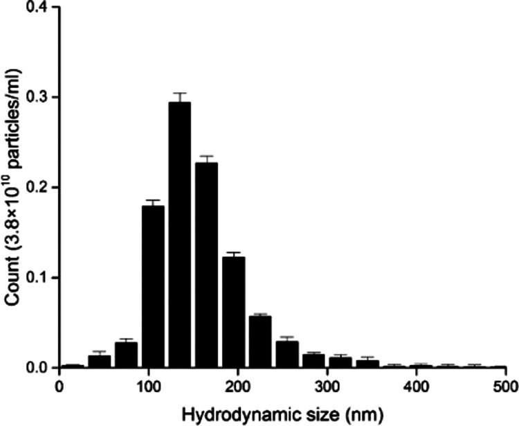
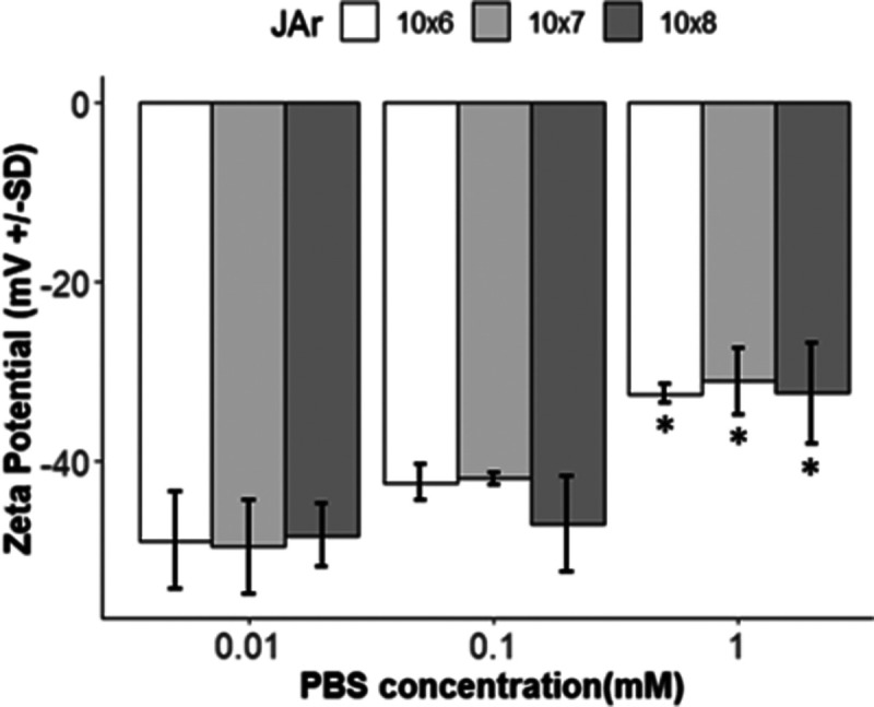
Extracellular vesicles (EVs), including exosomes and microvesicles (<200 nm), play a vital role in intercellular communication and carry a net negative surface charge under physiological conditions. Zeta potential (ZP) is a popular method to measure the surface potential of EVs, while used as an indicator of surface charge, and colloidal stability influenced by surface chemistry, bioconjugation, and the theoretical model applied. Here, we investigated the effects of such factors on ZP of well-characterized EVs derived from the human choriocarcinoma JAr cells. The EVs were suspended in phosphate-buffered saline (PBS) of various phosphate ionic concentrations (0.01, 0.1, and 1 mM), with or without detergent (Tween-20), or in the presence (10 mM) of different salts (NaCl, KCl, CaCl2, and AlCl3) and at different pH values (4, 7, and 10) while the ZP was measured. The ZP changed inversely with the buffer concentration, while Tween-20 caused a significant (p < 0.05) lowering of the ZP. Moreover, the ZP was significantly (p < 0.05) less negative in the presence of ions with higher valency (Al3+/Ca2+) than in the presence of monovalent ones (Na+/K+). Besides, the ZP of EVs became less negative at acidic pH, and vice versa. The integrated data underpins the crucial role of physicochemical attributes that influence the colloidal stability of EVs.


Conflict of interest statement

The authors declare no competing financial interest.

Figures

Figure 1
Figure 1
Physiology of exosomes: fusion of the multivesicular bodies (MVBs) with the cell membrane releases the exosomes decorated with markers such as CD9, CD63, CD81, and HSP70 and composed of intraluminal vesicles (ILVs) derived from the endoplasmic reticulum (ER) as part of the secretory and/or endocytic pathways. Exosomes carry nucleic acids, cytokines, and proteins (α-synuclein, superoxide dismutase/SOD1, PrP) and achieve intercellular communication via a range of mechanisms, including uptake by the receptor cell by clathrin-mediated pathway or pinocytosis, while the toxic materials (e.g., β-amyloid) in the exosomes are cleared by the microglia and macrophages. Adapted as a freely available open access material under Creative Commons Attribution License (CC BY) from Soria et al., 2017.
Figure 2
Figure 2
Morphology of JAr EVs was observed using SEM (left panel) and TEM (right panel, inset: zoomed view of an EV elucidating structural details). Scale bars are 200 nm for both the panels.
Figure 3
Figure 3
NTA-based hydrodynamic size distribution expressed as the mean ± standard deviation (SD) of the JAr EVs (n = 3). The PDI was calculated to be 1.11.
Figure 4
Figure 4
Western blot analysis with EV-specific markers: EVs isolated from the conditioned medium of human choriocarcinoma (JAr) cells were probed/immunoblotted with CD9, CD63, CD81, and HSP70.
Figure 5
Figure 5
ZP of JAr EVs (mean ± SD, n = 9) expressed as a function of different concentrations of EVs (106/mL (□), 107/mL (light gray boxes), and 108/mL (dark gray boxes)) and PBS (0.01, 0.1, and 1 mM) at pH 6.9. All of the data points at 1 mM were significantly different (p < 0.05) than those for the most dilute, that is, 0.01 mM, dispersions carrying 0.138 M and 0.0027 M NaCl and KCl, respectively, and thus were marked with an asterisk (*) symbol.
Figure 6
Figure 6
ZP (mean ± SD, n = 9) of JAr EVs under different buffer conditions. The EVs were suspended in PBS of various concentrations (0.01, 0.1, and 1 mM), without (gray boxes) or with (□) Tween-20 (0.03%), at pH 6.9 under three different particle concentrations (106/mL, 107/mL, and 108/mL).
Figure 7
Figure 7
ZP (mean ± SD, n = 9) of JAr EVs: (left panel) in the presence of 10 mM different chloride salts (NaCl, KCl, CaCl2, and AlCl3) in 0.01 mM PBS (pH 6.9) and (right panel) at various ionic strengths. The ZP values in the presence of CaCl2 and AlCl3 (ionic strength 6 × 10³) were significantly different (p < 0.05) than the control and, thus, were marked with an asterisk (*) symbol.
Figure 8
Figure 8
ZP measurements (mean ± SD, n = 9) of 108 EVs/mL expressed as a function of pH (acidic pH 4, neutral pH 7, and basic pH 10) in the presence of 0.01 mM (■), 0.1 mM (gray boxes), and 1 mM (□) PBS. All of the data points at 1 mM were significantly different (p < 0.05) than those of the most dilute, that is, 0.01 mM, dispersions and thus were marked with asterisk (*) symbols.

References

    1. Jella K. K.; Nasti T. H.; Li Z. T.; Malla S. R.; Buchwald Z. S.; Khan M. K. Exosomes, their biogenesis and role in inter-cellular communication, tumor microenvironment and cancer immunotherapy. Vaccines 2018, 6, 69.10.3390/vaccines6040069. - DOI - PMC - PubMed
    1. Wang J. Q.; Sun X. Y.; Zhao J. Y.; Yang Y.; Cai X. T.; Xu J. G.; Cao P. Exosomes: a novel strategy for treatment and prevention of diseases. Front. Pharmacol. 2017, 8, 300.10.3389/fphar.2017.00300. - DOI - PMC - PubMed
    1. Mathivanan S.; Fahner C. J.; Reid G. E.; Simpson R. J. ExoCarta 2012: database of exosomal proteins, RNA and lipids. Nucleic Acids Res. 2012, 40, D1241–D1244. 10.1093/nar/gkr828. - DOI - PMC - PubMed
    1. Mathivanan S.; Simpson R. J. ExoCarta: A compendium of exosomal proteins and RNA. Proteomics 2009, 9, 4997–5000. 10.1002/pmic.200900351. - DOI - PubMed
    1. Oksvold M. P.; Kullmann A.; Forfang L.; Kierulf B.; Li M.; Brech A.; Vlassov A. V.; Smeland E. B.; Neurauter A.; Pedersen K. W. Expression of B-cell surface antigens in subpopulations of exosomes released from B-cell lymphoma cells. Clin. Ther. 2014, 36, 847–862. 10.1016/j.clinthera.2014.05.010. - DOI - PubMed