Review
. 2020 Jul 6;4(5):774-788.
doi: 10.1002/rth2.12406. eCollection 2020 Jul.

Antiviral anticoagulation

Affiliations
Review

Antiviral anticoagulation

Edward L G Pryzdial et al. Res Pract Thromb Haemost. .

Abstract

Severe acute respiratory syndrome coronavirus 2 (SARS-CoV-2) is a novel envelope virus that causes coronavirus disease 2019 (COVID-19). Hallmarks of COVID-19 are a puzzling form of thrombophilia that has elevated D-dimer but only modest effects on other parameters of coagulopathy. This is combined with severe inflammation, often leading to acute respiratory distress and possible lethality. Coagulopathy and inflammation are interconnected by the transmembrane receptor, tissue factor (TF), which initiates blood clotting as a cofactor for factor VIIa (FVIIa)-mediated factor Xa (FXa) generation. TF also functions from within the nascent TF/FVIIa/FXa complex to trigger profound changes via protease-activated receptors (PARs) in many cell types, including SARS-CoV-2-trophic cells. Therefore, aberrant expression of TF may be the underlying basis of COVID-19 symptoms. Evidence suggests a correlation between infection with many virus types and development of clotting-related symptoms, ranging from heart disease to bleeding, depending on the virus. Since numerous cell types express TF and can act as sites for virus replication, a model envelope virus, herpes simplex virus type 1 (HSV1), has been used to investigate the uptake of TF into the envelope. Indeed, HSV1 and other viruses harbor surface TF antigen, which retains clotting and PAR signaling function. Strikingly, envelope TF is essential for HSV1 infection in mice, and the FXa-directed oral anticoagulant apixaban had remarkable antiviral efficacy. SARS-CoV-2 replicates in TF-bearing epithelial and endothelial cells and may stimulate and integrate host cell TF, like HSV1 and other known coagulopathic viruses. Combined with this possibility, the features of COVID-19 suggest that it is a TFopathy, and the TF/FVIIa/FXa complex is a feasible therapeutic target.

Keywords: COVID‐19; coagulation; herpes simplex virus; inflammation; protease‐activated receptor; tissue factor.


Figures

Figure 1
Figure 1
Tissue factor (TF) in viral D‐dimer production. TF activity localized on the stimulated cell or on the envelope virus surface combines with the protease factor VII (FVIIa) to accelerate factor Xa (FXa) generation in the presence of anionic phospholipid (green polar head groups) and calcium. Release of FXa from the nascent TF/FVIIa/FXa complex facilitates thrombin production (factor IIa [FIIa]). Thrombin is the pivotal effector of fibrin clot formation by proteolytic excision of fibrinopeptides (green) from fibrinogen triggering noncovalent (red lines) polymerization of soluble fibrin. Thrombin also activates the transglutaminase factor XIII (FXIII), which crosslink‐stabilizes the interfibrin associations (green bars). Both the TF/FVIIa/FXa complex and thrombin are potent protease‐activated receptor 2 (PAR2) agonists, which may induce the release of tissue‐type plasminogen activator (t‐PA) from cells to enhance plasminogen (Pg) to plasmin (Pn) activation, resulting in D‐dimer and fibrin degradation product formation. Thus, inhibition of FXa with small molecule inhibitors (eg apixaban) may attenuate both signaling and procoagulant branches of TF function toward D‐dimer formation
Figure 2
Figure 2
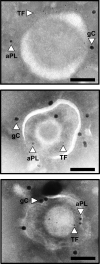
Coagulation initiators tissue factor (TF) and anionic phospholipid (aPL) are available on the herpes simplex virus type 1 (HSV1) surface 150 . Representative triple‐labeled immunogold electron micrographs simultaneously identifying the HSV1 marker, glycoprotein C (gC; 15 nm gold bead), aPL (10 nm bead), and TF (6 nm gold bead). (Scale bars = 100 nm. n = 3)
Figure 3
Figure 3
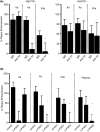
Viral tissue factor (TF) and hemostatic proteases enhance infection via protease‐activated receptors (PARs) in vitro. 73 , 74 (A) Human umbilical vein endothelial cells (HUVECs) were incubated with a constant amount of TF+ (left panel) or TF– (right panel) herpes simplex virus type 1 (HSV1; 4.5 × 105 vp/mL) and thrombin (IIa; 10nM), factor Xa (FXa; 1nM), or factor VIIa (FVIIa; 2.5 nM) with mouse IgG (55 nM) plus enzyme (IgG) or enzyme plus anti‐TF (55nM). The data were corrected for the amount of infection detected without added protease (n = 4; data are presented as mean ± SEM). *P ≤ .05 compared with mouse IgG plus enzyme. (B) HUVECs were incubated with TF + HSV1 (4.5 × 105 vp/mL) and thrombin (IIa; 10nM), FXa (1nM), FVIIa (2.5 nM) or plasmin (50 nM) with control mouse IgG (control; 50 M) plus enzyme, anti‐ PAR1 (α‐PAR1; 150 nM) plus enzyme, or anti‐PAR2 (α‐PAR2; 50 nM) plus enzyme. The data were corrected for the amount of infection without added protease in the presence of control IgG (n = 4; data are presented as mean ± SEM). *P ≤ .05 compared with control IgG plus enzyme
Figure 4
Figure 4
Tissue factor (TF) on herpes simplex virus type 1 (HSV1) enhances infectious virus production in mice. 158 (A) Eight‐week‐old female BALB/c mice were inoculated intravenously with 5 × 105 plaque‐forming units (PFUs) of either TF‐competent (TF+; n = 24) or TF‐deficient (TF−; n = 13) herpes simplex virus type 1 (HSV1) via the tail vein. Three days after infection, the mice were processed and the amounts of infectious virus (plaque‐forming units/mg) were determined. (B) Additional experiments were conducted after preimmunization of mice with mouse IgG or a mixture of three anti‐TF IgG1 monoclonal antibodies (5G9, 9C3, and 6B4; 0.33 mg each per mouse; n = 10), 4 h prior to injection of the virus. In all panels, data are expressed as mean ± SEM. As determined with Student’s t test, *P ≤ .05 when compared with the TF + virus alone
Figure 5
Figure 5
Infection of BALB/c mice is inhibited by anticoagulation. 158 Eight‐week‐old female BALB/c mice were inoculated intravenously with 5 × 105 plaque‐forming units (PFUs) of tissue factor (TF)‐competent (TF+; n = 24) HSV1 alone or simultaneously with hirudin (1 mg/kg, n = 9), nematode anticoagulant protein c2 (NAPc2) (1 mg/kg, n = 18), or apixaban (1 mg/kg, n = 18), via the tail vein. In each case, 3 days after infection, the mice were processed, and the amount of infectious virus (PFUs/mg) was determined in each organ. In all panels, data are expressed as mean ± SEM. As determined with Student’s t test, P ≤ .05 as compared with the TF + virus alone for all data points except liver treated with hirudin
Figure 6
Figure 6
Viral tissue factor (TF) in infection. A model envelope virus is depicted showing a phospholipid bilayer. Several pools of TF may be resent during virus infection including, cellular, viral or associated with extracellular vesicles. Based on studies with herpes simplex virus type 1 (HSV1), TF is embedded in the envelope and assembled with factor VIIa (FVIIa). The known domain organization of proteins is depicted including an active site on respective protease domains (cleft). The TF/FVIIa tenase activates factor X (FX) to FXa bound to viral anionic phospholipid polar headgroups (green). The nascent FXa may either remain bound and engage in signaling through protease‐activated receptor 2 (PAR2) or dissociate and participate in downstream thrombin (factor IIa [FIIa]) generation. When early events of infection were monitored in the absence of the immune system in vitro, both PAR2 via TF/FVIIa/FXa or protease‐activated receptor 1 (PAR1) via thrombin‐enhanced infection. In mice, the absence of envelope TF prevented infection of HSV1 in all organs evaluated. In these in vivo experiments, PAR1 continued to enhance HSV1/TF + virus infection. Highlighting a switch in the role of PAR2 function compared to only evaluating the early stages of infection in vitro (eg, cell attachment and entry), PAR2 reduced virus infection in vivo, presumably through innate immune cell recruitment

References

    1. Huang C, Wang Y, Li X, Ren L, Zhao J, Hu Y, et al. Clinical features of patients infected with 2019 novel coronavirus in Wuhan, China. Lancet. 2020;395:497–506. - PMC - PubMed
    1. Coronaviridae Study Group of the International Committee on Taxonomy of Viruses . The species Severe acute respiratory syndrome‐related coronavirus: classifying 2019‐nCoV and naming it SARS‐CoV‐2. Nat Microbiol. 2020;5:536–44. - PMC - PubMed
    1. Schoeman D, Fielding BC. Coronavirus envelope protein: current knowledge. Virol J. 2019;16:69. - PMC - PubMed
    1. Danzi GB, Loffi M, Galeazzi G, Gherbesi E. Acute pulmonary embolism and COVID‐19 pneumonia: a random association? Eur Heart J. 2020;41(19):1858. - PMC - PubMed
    1. Luo W, Yu H, Guo J, Li X, Sun Y, Li J, Liu L. Clinical pathology of critical patient with novel coronavirus pneumonia (COVID‐19). Pre‐prints. 2020. 2020020407.