. 2020 Oct;252(2):189-200.
doi: 10.1002/path.5510. Epub 2020 Aug 28.

The choroid plexus stroma constitutes a sanctuary for paediatric B-cell precursor acute lymphoblastic leukaemia in the central nervous system

Affiliations

The choroid plexus stroma constitutes a sanctuary for paediatric B-cell precursor acute lymphoblastic leukaemia in the central nervous system

Lidia M Fernández-Sevilla et al. J Pathol. 2020 Oct.

Abstract

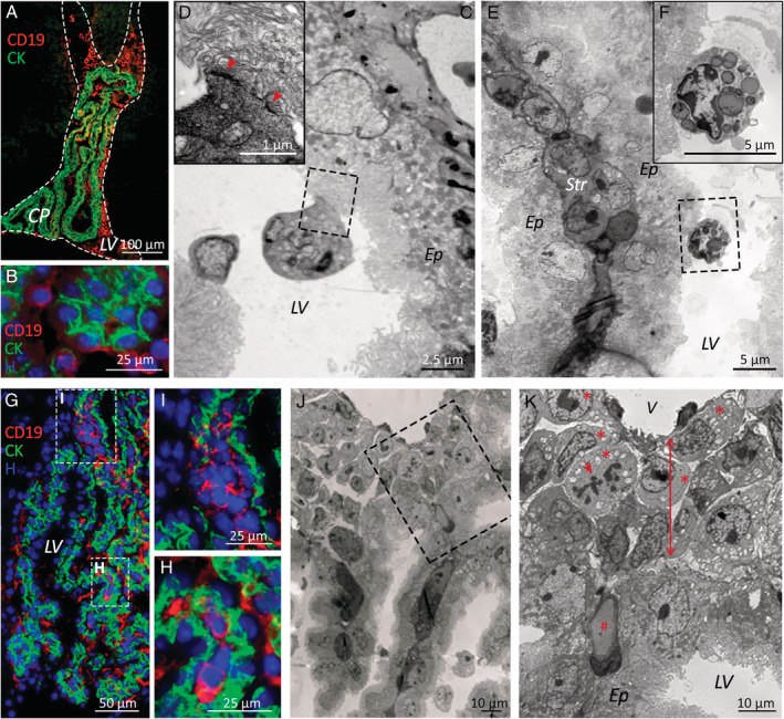
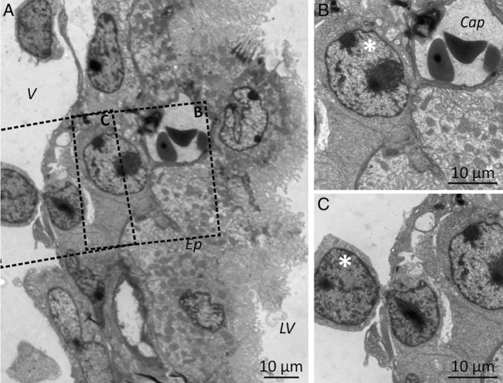
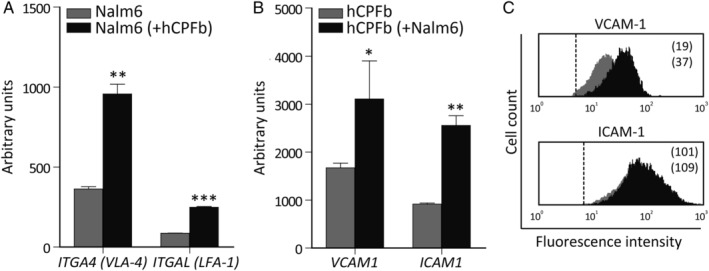
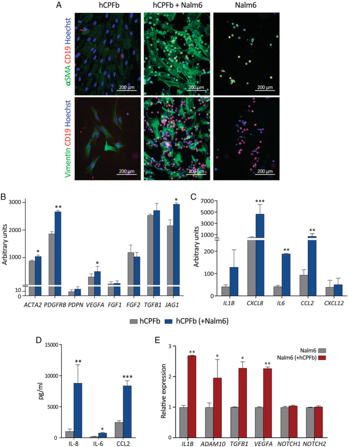
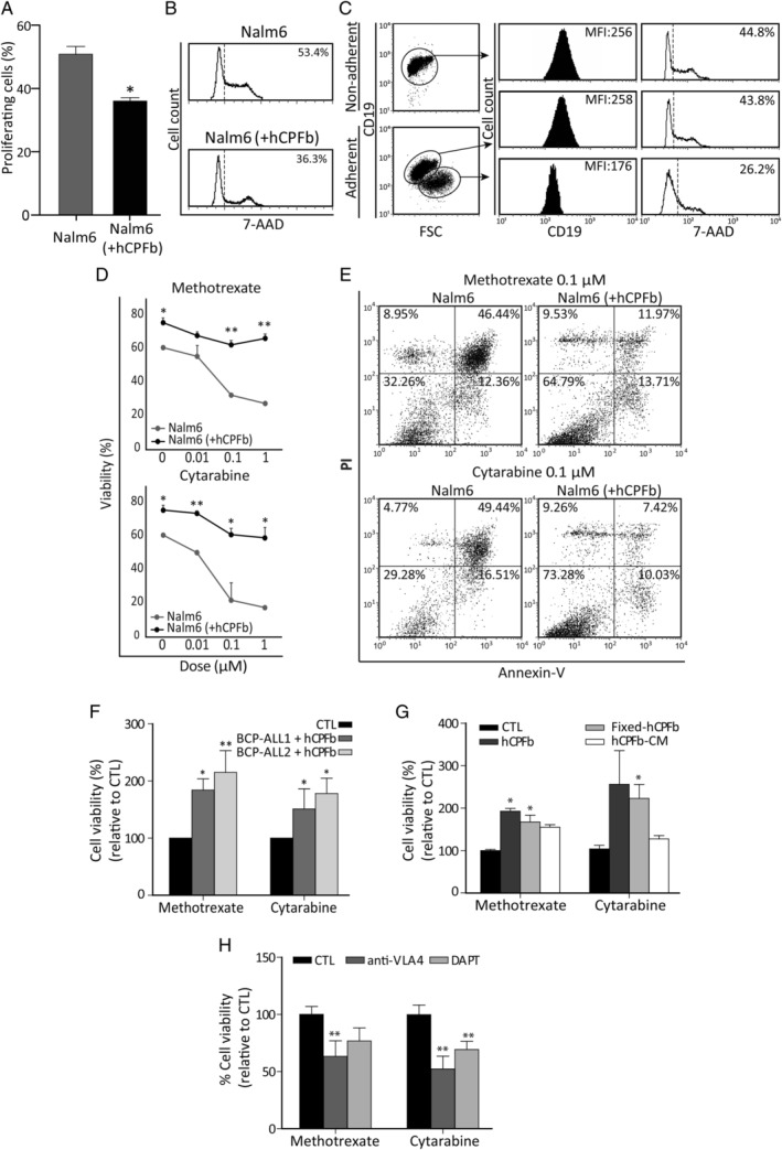
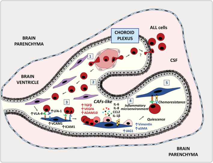
Despite current central nervous system-directed therapies for childhood B-cell precursor acute lymphoblastic leukaemia, relapse at this anatomical site still remains a challenging issue. Few reports have addressed the study of the specific cellular microenvironments which can promote the survival, quiescence, and therefore chemoresistance of B-cell precursor acute lymphoblastic leukaemia cells in the central nervous system. Herein, we showed by immunofluorescence and electron microscopy that in xenotransplanted mice, leukaemic cells infiltrate the connective tissue stroma of the choroid plexus, the brain structure responsible for the production of cerebrospinal fluid. The ultrastructural study also showed that leukaemia cells are able to migrate through blood vessels located in the choroid plexus stroma. In short-term co-cultures, leukaemic cells established strong interactions with human choroid plexus fibroblasts, mediated by an increased expression of ITGA4 (VLA-4)/ITGAL (LFA-1) and their ligands VCAM1/ICAM1. Upon contact with leukaemia cells, human choroid plexus fibroblasts acquired a cancer-associated fibroblast phenotype, with an increased expression of α-SMA and vimentin as well as pro-inflammatory factors. Human choroid plexus fibroblasts also have the capacity to reduce the proliferative index of leukaemic blasts and promote their survival and chemoresistance to methotrexate and cytarabine. The inhibition of VLA-4/VCAM-1 interactions using anti-VLA-4 antibodies, and the blockade of Notch signalling pathway by using a γ-secretase inhibitor partially restored chemotherapy sensitivity of leukaemia cells. We propose that the choroid plexus stroma constitutes a sanctuary for B-cell precursor acute lymphoblastic leukaemia cells in the central nervous system. © 2020 The Authors. The Journal of Pathology published by John Wiley & Sons, Ltd. on behalf of The Pathological Society of Great Britain and Ireland.

Keywords: central nervous system; choroid plexus; haematopoiesis; lymphocytes; paediatric B-cell precursor acute lymphoblastic leukaemia.


Figures

Figure 1
Figure 1
B‐cell precursor acute lymphoblastic leukaemia cells infiltrate the connective stroma of the choroid plexus in a xenograft model. (A) Immunofluorescence microscopy images show BCP‐ALL cells (hCD19 staining, red) floating in the CSF‐filled lateral brain ventricles (LV, dashed lines) and (B) in contact with CP epithelial cells (cytokeratin staining, green). (C, D) Electron microscopy images of leukaemic cells tightly attached (red arrowheads) to the apical surface of the CP epithelium (Ep) and (E, F) Kolmer cells containing CSF‐filled vesicles also in contact with CP epithelial cells. A stromal core of connective tissue (Str) surrounded by two epithelial linings can be seen in E. (G–I) Small groups of BCP‐ALL cells (hCD19 staining, red) located within the CP stroma, (I) mainly in the attachments of the CP. (J, K) Electron micrographs showing leukaemic blasts (red asterisks), some of them in mitosis (red arrow), housed in the space (red two‐sided arrow) between basal laminae of the CP epithelium and CP blood vessels (V). # denotes a CP capillary. Samples from patients 4 and 5 were used for electron microscopy studies, and samples from patients 1–3 and 6–11 for immunofluorescence studies.
Figure 2
Figure 2
Leukaemia cells can reach the choroid plexus stroma through the choroid plexus blood vessels. (A) Electron microscopy images showing lymphoid blasts (white asterisks) in the CP stroma, both (B) in the vicinity of capillaries (Cap) and (C) crossing venule walls. V: venule; Ep: CP epithelium; LV: lateral ventricle. Samples from patients 4 and 5 were used for electron microscopy studies.
Figure 3
Figure 3
Leukaemia–choroid plexus fibroblast interaction promotes the reciprocal expression of adhesion molecules. (A) RT‐qPCR quantification of mRNA levels of ITGA4 (VLA‐4) and ITGAL (LFA‐1) integrins in Nalm‐6 leukaemic cells co‐cultured for 12 h in the presence (black bars) or absence (grey bars) of human CP fibroblasts (hCPFb). (B) mRNA levels of the integrin ligands VCAM1 and ICAM1 in hCPFb co‐cultured with (black bars) or without (grey bars) BCP‐ALL cells. Mean ± SD of three independent experiments (*p ≤ 0.05, **p ≤ 0.01, ***p ≤ 0.001; Mann–Whitney test). (C) Representative flow cytometry histograms showing VCAM‐1 and ICAM‐1 expression in hCPFb co‐cultured in the presence (black histograms) or absence (grey histograms) of leukaemic cells. Mean fluorescence intensity values are shown.
Figure 4
Figure 4
B‐cell precursor acute lymphoblastic leukaemia cells induce a cancer‐associated fibroblast (CAF) phenotype in human choroid plexus fibroblasts. (A) Analysis of α‐SMA and vimentin expression (green staining) in human choroid plexus fibroblasts (hCPFb) co‐cultured for 12 h in direct contact with or without Nalm‐6 cells (red staining). (B) RT‐qPCR quantification of mRNA levels for different CAF markers, as well as the Notch ligand JAG1 (Jagged1), in hCPFb cultured for 12 h in the presence (blue bars) or absence (grey bars) of BCP‐ALL cells in a Transwell system. (C) mRNA expression levels for different cytokines and chemokines in hCPFb co‐cultured with (blue bars) or without (grey bars) BCP‐ALL cells. (D) The concentrations of IL‐8, IL‐6, and CCL2 in supernatants collected from leukaemia–CP fibroblast co‐cultures after 72 h were determined by ELISA and CBA systems. (E) Changes in the mRNA expression profile of Nalm‐6 leukaemic cells co‐cultured in the presence (red bars) or absence (grey bars) of hCPFb, assessed using RT‐qPCR. Mean ± SD of three independent experiments (*p ≤ 0.05, **p ≤ 0.01, ***p ≤ 0.001; Mann–Whitney test).
Figure 5
Figure 5
Human choroid plexus fibroblasts protect leukaemia cells from chemotherapy through VCAM‐1/VLA‐4 interactions and Notch signalling. (A) The proliferation rate of Nalm‐6 cells cultured in the presence or absence of human CP fibroblasts (hCPFb) for 48 h was determined by 7‐AAD staining and analysed by flow cytometry. Mean ± SD of proliferating cells from three independent experiments. (B) Representative flow cytometry histograms showing the fraction of cells in S + G2 + M phases. (C) Analysis of leukaemic cell size and CD19 expression when co‐cultured with hCPFb. Dot plots show forward scatter (FSC) properties and CD19 expression of Nalm‐6 cells from non‐adherent (in suspension) and adherent (attached to fibroblasts) fractions. Representative flow cytometry histograms show CD19 expression (solid black histograms) and 7‐AAD staining (open histograms). Mean fluorescence intensity (MFI) and percentage of proliferating cells, respectively, are indicated. (D) The cell viability of Nalm‐6 leukaemic cells exposed to increasing concentrations (0.01, 0.1, and 1 μm) of chemotherapeutic agents (methotrexate and cytarabine) in the presence (black lines) or absence (grey lines) of hCPFb for 72 h was assessed by flow cytometry. Results show the percentage of propidium iodide‐negative and annexin V‐negative viable leukaemic cells, and are the mean ± SD of four independent experiments. (E) Representative dot plots showing the viability of leukaemic cells treated with methotrexate and cytarabine (0.1 μm) in the presence of hCPFb or in control cultures analysed by annexin V and propidium iodide staining. (F) Primary samples from two BCP‐ALL patients (Nos 1 and 2) were exposed to methotrexate and cytarabine (0.1 μm) in the presence or absence of hCPFb. Results from three independent experiments are expressed as the mean ± SD of the relative viability compared with control cultures performed in the absence of hCPFb. (G) Relative viability of Nalm‐6 leukaemic cells co‐cultured with hCPFb, paraformaldehyde‐fixed hCPFb or cultured in the presence of hCPFb‐derived conditioned media and treated with methotrexate and cytarabine at a concentration of 0.1 μm. Data from three independent experiments are expressed as the mean ± SD of the relative viability compared with Nalm‐6 cell control cultures. (H) Cell viability of Nalm‐6 cells treated with chemotherapeutic agents and co‐cultured with hCPFb in the presence of anti‐VLA‐4 blocking antibodies or the Notch inhibitor, DAPT, compared with control cultures (CTL: anti‐IgG or DMSO). Results represent the mean ± SD of three to four independent experiments (*p ≤ 0.05, **p ≤ 0.01; Mann–Whitney test).
Figure 6
Figure 6
The choroid plexus stroma could act as a sanctuary for B‐cell precursor acute lymphoblastic leukaemia cells. Schematic representation of the involvement of the choroid plexus in acute lymphoblastic leukaemia: BCP‐ALL cells could reach the CP via its fenestrated vasculature that can become more permissive under the influence of leukaemic cells (1). Once the CP is colonized, BCP‐ALL cells would preferentially stay in the connective stroma and occasionally move towards the CSF crossing the CP epithelial lining which forms the BCSFB (2). BCP‐ALL cells would then interact with the CP stromal fibroblasts through the upregulated expression of VCAM‐1/VLA‐4 and ICAM‐1/LFA‐1 ligand–receptor pairs (3) and induce a CAF phenotype which would promote a pro‐tumoural inflammatory microenvironment (4). BCP‐ALL cells would remain attached to the CP CAF‐like cells and could acquire quiescence and chemoresistance, a process partially dependent on the VLA‐4 and Notch signalling pathways (5).

References

    1. Hunger SP, Mullighan CG. Acute lymphoblastic leukemia in children. N Engl J Med 2015; 373 : 1541–1552. - PubMed
    1. Ward E, DeSantis C, Robbins A, et al Childhood and adolescent cancer statistics, 2014. CA Cancer J Clin 2014; 64 : 83–103. - PubMed
    1. Pui CH, Carroll WL, Meshinchi S, et al Biology, risk stratification, and therapy of pediatric acute leukemias: an update. J Clin Oncol 2011; 29 : 551–565. - PMC - PubMed
    1. Pui CH, Robison LL, Look AT. Acute lymphoblastic leukaemia. Lancet 2008; 371 : 1030–1043. - PubMed
    1. Thomas LB. Pathology of leukemia in the brain and meninges: postmortem studies of patients with acute leukemia and of mice given inoculations of L1210 leukemia. Cancer Res 1965; 25 : 1555–1571. - PubMed

Publication types