. 2020 Oct;30(10):885-901.
doi: 10.1038/s41422-020-0370-1. Epub 2020 Jul 20.

Wnt signaling and Loxl2 promote aggressive osteosarcoma

Affiliations

Wnt signaling and Loxl2 promote aggressive osteosarcoma

Kazuhiko Matsuoka et al. Cell Res. 2020 Oct.

Abstract

Osteosarcoma (OS) is the most frequent primary malignant bone tumor in urgent need of better therapies. Using genetically modified mouse models (GEMMs), we demonstrate that Wnt signaling promotes c-Fos-induced OS formation via the actions of the collagen-modifying enzyme Loxl2. c-Fos/AP-1 directly regulates the expression of the Wnt ligands Wnt7b and Wnt9a in OS cells through promoter binding, and Wnt7b and Wnt9a in turn promote Loxl2 expression in murine and human OS cells through the transcription factors Zeb1 and Zeb2. Concordantly, inhibition of Wnt ligand secretion by inactivating the Wnt-less (Wls) gene in osteoblasts in c-Fos GEMMs either early or in a therapeutic setting reduces Loxl2 expression and progression of OS. Wls-deficient osteosarcomas proliferate less, are less mineralized and are enriched in fibroblastic cells surrounded by collagen fibers. Importantly, Loxl2 inhibition using either the pan-Lox inhibitor BAPN or a specific inducible shRNA reduces OS cell proliferation in vitro and decreases tumor growth and lung colonization in murine and human orthotopic OS transplantation models. Finally, OS development is delayed in c-Fos GEMMs treated with BAPN or with specific Loxl2 blocking antibodies. Congruently, a strong correlation between c-FOS, LOXL2 and WNT7B/WNT9A expression is observed in human OS samples, and c-FOS/LOXL2 co-expression correlates with OS aggressiveness and decreased patient survival. Therefore, therapeutic targeting of Wnt and/or Loxl2 should be considered to potentiate the inadequate current treatments for pediatric, recurrent, and metastatic OS.


Conflict of interest statement

A.M.V. is an employee of Gilead Sciences. The remaining authors declare no competing interests.

Figures

Fig. 1
Fig. 1. Wnt-less (Wls) deletion in osteoblast progenitors reduces c-Fos-induced OS.
a Experimental procedure to prevent Wnt secretion by deleting the Wls gene in osteoblast progenitors. Dox is removed at the onset of tumor formation (3 weeks of age, 1 week after Fos transgene expression is detected) and tumors are monitored at 5 and 15 weeks of age. b qPCR analysis of Wls, Wnt target genes and Fos mRNA species (endo, endogenous, Tg, transgene) in WlsWT and WlsΔOB-OS tumor-bearing bones at 5 weeks of age. cg Micro-CT analysis of WlsWT and WlsΔOB-OS mice. Representative Micro-CT: 3D reconstructions (Top) and 2D virtual sections (bottom) of tumor-bearing bones. Red arrows point to OS (c). Tumor number per mouse (d), average tumor volume (e) and average tumor burden per mouse (f) at 5 and 15 weeks in WlsWT-OS mice (n = 21) and WlsΔOB-OS mice (n = 15). g Volumetric follow-up over time for 41 WlsWT-OS tumors and 23 WlsΔOB-OS tumors. h EdU incorporation by WlsWT-OS (n = 13) and WlsΔOB-OS (n = 21) tumors at 15 weeks of age. The left panels are representative images of EdU-labeled cells 4 h after EdU injection (white arrows, nuclei counterstained with DAPI) while % EdU-labeled cells per tumor are plotted on the right. Bar graphs and plots represent or include mean ± SEM, respectively. *P < 0.05, **P < 0.01.
Fig. 2
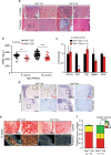
Fig. 2. Wls deficiency in osteoblast progenitors affects OS pathology.
a Representative H&E stainings of WlsWT-OS and WlsΔOB-OS tumors at 5 and 15 weeks. b Micro-CT measurement of vBMD in WlsWT-OS and WlsΔOB-OS tumors at 5 and 15 weeks of age. The total number of analyzed tumors is indicated between parentheses (WT/ΔOB) beside each time point. c qPCR analysis of osteoblast/osteocyte marker in tumor-bearing bones from 5 week-old WlsWT-OS and WlsΔOB-OS mice. d IHC analysis of Osteocalcin and Sclerostin at 15 weeks. e Picrosirius red staining of WlsWT-OS and WlsΔOB-OS tumors photographed under optical (Top) and polarized (Bottom) light. f Quantification of collagen packing density in the tumors using picrosirius red staining and polarized light at 15 weeks (n = 3). Bar graphs and plots represent or include mean ± SEM, respectively. *P < 0.05, **P < 0.01.
Fig. 3
Fig. 3. Wls deletion during tumor progression decreases OS in H2-c-fosLTR mice.
a Experimental procedure. Dox is removed at 5 weeks of age when tumors are detectable by Micro-CT and the mice are additionally subjected to Micro-CT at 15 and 20 weeks. bf Micro-CT analysis of WlsWT and WlsΔOB-OS mice. Tumor number per mouse (b), average tumor volume (c) and average tumor burden per mouse (d) at 5, 15 and 20 weeks in WlsWT-OS mice (n = 7) and WlsΔOB-OS mice (n = 5). e Volumetric follow-up over time for 43 WlsWT-OS tumors and 17 WlsΔOB-OS tumors. f vBMD in WlsWT-OS and WlsΔOB-OS tumors over time. The number of analyzed tumors is indicated in parentheses (WT/ΔOB) beside each time point. g Representative tumor histology at 30 weeks. Bar graphs and plots represent or include mean ± SEM, respectively. Black asterisks: *P < 0.05 and **P < 0.01 by two-way ANOVA with Bonferroni post hoc test. Red asterisks: ns not significant, *P < 0.05, **P < 0.01 and ***P < 0.001 by two-tailed t-test between each genotype at 20 weeks and all mice at 5 weeks.
Fig. 4
Fig. 4. c-Fos/AP-1 directly promotes the expression of Wnt7b and Wnt9a in OS.
ab qPCR analysis of Wnt7b, Wnt9a and Wnt3a in H2-c-fosLTR bones, tumor-bearing bones and dissected tumors (a) and primary OS cells isolated from WlsWT-OS and WlsΔOB-OS mice (b). Bones from WT littermates (a) and MC3T3-E1 osteoblastic cells (b) were included for comparison. N.D., not detectable. c, d Gene expression in MC3T3-E1 cells expressing c-Fos-ER or mutant (inactive) c-Fos*-ER in the presence/absence of tamoxifen (c) and in the H2-c-fosLTR OS cell line (FosTg-C3) expressing IPTG-inducible c-fos shRNA or non-target shRNA in the presence/absence of IPTG (d) was determined by qPCR. FosL1 (encoding Fra-1 and a bona fide Fos-target gene) is included as a control. e ChIP-qPCR quantification of c-Fos/AP-1 binding to Wnt7b and Wnt9a promoters in H2-c-fosLTR OS cells expressing IPTG-inducible c-fos shRNA or non-target shRNA in the presence/absence of IPTG. The FosL1 genomic region with a c-Fos/AP-1 binding site is included as a control. f Luciferase reporter assay in MC3T3-E1 cells cotransfected with increased amounts of c-Fos expressing vector (CMV-c-Fos) and Wnt7b and Wnt9a reporter constructs containing the ChIP sites from e. A Fosl1 reporter construct is included as a control. Bar graphs represent mean ± SEM. *P < 0.05 and **P < 0.01.
Fig. 5
Fig. 5. Loxl2 expression is modulated by Wnt and is essential for OS proliferation.
a, b qPCR analysis of collagen modifying enzymes in tumor-bearing bone (a) and primary OS cells (b) isolated from WlsWT-OS and WlsΔOB-OS mice. c Loxl2 immunofluorescence (red) in WlsΔOB-OS and WlsWT-OS tumors at 15 weeks. Nuclei are counterstained with DAPI. d Loxl2 immunoblot in primary tumor cells isolated from WlsWT-OS and WlsΔOB-OS mice. e qPCR analysis of Loxl2 and the Wnt targets Axin2 and Alp in FosTg-C3 cells stimulated with Wnt7b, Wnt9a, Wnt3a, Wnt5a or control conditioned medium (CM) for 48 h. f Loxl2 immunoblot in primary tumor cells isolated from H2-c-fosLTR OS cells treated with control or Wnt7b-CM for 48 h. g Immunoblot for Zeb1 and Zeb2 in in primary tumor cells isolated from WlsWT-OS and WlsΔOB-OS mice, in FosTg-C3 cells treated with Wnt7b CM and in LM7 cells ectopically expressing WNT7B. h Bone tumor burden 21 days post cell implantation in NSG mice orthotopically injected with FosTg-C3 cells expressing IPTG-inducible Loxl2 shRNA or non-target shRNA and treated with IPTG or vehicle (PBS) during 18 days. Actin is used as a loading control for immunoblot. Bar graphs and plots represent or include mean ± SEM, respectively. *P < 0.05 and **P < 0.01.
Fig. 6
Fig. 6. Inhibition of Lysyl oxidase activity or Loxl2 reduces OS growth.
a Experimental procedure: 5-week-old H2-c-fosLTR mice were injected three times/week during 15 weeks with 250 mg/kg BAPN to block lysyl oxidase activity or with vehicle (PBS) and tumors were monitored longitudinally by Micro-CT. b Average tumor burden per mouse. c Volumetric follow-up over time for 20 and 18 tumors from PBS-treated and BAPN-treated mice, respectively at 5, 10, 15 and 20 weeks in PBS- or BAPN-treated mice. d, e Representative images of Picrosirius red/polarized light staining (d) and quantification of tumor collagen packing density (e) from tumor-bearing PBS- and BAPN-treated mice at 20 weeks. fh Experimental procedure: 5-week old H2-c-fosLTR mice were injected two times/week during 15 weeks with anti-Loxl2 blocking antibody (30 mg/kg) or with IgG and tumors were monitored longitudinally by Micro-CT (f) for tumor number per mouse (g) and average tumor volume (h) at 5 and 20 weeks. Bar graphs and plots represent or include mean ± SEM, respectively. Black asterisks: *P < 0.05 and **P < 0.01 by two-way ANOVA with Bonferroni post hoc test. Red asterisks: *P < 0.05, **P < 0.01 and ***P < 0.001 by two-tailed t-test between PBS- and BAPN-treated at end point and all mice at start.
Fig. 7
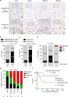
Fig. 7. LOXL2 is co-expressed with WNT7A/WNT9A and FOS in human OS and FOS/LOXL2 double positivity is predictive of OS patient survival.
Two independent tissue microarrays (TMA)/patient cohorts were employed. US Biomax TMA (OS802c) (ad), University Clinic of Navarra TMA (UCN) (e, f). a Representative IHC images of FOS/LOXL2/WNT9A/WNT7B-quadruple negative (N/N/N/N) and -quadruple positive (P/P/P/P) in human OS TMA. Positive cells are indicated by red arrows. b Quantification of FOS-positive and -negative OS that are either WNT7B/WNT9A double-positive or -negative in a human OS TMA (Biomax, n = 49). c Quantification of FOS-positive and negative-OS that are either LOXL2-positive or -negative in a human OS TMA (Biomax, n = 49). d Correlation of FOS and LOXL2 IHC groups with OS clinical stage. For bd, **P < 0.01, ***P < 0.001 and P = 0.08 by Fisher’s exact test. Brackets indicate the number of patients. e Relative abundance of FOS/LOXL2 positivity in OS analyzed by IHC in the UCN cohort according to patient diagnosis (primary tumor, post-treatment, metastatic or recurrent osteosarcoma). Bone, normal bone; Primary, primary tumor; Post-T, post-treatment; Mets, metastases; Relapse, relapsed tumor. P value is determined by χ2 statistics. f Overall survival of OS patients according to FOS/LOXL2 positivity (all patients included). Data are analyzed by Log-rank test and patient number is in brackets. P positive, N negative, N/N double-negative, P/N and N/P single positive, P/P double-positive.
Fig. 8
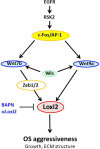
Fig. 8. Schematic representation of the Fos/AP-1-Wnt7b/Wnt9a-Loxl2 axis operating in OS formation.
Wnt9a and Wnt7b are two novel c-Fos/AP-1 target genes operating in an autocrine, and possibly paracrine, fashion in mouse and human OS to induce the expression of the collagen cross-linking enzyme Loxl2, thereby modulating collagen packing and impacting on osteoblast differentiation and tumor aggressiveness. c-Fos protein expression and stability is maintained through EGFR and its downstream kinase RSK2 and the new regulatory pathway can be therapeutically targeted by inhibiting Wnt secretion (Wls) and/or Loxl2 activity (BAPN or blocking antibodies), to benefit OS patients.

Similar articles

Cited by

References

    1. Heaney RP, et al. Peak bone mass. Osteoporos. Int. 2000;11:985–1009. - PubMed
    1. Mirabello L, Troisi RJ, Savage SA. Osteosarcoma incidence and survival rates from 1973 to 2004: data from the surveillance, epidemiology, and end results program. Cancer. 2009;115:1531–1543. - PMC - PubMed
    1. Fernandes DA, Costa AA, Lahdenperä P. Osteosarcoma genetics and epigenetics: emerging biology and candidate therapies. Int. J. Constr. Manag. 2018;18:482–496.
    1. Lindsey BA, Markel JE, Kleinerman ES. Osteosarcoma overview. Rheumatol. Ther. 2017;4:25–43. - PMC - PubMed
    1. Link MP, et al. The effect of adjuvant chemotherapy on relapse-free survival in patients with osteosarcoma of the extremity. N. Engl. J. Med. 1986;314:1600–1606. - PubMed

Publication types

Substances