

doi: 10.3201/eid2608.191644.
Disseminated Echinococcus multilocularis Infection without Liver Involvement in Child, Canada, 2018
- PMID: 32687039
- PMCID: PMC7392456
- DOI: 10.3201/eid2608.191644
Item in Clipboard
Disseminated Echinococcus multilocularis Infection without Liver Involvement in Child, Canada, 2018
Emerg Infect Dis.
2020 Aug.
Abstract
An immunocompetent child in Canada received a diagnosis of disseminated alveolar Echinococcus multilocularis infection. The case lacked typical features of liver involvement and was possibly related to a rare congenital portosystemic shunt. We summarize the rapidly evolving epidemiology of E. multilocularis parasites in Canada.
Keywords: Canada; Echinococcus multilocularis; alveolar; children; hydatid; liver; parasites; zoonoses.
Figures

Coronal contrast enhanced CT (computed tomography) of the abdomen of a child with disseminated Echinococcus multilocularis infection without liver involvement, Canada, 2018. There is a large irregular hypodense left renal lesion (red arrow). A large porto-systemic shunt is partially visualized (white arrow).
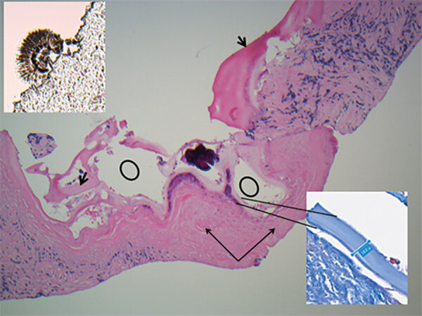
Kidney core biopsy of a child with disseminated Echinococcus multilocularis infection without liver involvement, Canada, 2018. Shown are folded laminated membrane (short black arrows) encircling variable-sized cystic structures (black circles) containing calcified and necrotic debris and dense periparasitic fibrosis (long black arrows), in a background of chronic inflammation and fibrosis. No residual normal kidney parenchyma was seen (hematoxylin and eosin stain, original magnification × 40). Inset at lower right shows laminar membrane, 18–19.4 μm in thickness in a background of fibrosis (Masson trichrome, original magnification ×40). Insert at upper left shows scolex attached to the paraffin edge of the block (original magnification ×40).
References
-
- Reinehr M, Micheloud C, Grimm F, Kronenberg PA, Grimm J, Beck A, et al. Pathology of echinococcosis: a morphologic and immunohistochemical study on 138 specimens with focus on the differential diagnosis between cystic and alveolar echinococcosis. Am J Surg Pathol. 2020;44:43–54. 10.1097/PAS.0000000000001374 - DOI - PubMed
MeSH terms
Supplementary concepts
LinkOut - more resources
Full Text Sources
