. 2020 Nov 1;77(11):1390-1397.
doi: 10.1001/jamaneurol.2020.2349.

A Pooled Analysis of Diffusion-Weighted Imaging Lesions in Patients With Acute Intracerebral Hemorrhage

Affiliations

A Pooled Analysis of Diffusion-Weighted Imaging Lesions in Patients With Acute Intracerebral Hemorrhage

Santosh B Murthy et al. JAMA Neurol. .

Abstract

Importance: The etiology and significance of diffusion-weighted imaging (DWI) lesions in patients with acute intracerebral hemorrhage (ICH) remain unclear.

Objective: To evaluate which factors are associated with DWI lesions, whether associated factors differ by ICH location, and whether DWI lesions are associated with functional outcomes.

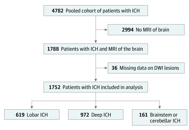
Design, setting, and participants: This analysis pooled individual patient data from 3 randomized clinical trials (Minimally Invasive Surgery Plus Alteplase for Intracerebral Hemorrhage Evacuation phase 3 trial, Antihypertensive Treatment of Acute Cerebral Hemorrhage trial, and Intracerebral Hemorrhage Deferoxamine phase 2 trial) and 1 multicenter prospective study (Ethnic/Racial Variations of Intracerebral Hemorrhage). Patients were enrolled from August 1, 2010, to September 30, 2018. Of the 4782 patients, 1788 who underwent magnetic resonance imaging scans of the brain were included. Data were analyzed from July 1 to December 31, 2019.

Main outcomes and measures: The primary outcome consisted of factors associated with DWI lesions. Secondary outcomes were poor functional outcome, defined as a modified Rankin score (mRS) of 4 to 6, and mortality, both assessed at 3 months. Mixed-effects logistic regression was used to evaluate the association between exposures and outcomes. Subgroup analyses stratified by hematoma location were performed.

Results: After exclusion of 36 patients with missing data on DWI lesions, 1752 patients were included in the analysis (1019 men [58.2%]; mean [SD] age, 60.8 [13.3] years). Diffusion-weighted imaging lesions occurred in 549 patients (31.3%). In mixed-effects regression models, factors associated with DWI lesions included younger age (odds ratio [OR] per year, 0.98; 95% CI, 0.97-0.99), black race (OR, 1.64; 95% CI, 1.17-2.30), admission systolic blood pressure (OR per 10-mm Hg increase, 1.13; 95% CI, 1.08-1.18), baseline hematoma volume (OR per 10-mL increase, 1.12; 95% CI, 1.02-1.22), cerebral microbleeds (OR, 1.85; 95% CI, 1.39-2.46), and leukoaraiosis (OR, 1.59; 95% CI, 1.67-2.17). Diffusion-weighted imaging lesions were independently associated with poor mRS (OR, 1.50; 95% CI, 1.13-2.00), but not with mortality (OR, 1.11; 95% CI, 0.72-1.71). In subgroup analyses, similar factors were associated with DWI lesions in lobar and deep ICH. Diffusion-weighted imaging lesions were associated with poor mRS in deep but not lobar ICH.

Conclusions and relevance: In a large, heterogeneous cohort of prospectively identified patients with ICH, results were consistent with the hypothesis that DWI lesions represent acute sequelae of chronic cerebral small vessel disease, particularly hypertensive vasculopathy. Diffusion-weighted imaging lesions portend a worse prognosis after ICH, mainly deep hemorrhages.


Conflict of interest statement

Conflict of Interest Disclosures: Dr Murthy reported receiving funding from the Leon Levy Foundation. Dr Navi reported receiving grant K23NS091395 from the National Institutes of Health (NIH) and support from the Florence Gould Endowment for Discovery in Stroke; serving as a member of the data and safety monitoring board for the Patient-Centered Outcomes Research Institute–funded TRAVERSE trial; and receiving personal fees for medicolegal consulting on stroke. Dr Mandava reported serving as a co–principal investigator in a Government of India Ministry of Human Resources Development–funded project to develop a tool to apply machine learning techniques to central nervous system tumor detection and receiving personal fees for medicolegal consulting on stroke. Dr Iadecola reported serving on the scientific advisory board of Broadview Ventures. Dr Falcone reported receiving grants K76AG059992 and R03NS112859 from the NIH, grant 18IDDG34280056 from the American Heart Association, Yale Pepper Scholar Award P30AG021342, and the Neurocritical Care Society Research Fellowship. Dr Sheth reported receiving grants U24NS107215, U24NS107136, RO1NR018335, and U01NS106513 from the NIH, support from Novartis International AG and Bard Pharmaceuticals, and grants from Hyperfine, Biogen, Inc, and Astrocyte Pharmaceuticals unrelated to this work. Dr Biffi reported receiving grant K23NS100816 from the NIH. Dr Rosand reported receiving grants R01NS036695, UM1HG008895, R01NS093870, and R24NS092983 from the NIH, support from One Mind, and consulting fees from Boehringer Ingelheim, Pfizer, Inc, and New Beta Innovation Limited. Dr Selim reported receiving grants U01NS074425 and U01NS102289 from the NIH and serving on the scientific advisory board of MedRhythms Inc. Dr Hanley reported receiving grants U01NS080824 and U24TR001609 from the NIH, personal fees from op2lysis, BrainScope Company, Inc, and Neurotrope, Inc, and nonfinancial support from Genentech, Inc, outside the submitted work. Dr Kamel reported receiving grants U01NS095869 and R01NS097443 from the NIH and support from the Michael Goldberg Research Fund; serving as the co–principal investigator for the NIH-funded ARCADIA trial, which receives in-kind study drug from the BMS-Pfizer Alliance and in-kind study assays from Roche Diagnostics; serving as a steering committee member of Medtronic plc’s Stroke AF trial (uncompensated); serving on an end point adjudication committee for a trial of empagliflozin for Boehringer Ingelheim; and serving on an advisory board for Roivant Sciences, Ltd, related to factor XI inhibition. Dr Ziai reported receiving grant 1U01NS080824 from the NIH and consulting fees from C.R. Bard, Inc, outside the present study. No other disclosures were reported.

Figures

Figure.
Figure.. Flowchart Showing Selection of Patients in the Study
DWI indicates diffusion-weighted imaging; ICH, intracerebral hemorrhage; and MRI, magnetic resonance imaging.

References

    1. Kimberly WT, Gilson A, Rost NS, et al. . Silent ischemic infarcts are associated with hemorrhage burden in cerebral amyloid angiopathy. Neurology. 2009;72(14):1230-1235. doi:10.1212/01.wnl.0000345666.83318.03 - DOI - PMC - PubMed
    1. Garg RK, Liebling SM, Maas MB, Nemeth AJ, Russell EJ, Naidech AM. Blood pressure reduction, decreased diffusion on MRI, and outcomes after intracerebral hemorrhage. Stroke. 2012;43(1):67-71. doi:10.1161/STROKEAHA.111.629493 - DOI - PMC - PubMed
    1. Auriel E, Gurol ME, Ayres A, et al. . Characteristic distributions of intracerebral hemorrhage-associated diffusion-weighted lesions. Neurology. 2012;79(24):2335-2341. doi:10.1212/WNL.0b013e318278b66f - DOI - PMC - PubMed
    1. Prabhakaran S, Naidech AM. Ischemic brain injury after intracerebral hemorrhage: a critical review. Stroke. 2012;43(8):2258-2263. doi:10.1161/STROKEAHA.112.655910 - DOI - PubMed
    1. Kidwell CS, Rosand J, Norato G, et al. . Ischemic lesions, blood pressure dysregulation, and poor outcomes in intracerebral hemorrhage. Neurology. 2017;88(8):782-788. doi:10.1212/WNL.0000000000003630 - DOI - PMC - PubMed

Publication types

Substances