. 2020 Sep 1;465(1):79-87.
doi: 10.1016/j.ydbio.2020.07.003. Epub 2020 Jul 18.

Conjugated activation of myocardial-specific transcription of Gja5 by a pair of Nkx2-5-Shox2 co-responsive elements

Affiliations

Conjugated activation of myocardial-specific transcription of Gja5 by a pair of Nkx2-5-Shox2 co-responsive elements

Tianfang Yang et al. Dev Biol. .

Abstract

The sinoatrial node (SAN) is the primary pacemaker in the heart. During cardiogenesis, Shox2 and Nkx2-5 are co-expressed in the junction domain of the SAN and regulate pacemaker cell fate through a Shox2-Nkx2-5 antagonism. Cx40 is a marker of working myocardium and an Nkx2-5 transcriptional output antagonized by Shox2, but the underlying regulatory mechanisms remain elusive. Here we characterized a bona fide myocardial-specific Gja5 (coding gene of Cx40) distal enhancer consisting of a pair of Nkx2-5 and Shox2 co-bound elements in the regulatory region of Gja5. Transgenic reporter assays revealed that neither element alone, but the conjugation of both elements together, drives myocardial-specific transcription. Genetic analyses confirmed that the activation of this enhancer depends on Nkx2-5 but is inhibited by Shox2 in vivo, and its presence is essential for Gja5 expression in the myocardium but not the endothelial cells of the heart. Furthermore, chromatin conformation analysis showed an Nkx2-5-dependent loop formation between these two elements and the Gja5 promoter in vivo, indicating that Nkx2-5 bridges the conjugated activation of this enhancer by pairing the two elements to the Gja5 promoter.

Keywords: Enhancer; Gja5; Myocardium; Nkx2-5; Shox2; Sinoatrial node.


Conflict of interest statement

Declaration of competing interest The authors declare no competing or financial interests.

Figures
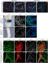
Figure 1.
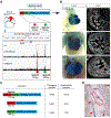
Figure 1.. Identification of Gja5-S1 and Gja5-S2 and generation of Gja5-eh-LacZ reporter mice.
(A) Schematic overview of ChIP-seq data collection, visualization, and identification of Gja5-S1 and Gja5-S2, and generation of Gja5-eh-LacZ transgenic construct. (B) Whole-mount X-gal staining and section immunostaining of β-galactosidase of E10.5, E11.5 and E12.5 Gja5-eh-LacZ embryos. (C) Schematic diagram summarizing the reporter constructs of Gja5-S1, Gja5-S2, and Gja5-eh. The left column indicates the design of each construct. The middle column indicates the number of transgene-positive F0 embryos as a fraction of the total number of F0 embryos. The right column indicates the number of F0 embryos showing cardiac-specific X-gal activity as a fraction of the total number of transgene-positive F0 embryos. (D) X-gal staining on a cryo-sectioned E10.5 Gja5-eh-LacZ embryo. Red arrowheads point to positive X-gal staining in the atrial tissue. Note that the staining is excluded from the SAN (circled in red). RSVC, right superior vena cava. SAN, sinoatrial node. Scale bars: 50 μm.
Figure 2.
Figure 2.. Gja5-eh enhancer activity recapitulates endogenous Gja5 expression in Nkx2–5+ myocardium, and is dependent on Nkx2–5 but is inhibited by Shox2.
(A-D) Triple immunofluorescent staining (Nkx2–5, Cx40, β-gal) on right atrium (A,B) and right ventricle (C,D) of the Gja5-eh-LacZ hearts at E12.5 and P60. CA, coronary arteries; VT, ventricular trabeculae; SAN, sinoatrial node. Scale bars: 100 μm. Also see Fig. S2. (E-J) Gja5-eh-LacZ transgenic mice were crossed onto Nkx2–5+/+ (E-G) and Nkx2–5Cre/Cre (H-J) backgrounds and enhancer activity was examined by whole-mount X-gal staining and fluorescent immunostaining for β-galactosidase at E10.5. Scale bars: 50 μm. (K-R) Gja5-eh-LacZ transgenic mice were crossed onto Shox2+/Cre;ROSA26mTmG/mTmG (K-N) and Shox2Cre/Cre;ROSA26mTmG/mTmG (O-R) backgrounds, and examined at E13.5 for expression of Nkx2–5, mGFP and β-galactosidase by triple fluorescent immunostaining. Blue arrowheads point to ectopic β-gal signal co-localized with Nkx2–5 and mGFP in the SAN junction domain. RSVC, right superior vena cava; Sh, SAN head; Sj, SAN junction. Scale bars: 50 μm.
Figure 3.
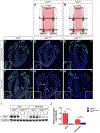
Figure 3.. Gja5-S1 and Gja5-S2 are essential for myocardial Gja5 expression.
(A, B) Schematic overview of CRISPR/Cas9-mediated mouse genome editing and generation of Gja5Δ14 and Gja5Δ12 alleles. (C-H) Double fluorescent immunostaining of Cx40 and Hcn4 on Gja5+/+ (C, F), Gja5Δ14/Δ14 (D, G), and Gja5Δ12/Δ12 (E, H) hearts at P0. White/yellow arrowheads pointed regions are highlighted in white/yellow outlined squares as inserts. SAN, sinoatrial node; VT, ventricular trabeculae; CA, coronary arteries. Scale bars: 250 μm. (I) Western blot of Cx40 and GAPDH on protein extracts from atria or ventricles of Gja5+/+ and Gja5Δ14/Δ14 hearts at P0. (J) RT-qPCR detecting Gja5 and Gapdh expression on atria or ventricles of Gja5+/+ and Gja5Δ14/Δ14 hearts at P0. Maximum Gja5 expression relative to Gapdh is set as one. ****: P < 0.0001. Biological replicates N = 3.
Figure 4.
Figure 4.. Nkx2–5 mediates the interaction between Gja5-S1/Gja5-S2 and the Gja5 promoter.
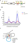
(A) Relative positions of primer sets used for 3C assays. (B) Relative interaction frequency (RIF) in 3C assay between a combination of forward and reverse primer sets in E10.5 hearts of wild type and Nkx2–5Cre/Cre mice. Maximum RIF is set as one and relative fold changes are shown. ****: P < 0.0001. Biological replicates N = 3. (C) a schematic model proposing that Gja5-S1 and Gja5-S2 act in conjugation as a myocardial-specific Gja5 distal enhancer by interacting with the Gja5 promoter, and the enhancer activity depends on Nkx2–5 but inhibited by Shox2.

References

    1. Ambrosetti DC, Basilico C, Dailey L, 1997. Synergistic activation of the fibroblast growth factor 4 enhancer by Sox2 and Oct-3 depends on protein-protein interactions facilitated by a specific spatial arrangement of factor binding sites. Mol. Cell. Biol 17, 6321–6329. 10.1128/mcb.17.11.6321 - DOI - PMC - PubMed
    1. Anderson CM, Hu J, Thomas R, Gainous TB, Celona B, Sinha T, Dickel DE, Heidt AB, Xu SM, Bruneau BG, Pollard KS, Pennacchio LA, Black BL, 2017. Cooperative activation of cardiac transcription through myocardin bridging of paired MEF2 sites. Development 144, 1235–1241. 10.1242/dev.138487 - DOI - PMC - PubMed
    1. Bagwe S, Berenfeld O, Vaidya D, Morley GE, Jalife J, 2005. Altered Right Atrial Excitation and Propagation in Connexin40 Knockout Mice. Circulation 112, 2245–2253. 10.1161/CIRCULATIONAHA.104.527325 - DOI - PMC - PubMed
    1. Bai D, 2014. Atrial fibrillation-linked GJA5/connexin40 mutants impaired gap junctions via different mechanisms. FEBS Lett. 588, 1238–1243. 10.1016/j.febslet.2014.02.064 - DOI - PubMed
    1. Barrington C, Georgopoulou D, Pezic D, Varsally W, Herrero J, Hadjur S, 2019. Enhancer accessibility and CTCF occupancy underlie asymmetric TAD architecture and cell type specific genome topology. Nat. Commun 10, 1–14. 10.1038/s41467-019-10725-9 - DOI - PMC - PubMed

Publication types

LinkOut - more resources