

Characterizing Microbiota from Sjögren's Syndrome Patients
- PMID: 32689841
- PMCID: PMC8209841
- DOI: 10.1177/2380084420940623
Characterizing Microbiota from Sjögren's Syndrome Patients
Abstract
Objective: To compare the oral microbiota of Sjögren's syndrome (SS) with that of healthy subjects (HS).
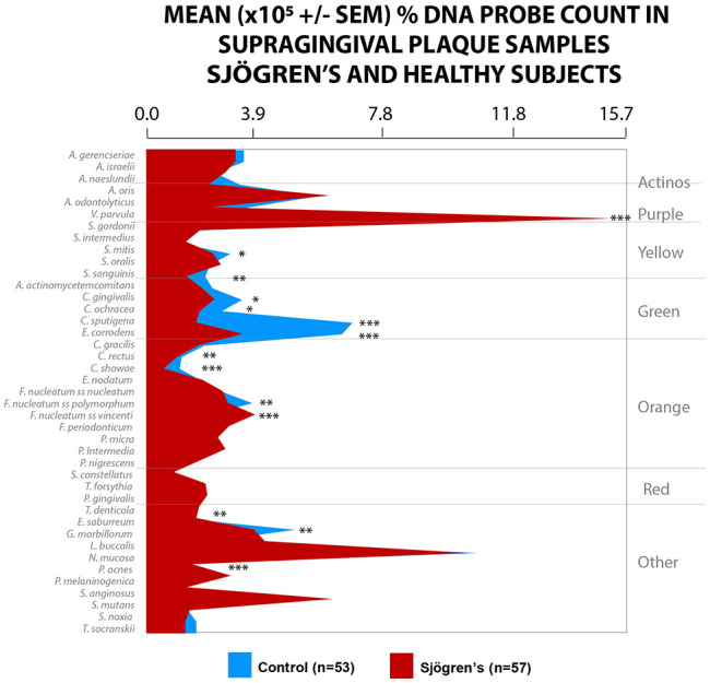
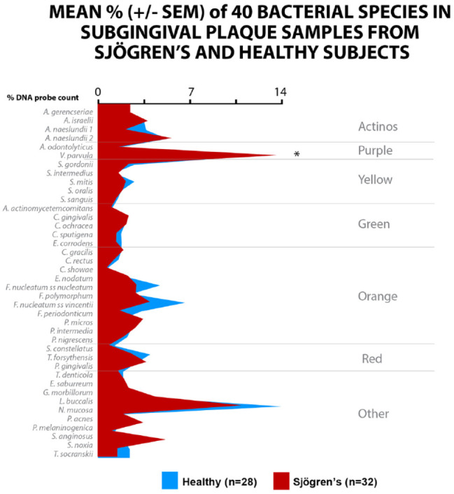
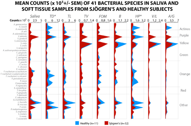
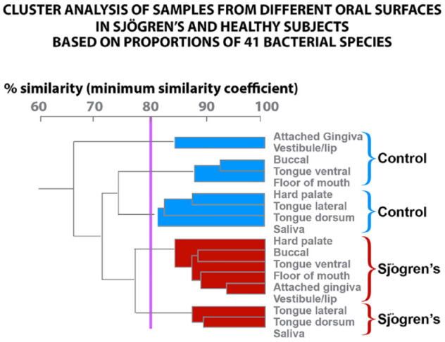
Methods: Supragingival and subgingival biofilm samples were collected from the mesial-buccal tooth surfaces of SS patients (n = 57) and age- and sex-matched HS (n = 53). Unstimulated saliva and 8 oral tissue samples were taken using a buccal brush. Caries and periodontal measures were recorded. All supragingival samples and a subgroup of 24 SS and 28 HS subgingival samples, as well as 32 SS and 11 HS saliva and oral tissue samples, were analyzed for their content of 41 bacterial species using checkerboard DNA-DNA hybridization. Mean levels (×105 ± SEM) and percentage of DNA probe counts of each species were determined for each sample site and averaged within subjects in the 2 clinical groups. Kruskal-Wallis tests, adjusting for multiple comparisons and cluster analysis, were used for soft tissue and microbial analysis, and the Mann-Whitney test was used to compare caries and periodontal measures.
Results: Mean (×105 ± SEM) total DNA probe counts in supragingival samples were significantly lower (P < 0.001) in the SS (13.3 ± .7) compared to the HS (44.1 ± 6.8) group. In supragingival samples, Veillonella parvula, Fusobacterium nucleatum ss vincenti, and Propionibacterium acnes were markedly elevated in the SS compared to the HS group in both mean (×105 ± SEM) and mean (± SEM) percentage DNA probe counts (P < 0.001). In subgingival samples of SS, V. parvula was significantly different compared to HS (P < 0.05). SS was characterized by high levels of purple and low levels of orange and red complexes. Cluster analysis of oral tissues and saliva demonstrated that the mean microbial profiles for SS patients and the HS group clustered separately. Active root caries (P < 0.003) and attachment loss were significantly higher (P < 0.029) in the SS group compared to the HS group.
Conclusion: These findings indicate that saliva is a major controlling factor of intraoral biofilm. V. parvula may be a unique microbial biomarker for Sjögren's syndrome.
Knowledge transfer statement: The microbiome characterized for Sjögren's syndrome in salivary hypofunction is shown to be under stress and reduced. Veillonella parvula can be a possible identification of a biomarker for Sjögren's syndrome.
Keywords: DNA-DNA hybridization; Veillonella parvula; bulk fluid; mean and percentages of DNA count; plaque; salivary hypofunction.
Conflict of interest statement
The authors declare no potential conflicts of interest with respect to the authorship and/or publication of this article.
Figures
References
-
- Almståhl A, Wikström M. 1999. Oral microflora in subjects with reduced salivary secretion. J Dent Res. 78(8):1410–1416. - PubMed
-
- Bhatti MA, Frank MO. 2000. Veillonella parvula meningitis: case report and review of veillonella infections. Clin Infect Dis. 31 (3): 839–840. - PubMed
-
- Dawes C. 1987. Physiological factors affecting salivary flow rate, oral sugar clearance, and the sensation of dry mouth in man. J Dent Res. 66 Spec No:648–653. - PubMed
-
- Delwiche EA, Pestka JJ, Tortorello ML. 1985. The veillonellae: gram-negative cocci with a unique physiology. Annu Rev Microbiol. 39:175–193. - PubMed
Publication types
MeSH terms
Substances
Supplementary concepts
Grants and funding
LinkOut - more resources
Full Text Sources
Medical
