Review
. 2020 Jul 20;20(1):107.
doi: 10.1186/s12894-020-00676-5.

Squamous cell carcinoma of the renal parenchyma presenting as hydronephrosis: a case report and review of the recent literature

Affiliations
Review

Squamous cell carcinoma of the renal parenchyma presenting as hydronephrosis: a case report and review of the recent literature

Xirong Zhang et al. BMC Urol. .

Abstract

Background: Primary squamous cell carcinoma of the renal parenchyma is extremely rare, only 5 cases were reported.

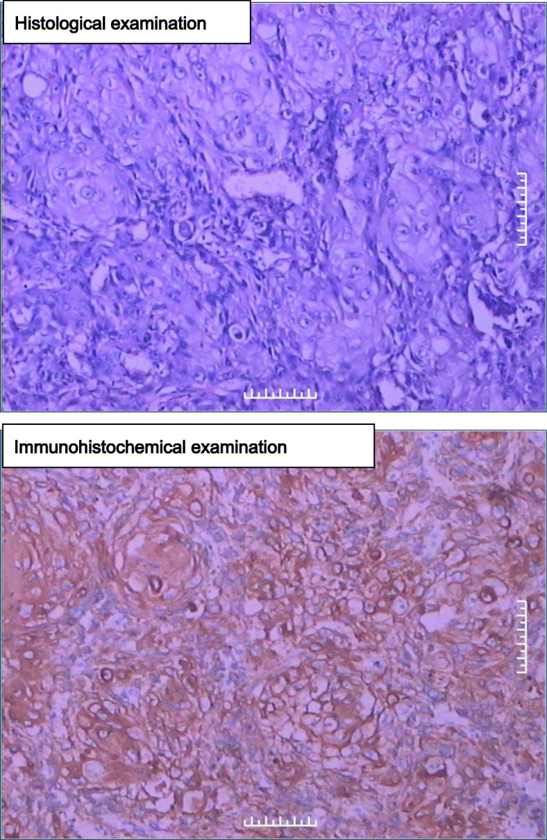
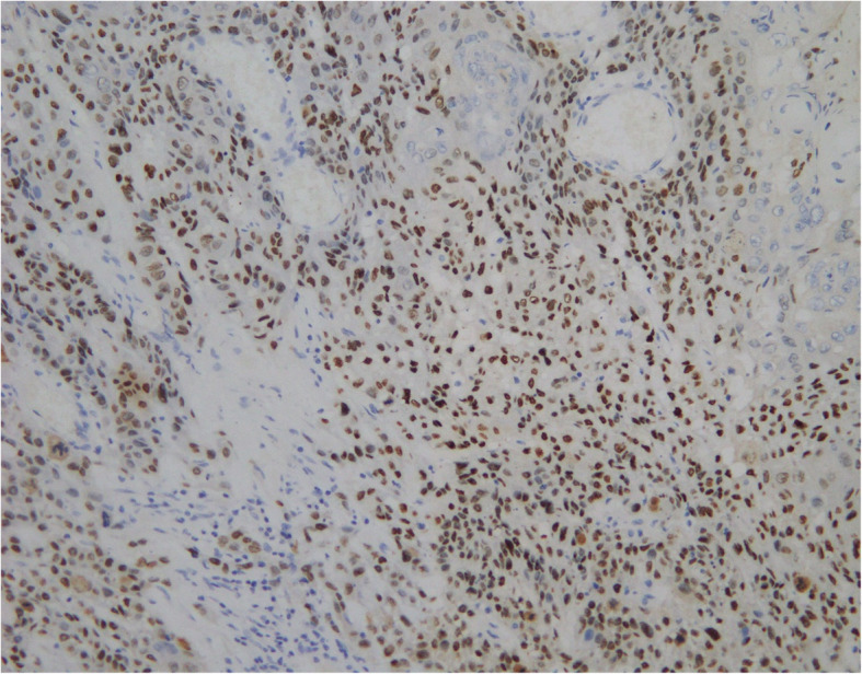
Case presentation: We probably report the fifth case of primary Squamous cell carcinoma (SCC) of the renal parenchyma in a 61-year-old female presenting with intermittent distending pain for 2 months. Contrast-enhanced computed tomography (CECT) revealed hydronephrosis of the right kidney, but a tumor cannot be excluded completely. Finally, nephrectomy was performed, and histological analysis determined that the diagnosis was kidney parenchyma squamous cell carcinoma involving perinephric adipose tissue.

Conclusions: The present case emphasizes that it is difficult to make an accurate preoperative diagnosis with the presentation of hidden malignancy, such as hydronephrosis.

Keywords: Hydronephrosis; Kidney; Malignancy; Renal parenchyma; Squamous cell carcinoma.


Conflict of interest statement

The authors declare that they have no competing interests.

Figures

Fig. 1
Fig. 1
a-c Contrast-enhanced computed tomography showed marked hydronephrosis of the right kidney and some perinephric exudates. d-f. Contrast-enhanced computed tomography revealed right gross hydronephrosis and right perinephric chronic inflammation; in the lower pole of the right kidney, a tumor could not be completely excluded with mild to moderate enhancement. g,h Plain scan of coronal and sagittal images
Fig. 2
Fig. 2
(upper) Histological examination, showing classical morphology of squamous carcinoma (HE staining, 200×). (Fig. 2 lower) immunohistochemistry, CK5/6 positive (200×)
Fig. 3
Fig. 3
immunohistochemistry of p63, positive (+++) (200×)

References

    1. Li MK, Cheung WL. Squamous cell carcinoma of the renal pelvis. J Urol. 1987;138(2):269–271. doi: 10.1016/S0022-5347(17)43116-8. - DOI - PubMed
    1. Terada T. Synchronous squamous cell carcinoma of the kidney, squamous cell carcinoma of the ureter, and sarcomatoid carcinoma of the urinary bladder: a case report. Pathol Res Pract. 2010;206(6):379–383. doi: 10.1016/j.prp.2009.07.021. - DOI - PubMed
    1. Kulshreshtha P, Kannan N, Bhardwaj R, Batra S. Primary squamous cell carcinoma of the renal parenchyma. Indian J Pathol Microbiol. 2012;55(3):370–371. doi: 10.4103/0377-4929.101747. - DOI - PubMed
    1. Pusiol T, Zorzi MG, Morini A. Comment on: primary squamous cell carcinoma of the renal parenchyma. Indian J Pathol Microbiol. 2013;56(1):70. doi: 10.4103/0377-4929.116160. - DOI - PubMed
    1. Ghosh P, Saha K. Primary Intraparenchymal squamous cell carcinoma of the kidney: a rare and unique entity. Case Rep Pathol. 2014;2014:1–3. doi: 10.1155/2014/256813. - DOI - PMC - PubMed