. 2020 Jul;8(2):e000999.
doi: 10.1136/jitc-2020-000999.

A heat-shocked melanoma cell lysate vaccine enhances tumor infiltration by prototypic effector T cells inhibiting tumor growth

Affiliations

A heat-shocked melanoma cell lysate vaccine enhances tumor infiltration by prototypic effector T cells inhibiting tumor growth

María Alejandra Gleisner et al. J Immunother Cancer. 2020 Jul.

Abstract

Background: Immune checkpoint blocker (ICB) therapy has shown survival benefits for some patients with cancer. Nevertheless, many individuals remain refractory or acquire resistance to treatment, motivating the exploration of complementary immunotherapies. Accordingly, cancer vaccines offer an attractive alternative. Optimal delivery of multiple tumor-associated antigens combined with potent adjuvants seems to be crucial for vaccine effectiveness.

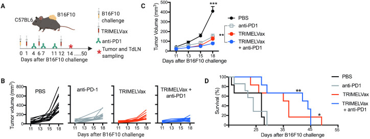
Methods: Here, a prototype for a generic melanoma vaccine, named TRIMELVax, was tested using B16F10 mouse melanoma model. This vaccine is made of heat shock-treated tumor cell lysates combined with the Concholepas concholepas hemocyanin as adjuvant.

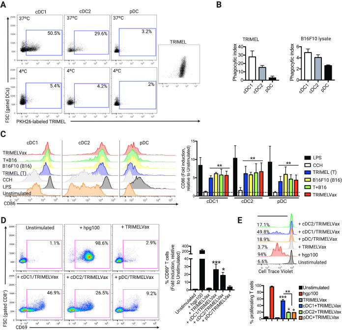
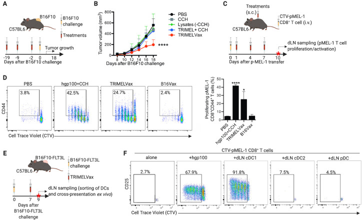
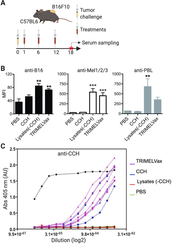
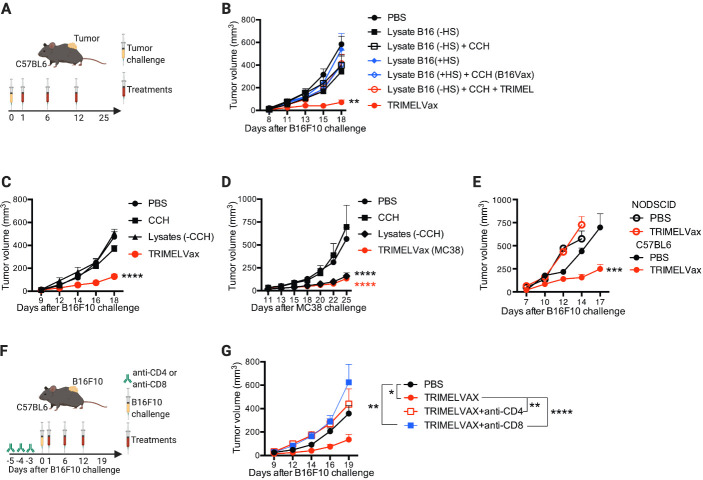
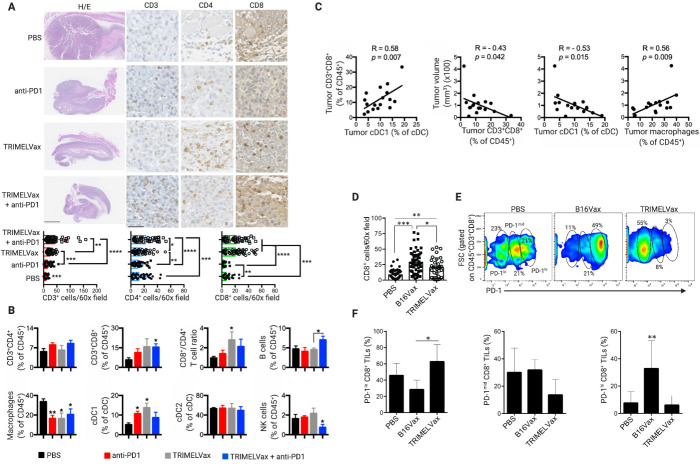
Results: While B16F10 lysate provides appropriate melanoma-associated antigens, both a generic human melanoma cell lysate and hemocyanin adjuvant contributes with danger signals promoting conventional dendritic type 1 cells (cDC1), activation, phagocytosis and effective antigen cross-presentation. TRIMELVax inhibited tumor growth and increased mice survival, inducing cellular and humoral immune responses. Furthermore, this vaccine generated an increased frequency of intratumor cDC1s but not conventional type 2 dendritic cells (cDC2s). Augmented infiltration of CD3+, CD4+ and CD8+ T cells was also observed, compared with anti-programmed cell death protein 1 (PD-1) monotherapy, while TRIMELVax/anti-PD-1 combination generated higher tumor infiltration of CD4+ T cells. Moreover, TRIMELVax promoted an augmented proportion of PD-1lo CD8+ T cells in tumors, a phenotype associated with prototypic effector cells required for tumor growth control, preventing dysfunctional T-cell accumulation.

Conclusions: The therapeutic vaccine TRIMELVax efficiently controls the weakly immunogenic and aggressive B16F10 melanoma tumor growth, prolonging tumor-bearing mice survival even in the absence of ICB. The strong immunogenicity shown by TRIMELVax encourages clinical studies in patients with melanoma.

Keywords: alarmins; immunogenicity, vaccine; immunotherapy, active; melanoma; therapies, investigational.


Conflict of interest statement

Competing interests: None declared.

Figures

Figure 1
Figure 1
Heatshock (HS)-conditioned melanoma cell lysates are phagocyted by conventional type 1 dendritic cells (cDC1) inducing cross-presentation of melanoma-associated antigen (MAA) in vitro. (A) Bone marrow dendritic cells differentiated with FLT3-L (FL-DCs) were incubated with PKH26-pre-stained TRIMEL (A, B) or B16F10 HS-conditioned cell lysates. Foward scatter (FSC) relative to CD69 expression was analyzed by flow cytometry (B). The percentages of cDC1, conventional type 2 dendritic cell (cDC2) and plasmacytoid dendritic cell (pDC) FL-DCs acquiring PKH26-labeled TRIMEL are shown in representative dot plots. (B) The bar graphs show the average phagocytic index for three independent experiments using TRIMEL (left) or B16F10 HS-conditioned cell lysate (right). (C) FL-DCs were stimulated with lipopolysaccharide (LPS), Concholepas concholepas hemocyanin (CCH), TRIMEL (T), B16F10 HS-conditioned cell lysate (B16), a 1:1 mixture of TRIMEL:B16F10 HS-conditioned cell lysate (T+B16), TRIMELVax or kept unstimulated. The expression level of CD86 was analyzed by flow cytometry on FL-DCs. The bar graphs (right) show CD86 expression as fold-change relative to unstimulated FL-DCs. (D) pMEL-1 CD8+ T cells were incubated with the peptide gp10025-33, TRIMELVax, with sorted and fixed TRIMELVax-loaded FL-DCs or kept unstimulated. CD69 expression was evaluated by flow cytometry. The bar graph (right) shows CD69 expression as fold-change relative to unstimulated T cells. (E) CellTrace Violet (CTV)-preloaded pMEL-1 CD8+ T cells were incubated with gp10025-33 peptide, TRIMELVax, sorted TRIMELVax-loaded FL-DCs or kept unstimulated. Representative histograms (upper) and graph bars (lower) show the percentage of proliferating CD8+ T cells. Statistical analysis was performed with two-way analysis of variance after Bonferroni correction. *p<0.05; **p<0.01; ***p<0.001.
Figure 2
Figure 2
TRIMELVax induces conventional type 1 dendritic cell (cDC1)-mediated CD8+ T-cell antimelanoma immune responses in vivo. (A) Schematic representation of prophylactic treatments. (B) Tumor growth curves of mice immunized with different treatments and challenged with B16F10 cells. Each point represents the mean tumor volume±SEM per group (n=7–10). (C) Schematic representation of antigen cross-presentation assay. (D) Proliferation (CellTrace Violet (CTV) dilution) and activation (% of CD44+) of CD8+ T cells were determined by flow cytometry. (E) Schematic representation of ex vivo antigen cross-presentation assay. (F) CTV-stained pMEL-1 CD8+ T cells were cocultured with draining lymph node dendritic cells (dLN-DCs), stimulated with hgp100 peptide or kept unstimulated. The proliferation and activation of the pMEL-1 CD8+ T cells was determined after 3 days of coculture. The percentage of proliferating T cells are shown in each representative density plot. Statistical analysis was performed with two-way analysis of variance after Bonferroni correction. *p<0.05; ****p<0.0001. CCH, Concholepas concholepas hemocyanin; cDC2, conventional type 2 dendritic cell; PBS, phosphate-buffered saline; pDC, plasmacytoid dendritic cell; s.c., subcutaneously.
Figure 3
Figure 3
TRIMELVax induces humoral antimelanoma immune responses in vivo. (A) C57BL6 mice (three per group) were challenged subcutaneously with B16F10 cells and then subcutaneously treated as shown. Serum samples were collected 6 days after the last immunization and the presence of IgGs against B16F10, Mel1/Mel2/Mel3 cells, human peripheralblood leucocyte (PBL) (B) or Concholepas concholepas hemocyanin (CCH) (C) were tested by flow cytometry (B) or ELISA (C). (C) The curves represent the anti-CCH antibodies present in different dilution of serum obtained from animals treated with TRIMELVax (purple), CCH alone (blue), lysates without CCH (red) or vehicle/phosphate-buffered saline (PBS) (green). The black line corresponds to the detection of purified CCH (ELISA positive control). **p<0.01; ***p<0.001. MFI, Mean of fluorescence. Abs, absorbance.
Figure 4
Figure 4
B16F10 tumor control by TRIMELVax requires heat shock (HS) pretreatment of melanoma cells and presence of CD4+ and CD8+ T cells. (A) Schematic representation of the therapeutic protocols used in (B–D). (B, C) Tumor growth curves of treated mice challenge with B16F10. Each point represents the mean tumor volume±SEM per group (n=7 (B) or=12 (C)). (D) Tumor growth curves of treated mice challenge with MC38. Each point represents the mean tumor volume±SEM per group (n=7). (E) Tumor growth curves of C57BL6 or NOD-SCID mice prophylactically treated with TRIMELVax or phosphate-buffered saline (PBS) and challenged with B16F10 cells. Each point represents the mean tumor volume±SEM per group (n=5). (F) Schematic representation of therapeutic protocols for CD4+ or CD8+ cell depletions and treatments used in (G). (G) Tumor growth curves of mice depleted or not for CD4+ or CD8+ cells challenged with B16F10 cells and treated with TRIMELVax. Statistical analysis was performed with two-way analysis of variance after Bonferroni correction. *p<0.05; **p<0.01; ***p<0.001; ****p<0.0001. CCH, Concholepas concholepas hemocyanin.
Figure 5
Figure 5
TRIMELVax controls tumor growth and increases survival of B16F10 tumor-bearing mice even in the absence of anti-PD-1 (programmed cell death protein 1) therapy. (A) Schematic representation of the therapeutic protocols for TRIMELVax/anti-PD-1 combinatory treatments. On day 14 after tumor challenging (asterisk), tumors and tumordraining lymph nodes (TdLNs) were sampled for 4–5 mice per group for further experiments. (B) Tumor growth curves of individual mice. (C) Each point represents the mean tumor volume±SEM per group (n=14). Statistical analysis was performed with two-way analysis of variance after Bonferroni correction. **p<0.01; ***p<0.001. (D) Kaplan-Meier curves for mice survival analysis (n=6–7 per group). The median survival time (in days after tumor challenge) per group is: phosphate-buffered saline (PBS), 27; anti-PD-1, 24; TRIMELVax, 36; TRIMELVax+anti-PD-1, 43. Statistical analysis was performed with log-rank (Mantel-Cox) test. *p<0.05; **p<0.01; ***p<0.001.
Figure 6
Figure 6
TRIMELVax promotes tumor infiltration of conventional type 1 dendritic cell (cDC1) and programmed cell death protein 1 (PD-1)lo CD8+ T cells. (A) Immunohistochemistry analysis for CD3+, CD4+ and CD8+ tumor infiltrating cells of animals treated as described in figure 5. H&E representative photomicrographs of full tumors (left panels; scale bar: 1 mm), and for CD3+, CD4+ and CD8+ immune staining of selected tumor areas (scale bar: 40 µM). Dot plot/bar graphs show the quantification of immune cell infiltration by 60× field of tumors. (B) Tumors were sampled from mice treated as described in figure 5 and immune cell frequencies (among CD45+ cells) were analyzed by flow cytometry. Bar graphs represent average±SEM per group (n=4–5). (C) Percentage of intratumor CD8+ T cells as a function of the percentage of intratumor cDC1 (left); and tumor volume as a function of the percentage of different immune cells in tumors. Pearson’s r correlation values and p values are shown. (D) Dot plot/bar graphs show the quantification of CD8+ cells by 60× field of tumors of animals therapeutically treated with TRIMELVax, B16Vax or phosphate-buffered saline (PBS). (E, F) Flow cytometry analysis of PD-1 expression on CD8+ tumor-infiltrating lymphocytes (TILs) of animals treated as in (D). The percentage of PD-1lo, PD-1mid and PD-1hi CD8+ TILs is showed (n=4–6 animals/group). Statistical analysis was performed with two-way analysis of variance after Bonferroni correction. *p<0.05; **p<0.01; ***p<0.001; ****p<0.0001.

Similar articles

Cited by

References

    1. Hodi FS, O'Day SJ, McDermott DF, et al. . Improved survival with ipilimumab in patients with metastatic melanoma. N Engl J Med 2010;363:711–23. 10.1056/NEJMoa1003466 - DOI - PMC - PubMed
    1. Sharma P, Allison JP. Immune checkpoint targeting in cancer therapy: toward combination strategies with curative potential. Cell 2015;161:205–14. 10.1016/j.cell.2015.03.030 - DOI - PMC - PubMed
    1. Khair DO, Bax HJ, Mele S, et al. . Combining immune checkpoint inhibitors: established and emerging targets and strategies to improve outcomes in melanoma. Front Immunol 2019;10:453. 10.3389/fimmu.2019.00453 - DOI - PMC - PubMed
    1. Ribas A, Dummer R, Puzanov I, et al. . Oncolytic virotherapy promotes intratumoral T cell infiltration and improves anti-PD-1 immunotherapy. Cell 2017;170:1109–19. 10.1016/j.cell.2017.08.027 - DOI - PMC - PubMed
    1. Bonaventura P, Shekarian T, Alcazer V, et al. . Cold tumors: a therapeutic challenge for immunotherapy. Front Immunol 2019;10:168. 10.3389/fimmu.2019.00168 - DOI - PMC - PubMed

Publication types

Substances