. 2020 Sep;12(9):832-837.
doi: 10.1038/s41557-020-0506-4. Epub 2020 Jul 20.

Single-molecule visualization of DNA G-quadruplex formation in live cells

Affiliations

Single-molecule visualization of DNA G-quadruplex formation in live cells

Marco Di Antonio et al. Nat Chem. 2020 Sep.

Abstract

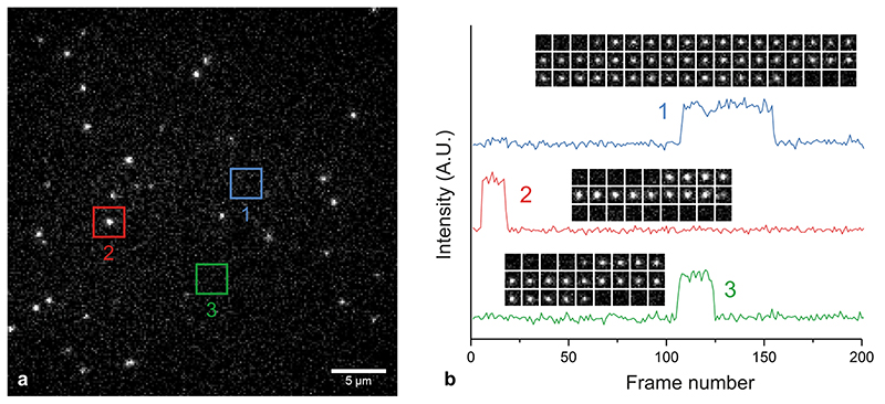
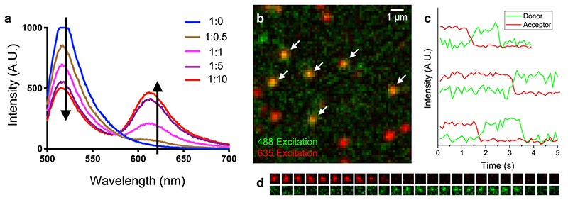
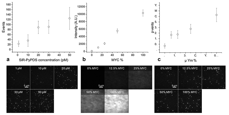
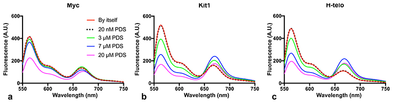
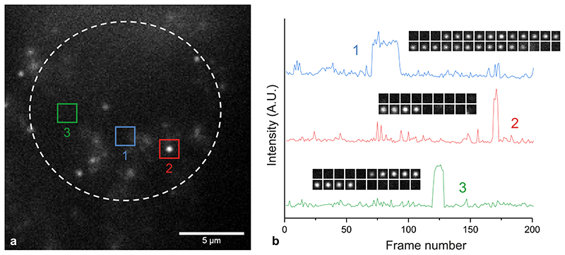
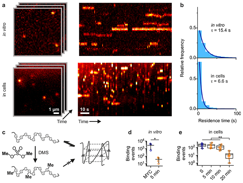
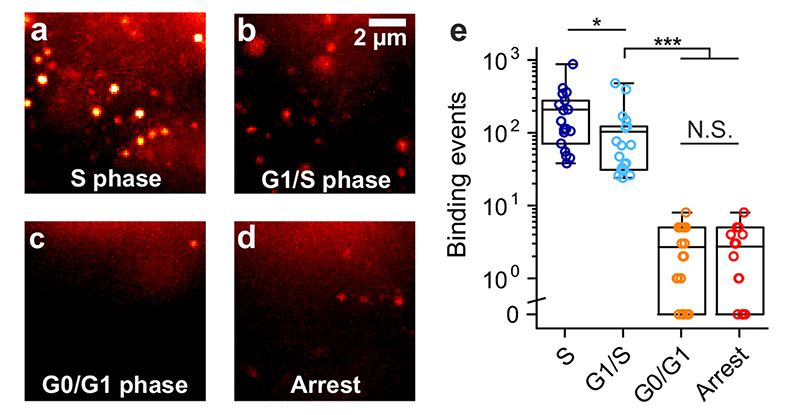
Substantial evidence now exists to support that formation of DNA G-quadruplexes (G4s) is coupled to altered gene expression. However, approaches that allow us to probe G4s in living cells without perturbing their folding dynamics are required to understand their biological roles in greater detail. Herein, we report a G4-specific fluorescent probe (SiR-PyPDS) that enables single-molecule and real-time detection of individual G4 structures in living cells. Live-cell single-molecule fluorescence imaging of G4s was carried out under conditions that use low concentrations of SiR-PyPDS (20 nM) to provide informative measurements representative of the population of G4s in living cells, without globally perturbing G4 formation and dynamics. Single-molecule fluorescence imaging and time-dependent chemical trapping of unfolded G4s in living cells reveal that G4s fluctuate between folded and unfolded states. We also demonstrate that G4 formation in live cells is cell-cycle-dependent and disrupted by chemical inhibition of transcription and replication. Our observations provide robust evidence in support of dynamic G4 formation in living cells.


Conflict of interest statement

Competing Interests

S.B. is a founder and shareholder of Cambridge Epigenetix Ltd.

Figures

Extended Data Fig. 1
Extended Data Fig. 1. SiR-PyPDS analogues synthesized
SiR-PyPDS analogues (1, 2, 3 and 4) with different chemical linkers between the PyPDS and the SiR scaffold synthesised in this study. TSTU was used as amide coupling reagent in the synthesis of all 3 analogues.
Extended Data Fig. 2
Extended Data Fig. 2. Single-step photobleaching confirms detection of individual probes.
(a) 25 pM SiR-PyPDS binding to MYC in vitro. The red square indicates a single binding event. (b) Intensity traces from three binding events in (a), showing probes undergoing single-step photobleaching. The insets show time lapses for each molecule. Similar single-step photobleaching could be consistently observed in all single-molecule video acquisitions.
Extended Data Fig. 3
Extended Data Fig. 3. FRET between SiR-PyPDS and Alexa Fluor 488-labelled MYC confirms direct binding to G4s.
(a) Emission spectrum of 488-MYC-G4 at 1 μM and SiR-PyPDS at various stoichiometric ratios. As the probe concentration increases, donor emission drops and acceptor emission increases, indicating FRET. (b) In vitro G4 FRET experiment. 250 pM of SiR-PyPDS (shown in red with acceptor excitation) interacting with Alexa Fluor 488-labelled MYC-G4 (with ~1% surface coverage). The green channel shows acceptor emission under donor excitation. FRET between MYC and SiR-PyPDS is highlighted with white arrows. (c) 10 nM SiR-PyPDS interacting with 488-MYC-G4 (0.001% surface coverage). Temporal intensity traces of donor (green) and acceptor (red) emission under donor excitation. Anti-correlated intensity fluctuation upon acceptor photobleaching indicates single-molecule FRET between PyPDS and MYC. (d) Example time lapse of acceptor (top, red) and donor (bottom, green) emission from (c). Experiments a-d were performed as 3 independent replicates all providing similar results.
Extended Data Fig. 4
Extended Data Fig. 4. Single-molecule imaging with SiR-PyPDS can be used to quantify MYC-G4 prevalence in vitro.
(a) Number of detected binding events increases with probe concentrations. (b) MYC fluorescence showing that the concentration of MYC on the surface can be controlled by mixing with a competing biotinylated oligomer. (c) Number of detected events increases with G4 concentration. Sample images for each condition is shown beneath each plot. Error bars indicate mean ± sd. n = 12 measurements taken from 2 independent replicates.
Extended Data Fig. 5
Extended Data Fig. 5. Induction of G4-folding by increasing concentrations of SiR-PyPDS measured with dually labelled FRET oligos:
(a-c) Fluorescence emission spectra under Cy3 excitation for each G4 sequence. Experiments a-c were performed as 3 independent replicates all providing similar results.
Extended Data Fig. 6
Extended Data Fig. 6. The effect on SiR-PyPDS binding on unfolding kinetics of G4 DNA sequences in vitro.
G4 two-phase unfolding kinetics were measured by introducing 10 μM of respective complimentary DNA oligonucleotide at t = 0 to trap the unfolded G4 oligonucleotide state. Data presented here are of best fit of a two-phase association model. Error indicates the standard error of the fit. n = 1 measurement for each condition. Each experiment has been repeated 3 times providing consistent results
Extended Data Fig. 7
Extended Data Fig. 7. Single-step photobleaching confirms detection of individual probes in cells
(a) 20 nM SiR-PyPDS binding to targets in a living cell. The red square indicates a single binding event. (b) Intensity traces from three binding events in A, showing probes undergoing single-step photobleaching. The insets show time lapses for each molecule. Similar single-step photobleaching could be observed in all single-molecule video acquisitions.
Extended Data Fig. 8
Extended Data Fig. 8. SiR-PyPDS mainly accumulates in lysosomes.
Representative confocal and HILO microscopy images obtained in the presence of SiR-PyPDS (1 μM in confocal and 40 nM in HILO) and LysoTracker Green (50 nM), confirming co-localisation of extranuclear staining with lysosomes. Experiments have been repeated 3 times providing similar results.
Extended Data Fig. 9
Extended Data Fig. 9. Total nuclear accumulation of SiR-PyPDS and SiR-iPyPDS in U2OS cells.
Total fluorescence intensity measured inside the nuclei of >300 U2OS cells after incubation with 10μM SiR-PyPDS or SiR-iPyPDS by standard confocal microscopy at 633 nm. Each point on the graph represents the total fluorescence of SiR measured at 633 nm per nuclei, data are plotted as the mean of >300 nuclei measured in 3 independent replicates. Total fluorescence measurement revealed comparable ability of the two molecules to accumulate in the nuclei. Error bars indicate mean ± sd.
Extended Data Fig. 10
Extended Data Fig. 10. Cellular displacement experiments of SiR-PyPDS with the established G4-ligands PDS and PhenDC3
Displacement of SiR-PyPDS in cells by competition with 10 μM of unlabelled G4-ligands PDS and PhenDC3. Cells were pre-incubated 30 minutes with PDS or PhenDC3 at 10 μM prior standard single-molecule imaging with SiR-PyPDS. Each point on the graph depicts the number of long-lived SiR-PyPDS event measured in independent replicates. Data are plotted as the mean of 3 or more independent replicates. Error bars indicate mean ± sd. * P < 0.05, two-sided Mann-Whitney U-test. n = 5,3 and 3 measurements taken from 3 independent replicates for no displacement, PDS displacement and PhenDC3 displacement respectively.
Figure 1
Figure 1. In vitro single-molecule fluorescence imaging of G-quadruplexes.
(a) Schematic representation of a G-tetrad (left) and a G4 structure (right). (b) Chemical structure of the selective G4-fluorogenic ligand SiR-PyPDS (1) and its inactive isomer, SiR-iPyPDS (2). (c) Schematic of methodology used for visualizing individual G4s. Pre-folded G4 MYC is attached to a coverslip via a biotin-neutravidin linker. The fluorescent G4-probe SiR-PyPDS (1) binds to G4 MYC, which can be visualized using single-molecule fluorescence imaging. (d) SiR-PyPDS will not bind single stranded mutated-MYC that cannot form a G4. (e) The inactive isomer SiR-iPyPDS (2) with its 10 times reduced binding affinity is less likely to bind G4 MYC. (f) Quantification of SiR-PyPDS (1) binding to the G4 MYC ii) SiR-PyPDS (1) binding to the mutated-MYC; iii) SiR-iPyPDS (2) binding to the G4 MYC; iv) SiR-PyPDS (1) binding to the G4 MYC in the presence of 10 μM unlabeled PhenDC3 competitor. Error bars indicate mean ± sd. *** P < 10-5, unpaired two sided t-test, n = 12 measurements form 3 independent replicates. (g) Representative images (500 ms exposure) of individual SiR-PyPDS (1) molecules (250 pM) binding to a surface coated with pre-folded MYC G4 oligonucleotide; individual fluorescent puncta indicate binding of single SiR-PyPDS (1) molecules. (H) SiR-PyPDS (1) (250 pM) binding to mutated-MYC. (i) SiR-iPyPDS (2) (250 pM) binding to pre-folded MYC. Experiments g-i were repeated 3 times independently with similar results. (j) Interactions of G4 ligands and G4s can alter the equilibrium between unfolded and folded G4s. Error bars indicate mean ± sd. n = 12 measurements, from 3 independent replicates. Changes in the FRET ratio can be observed at μM PDS concentrations for c-KIT1 and hTelo and larger concentrations for MYC, indicative of G4 induction, which does not occur at lower concentrations.
Figure 2
Figure 2. Single-molecule fluorescence imaging of G-quadruplexes in living cells using the fluorescent probe SiR-PyPDS (1).
(a) Schematic of G4s in the cell nucleus with a zoom-in showing G4s stained by SiR-PyPDS (1). (b) Representative background-subtracted image (max projection of 100 frames with 200 ms exposure) of SiR-PyPDS (1) binding events in a living U2OS cell treated with 20 nM SiR-PyPDS (1) for 30 min before imaging; fluorescent puncta indicate binding of single SiR-PyPDS (1) molecules. Blue color corresponds to nuclear staining with Hoechst 33342. Scale bar is 2 μm. Inset scale bar is 1 μm. (c) Representative image of SiR-iPyPDS (2) staining in living U2OS cell treated with 20 nM SiR-PyPDS (1) for 30 min before imaging. Experiments b-c were repeated 3 times independently with similar results. (d) Quantification of the binding events within the nucleus lasting more than one frame (100 ms per frame) per cell for SiR-PyPDS (1) and SiR-iPyPDS (2). Center lines indicate the median; boxes show interquartile range; whiskers denote 5th and 95th percentiles. *** P = 3.5×10⁻⁷, two-sided Mann-Whitney U-test, n = 18 measurements from 6 cells each time in 3 independent replicates.
Figure 3
Figure 3. G-quadruplexes in living cells undergo dynamic folding/unfolding.
(a) Single-molecule time-lapse imaging of SiR-PyPDS (1) in vitro (top) and in cells (bottom). Individual images from the time-lapse stack are shown on the left and kymographs on the right show the dynamic binding kinetics of SiR-PyPDS (1) to G4s. Experiments were repeated 3 times independently with similar results. (b) The histograms of dwell times for each experiment (3 positions on a cover slip for in vitro and 6 cells for the cell experiment) were fitted with a single-exponential fit to determine the binding lifetime in each condition. (c) Schematic of DMS-meditated chemical trapping of unfolded G4s. (d) Quantification of G4-binding events for untreated and 600 mM DMS-treated G4 MYC for 20 min. Error bars indicate mean ± sd. * P = 0.05, two-sided Mann-Whitney U-test. n = 3 (MYC) and n = 4 (DMS) measurements taken from 3 independent replicates. (e) Quantification of G4-binding events detected in living cells upon increased exposure to DMS (20 mM), showing a clear time-dependent depletion of G4s. Center lines indicate the median; boxes show interquartile range; whiskers denote 5th and 95th percentiles. ** P < 0.01, two-sided Mann-Whitney U-test. n = 5, 6, 7 and 8 cells for untreated, 5 min, 10 min and 20 min respectively, taken from 3 independent replicates.
Figure 4
Figure 4. The observation of G4s in live cells is altered by the cell cycle phase and transcription.
Representative single-molecule images of G4-binding events are shown for synchronized U2OS cells in (a) the S phase, (b) the G1/S phase, (c) the G0/G1 phase and (d) for unsynchronized cells treated with both the transcriptional inhibitor DRB and the replication inhibitor Aphidicolin. Experiments a-d were repeated 3 times independently with similar results. (e) Quantification of binding events lasting more than two frames (100 ms per frame) per cell in living U2OS cells at different cell-cycle phases and after transcription/replication arrest. Center lines indicate the median; boxes show interquartile range; whiskers denote 5th and 95th percentiles. *** P < 10-6, * P = 0.01, N.S P = 0.99, two-sided Mann-Whitney U test. n = 18, 19, 19 and 15 cells for S, G0, G1 and Arrest respectively, taken from 3 independent replicates.

References

    1. Sen D, Gilbert W. Formation of parallel four-stranded complexes by guanine-rich motifs in DNA and its implications for meiosis. Nature. 1988;334:364–366. - PubMed
    1. Hänsel-Hertsch R, Di Antonio M, Balasubramanian S. DNA G-quadruplexes in the human genome: detection, functions and therapeutic potential. Nat Rev Mol Cell Biol. 2017;18:279–284. - PubMed
    1. Chambers, et al. High-throughput sequencing of DNA G-quadruplex structures in the human genome. Nat Biotechnol. 2015;33:877–881. - PubMed
    1. Schaffitzel C, et al. In vitro generated antibodies specific for telomeric guanine-quadruplex DNA react with Stylonychia lemnae macronuclei. Proc Natl Acad Sci U S A. 2001;98:8572–8577. - PMC - PubMed
    1. Biffi G, Tannahill D, McCafferty J, Balasubramanian S. Quantitative visualization of DNA G-quadruplex structures in human cells. Nat Chem. 2013;3:182–186. - PMC - PubMed

Publication types

Substances