Review
. 2020 Aug 5;18(30):5747-5763.
doi: 10.1039/d0ob01131b.

Rational design of small molecule fluorescent probes for biological applications

Affiliations
Review

Rational design of small molecule fluorescent probes for biological applications

Joomyung V Jun et al. Org Biomol Chem. .

Abstract

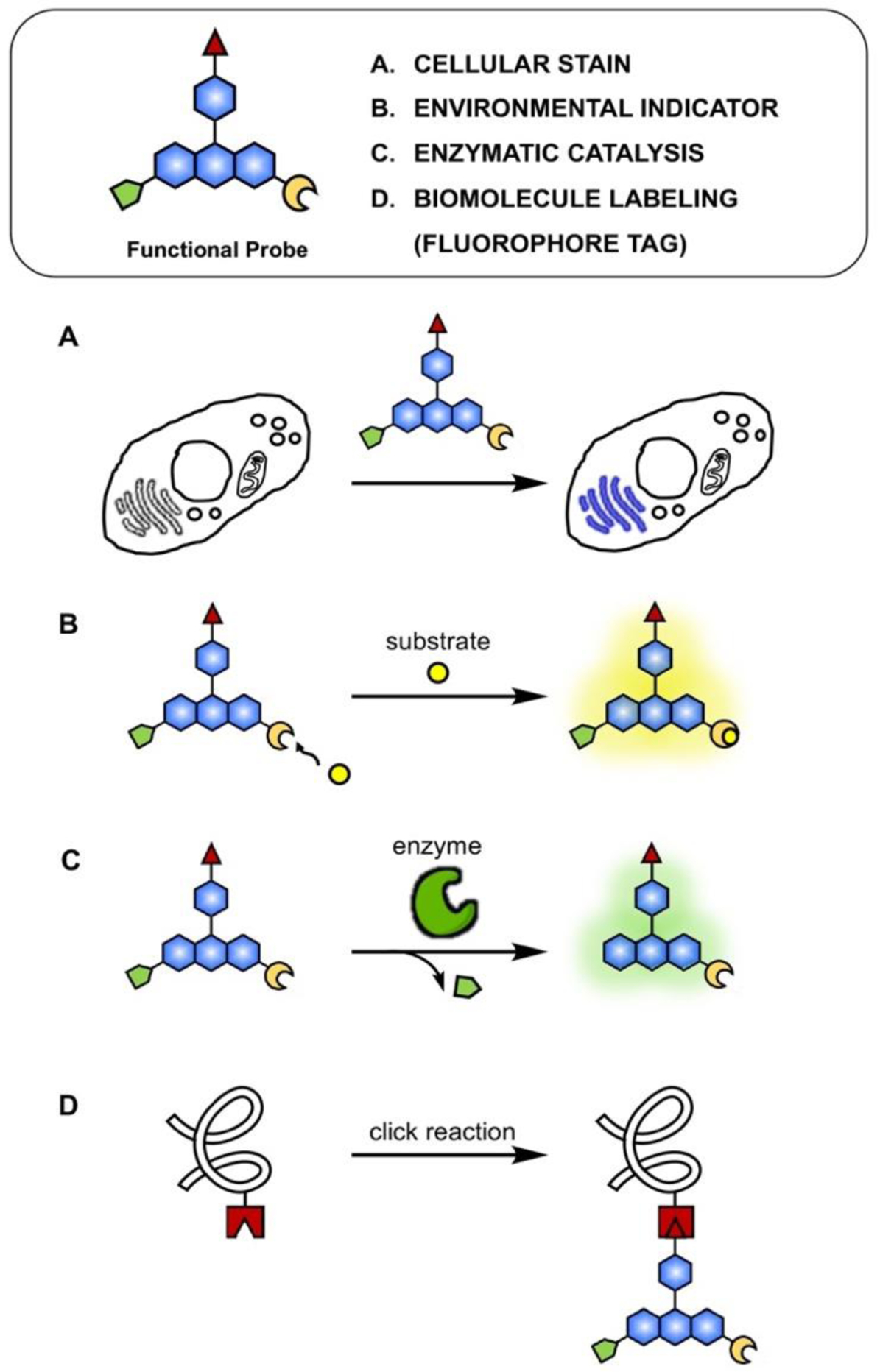
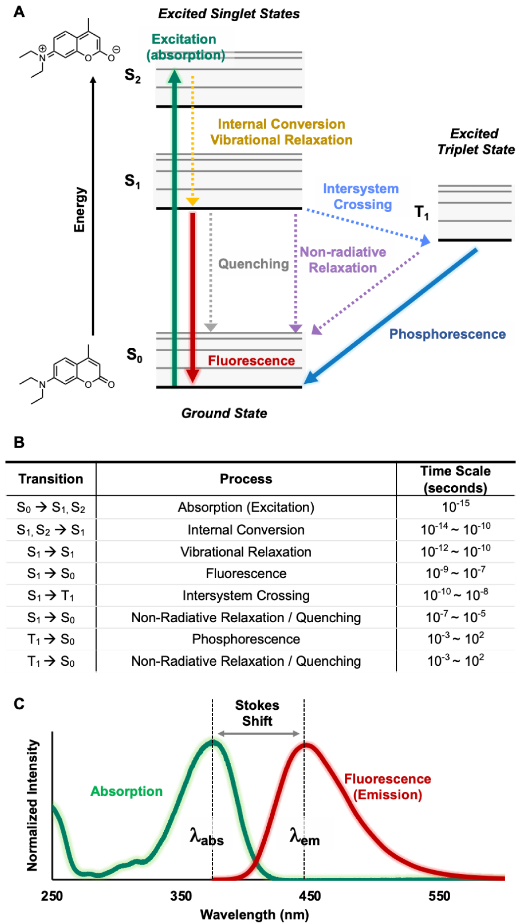
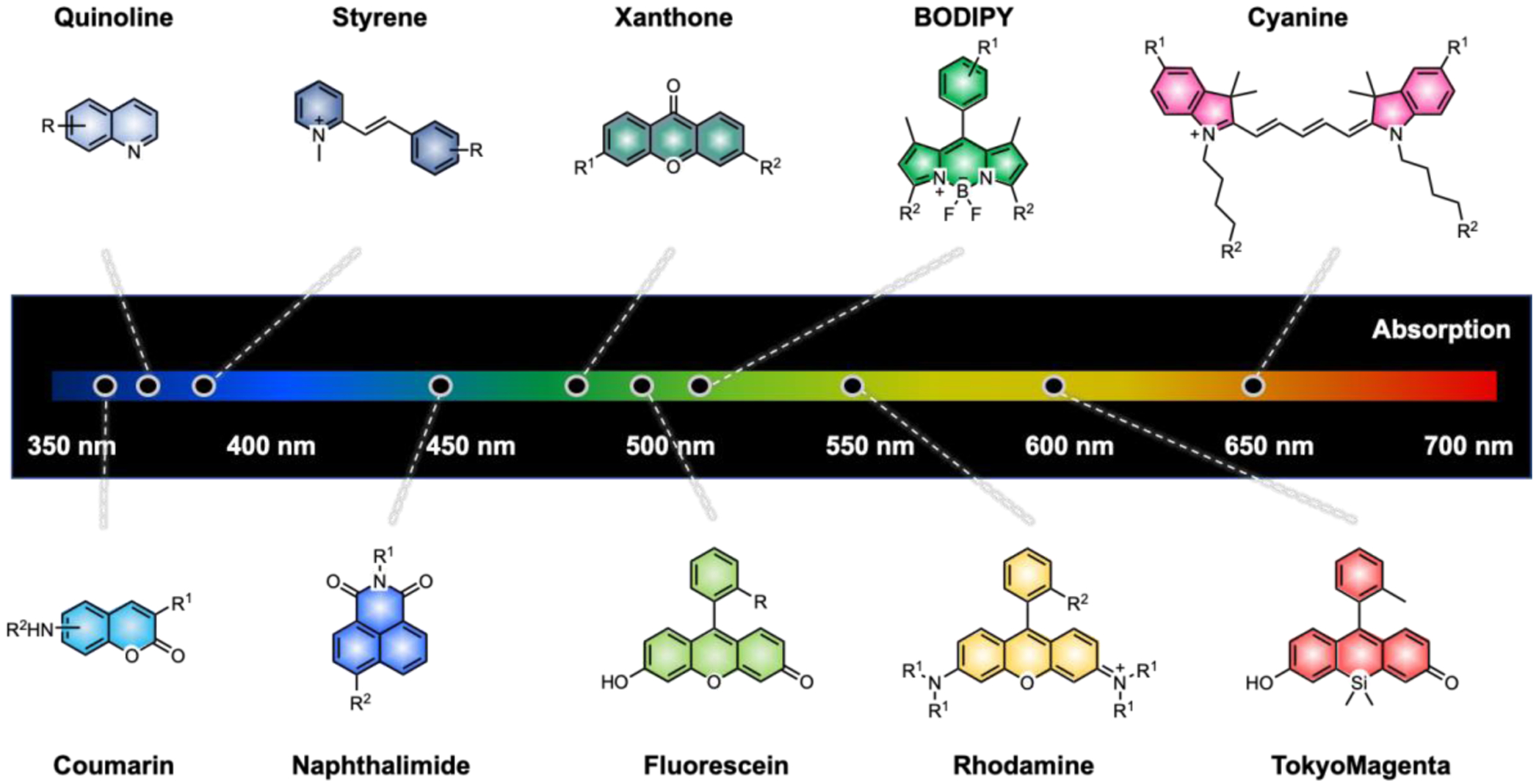
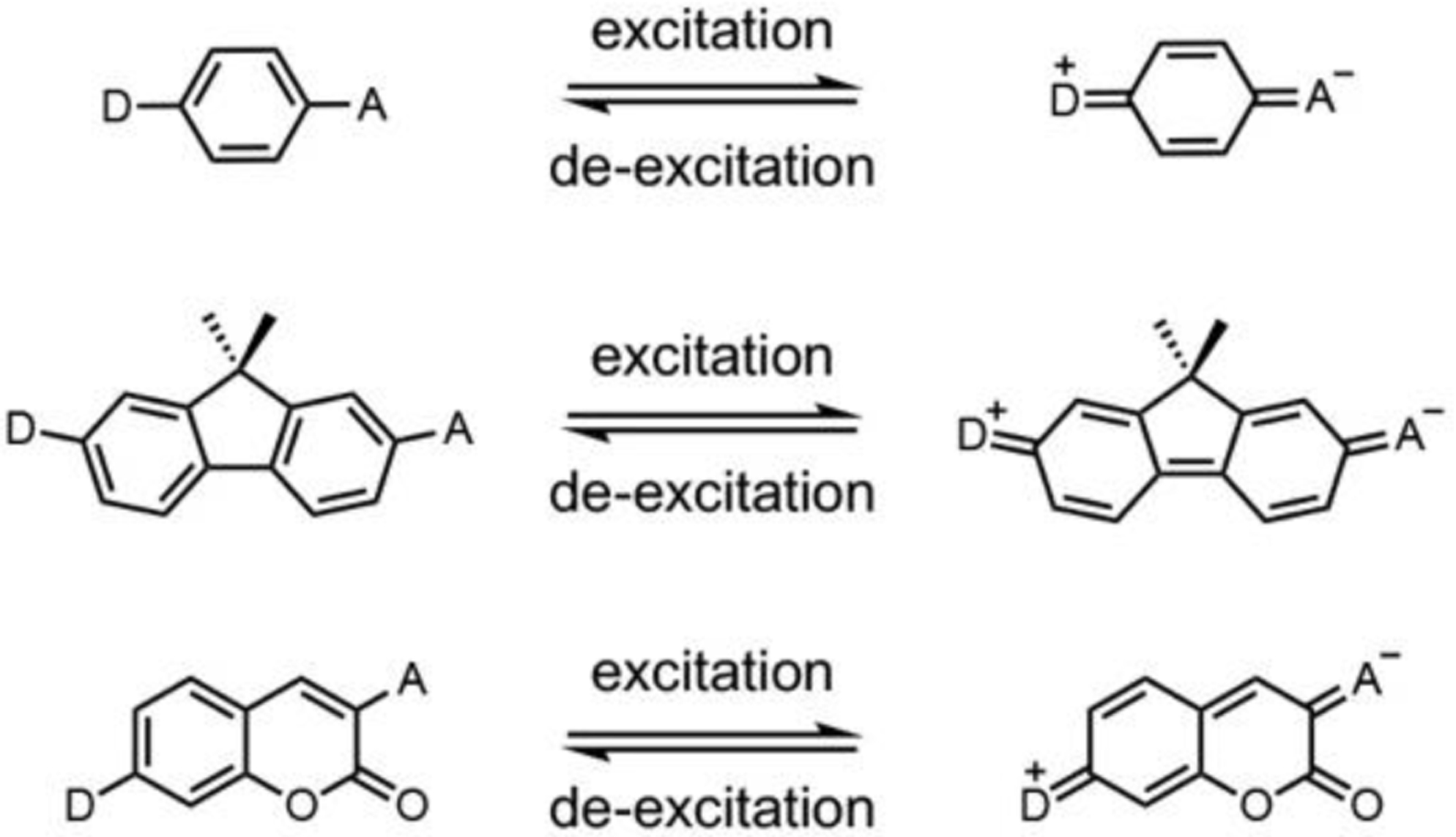
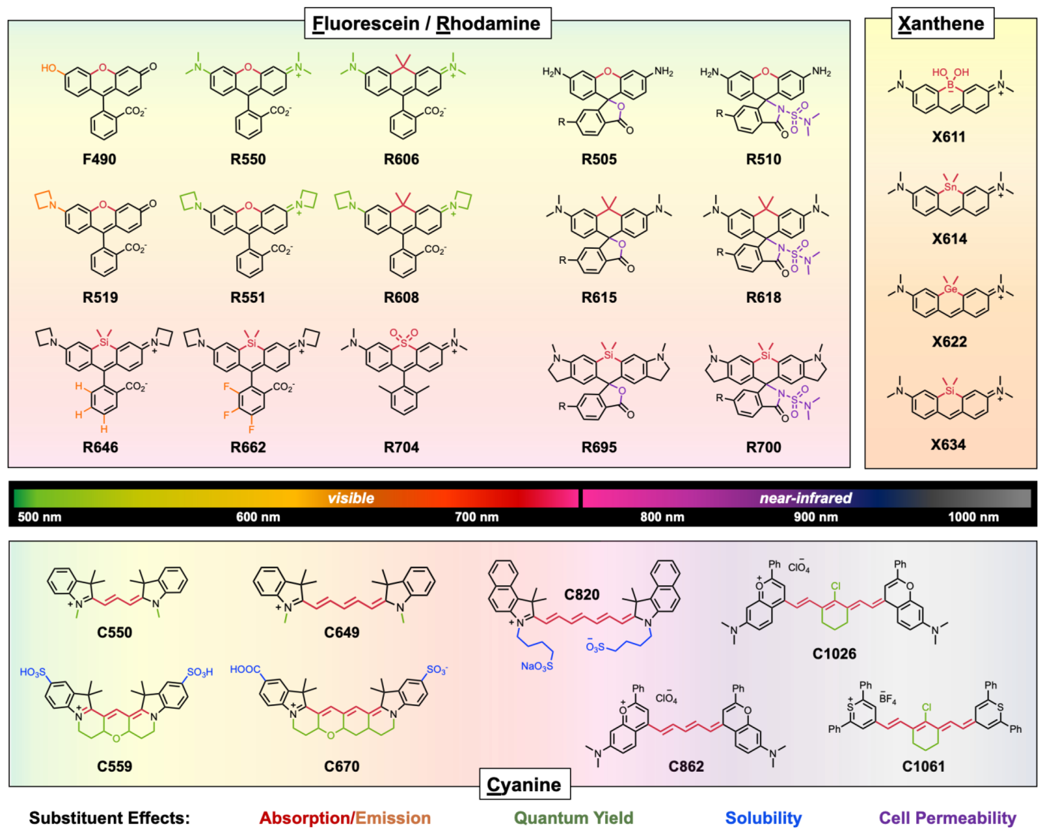
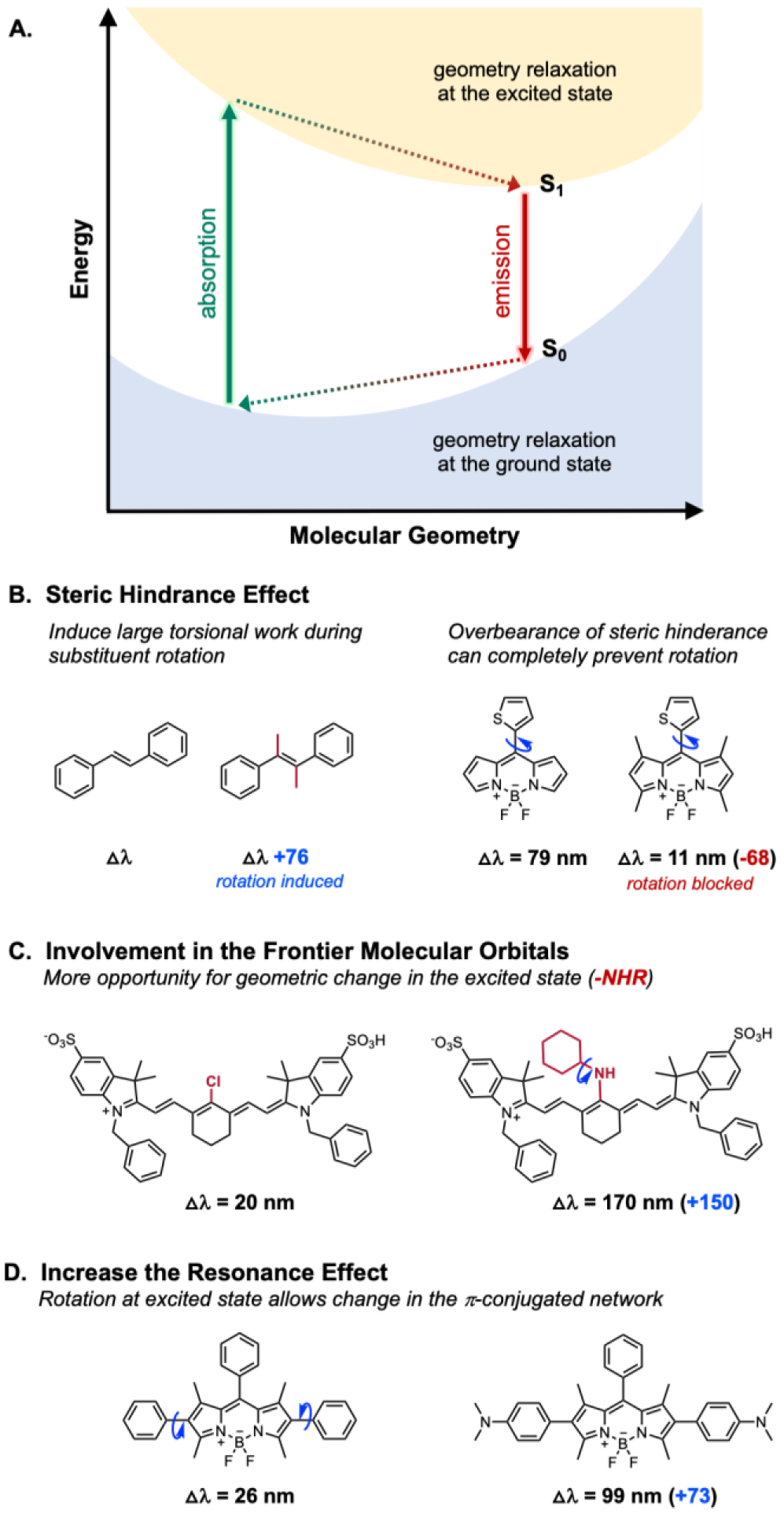
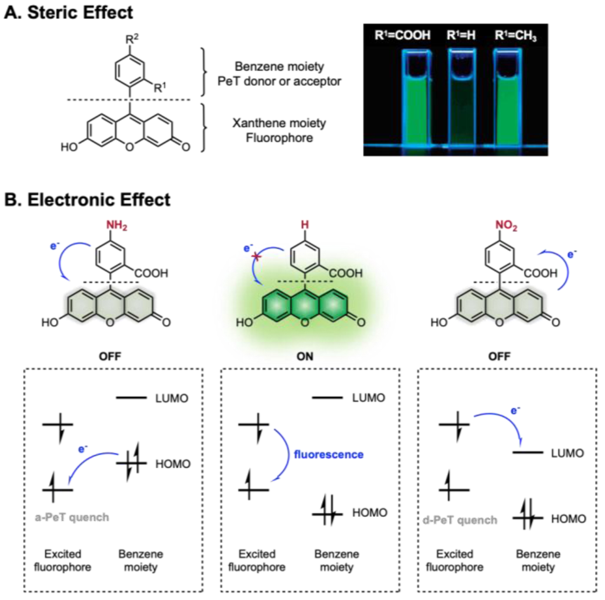
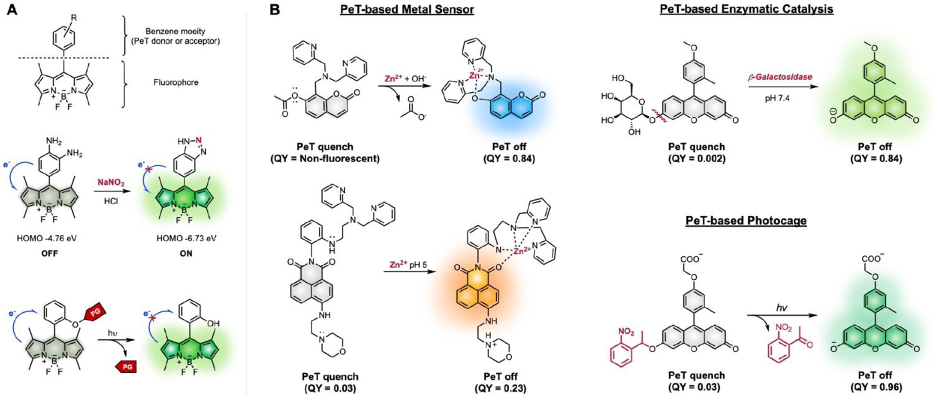
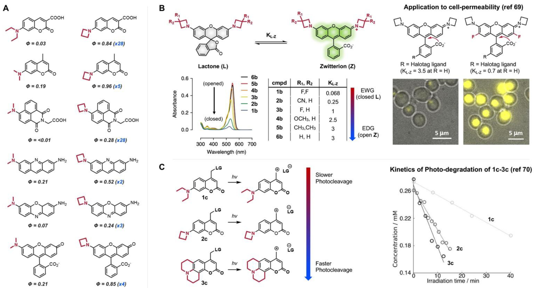
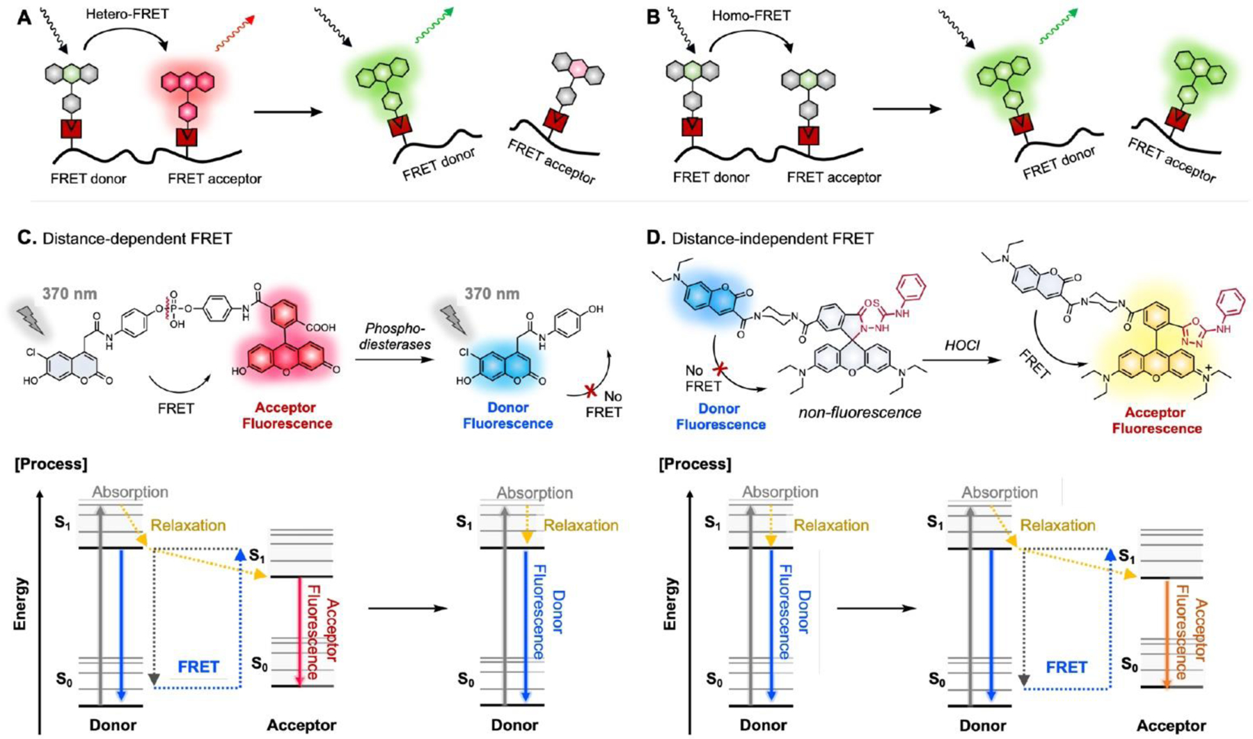
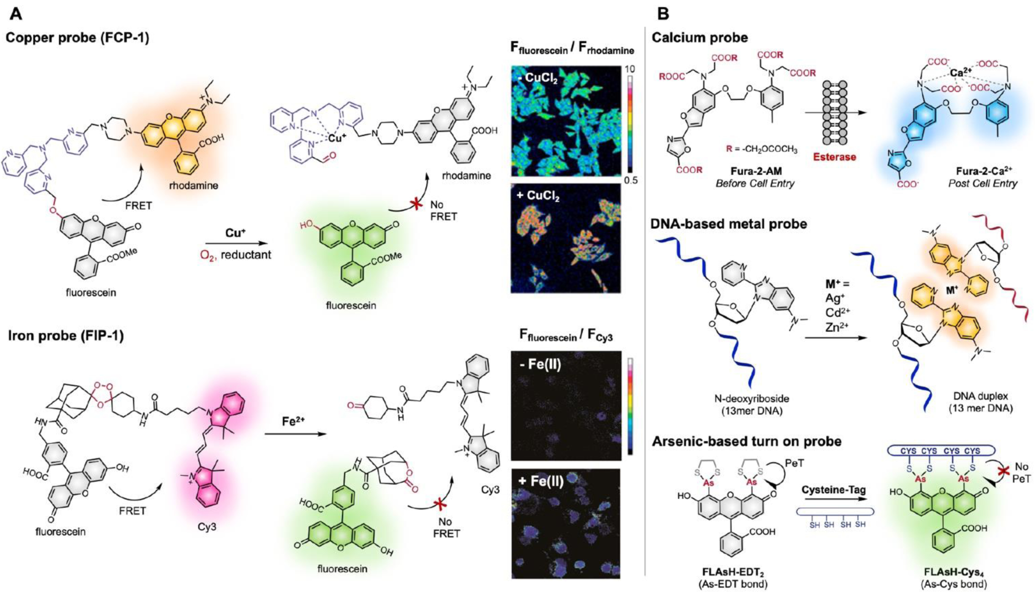
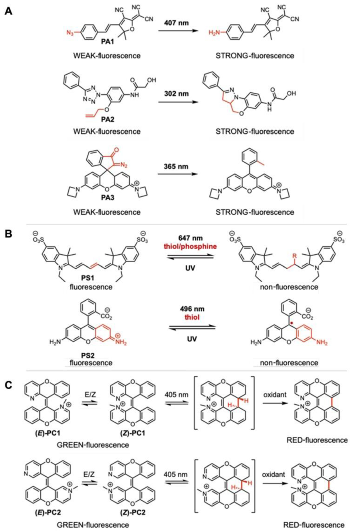
Fluorescent small molecules are powerful tools for visualizing biological events, embodying an essential facet of chemical biology. Since the discovery of the first organic fluorophore, quinine, in 1845, both synthetic and theoretical efforts have endeavored to "modulate" fluorescent compounds. An advantage of synthetic dyes is the ability to employ modern organic chemistry strategies to tailor chemical structures and thereby rationally tune photophysical properties and functionality of the fluorophore. This review explores general factors affecting fluorophore excitation and emission spectra, molar absorption, Stokes shift, and quantum efficiency; and provides guidelines for chemist to create novel probes. Structure-property relationships concerning the substituents are discussed in detail with examples for several dye families. We also present a survey of functional probes based on PeT, FRET, and environmental or photo-sensitivity, focusing on representative recent work in each category. We believe that a full understanding of dyes with diverse chemical moieties enables the rational design of probes for the precise interrogation of biochemical and biological phenomena.


Conflict of interest statement

Conflicts of interest

There are no conflicts to declare.

Figures

Fig. 1
Fig. 1
General types of fluorescent probe for biological applications. A. Cellular stains with specific localization; B. Environmental indicators that activate fluorescence upon reacting with substrates (e.g., protonation, intracellular metals); C. Indicators for enzymatic activity (e.g., esterase cleaving off ester tag in fluorophore); D. Biorthogonal tags (e.g., azide-alkyne 1,3-dipolar cycloaddition, oxime/hydrazine formation from aldehyde/ketones, tetrazine ligation, and photo-crosslinking).
Fig. 2
Fig. 2
Photophysical processes and properties. A. Jabłoński diagram; B. Time scale of each process in order of fastest to slowest; C. Fluorescence spectra (e.g., coumarin) with schematic view of Stokes shift, absorption maximum (λabs) and emission maximum (λem).
Fig. 3
Fig. 3
Core fluorescent scaffolds. Common scaffolds listed in order of increasing absorption values. R groups indicate sites for common functionalization.
Fig. 4
Fig. 4
Donor-π-Acceptor “Push-Pull” moiety in three fluorescent small molecule scaffolds. Donor (D) typically refers to aliphatic, aromatic, or saturated cyclic amines. Acceptor (A) frequently refers to formyl, succinyl, keto, or triazolyl electron withdrawing groups.
Fig. 5
Fig. 5
Structure-photophysical relationship chart of visible and NIR dyes. From ‘top to down’ or ‘left to right’ within the core, increase in absorption and emission (red-xanthene substituent /orange-phenyl ring substituent), Increase in quantum yield (green), increase in cell permeability (purple), and increase in solubility in water (blue). Compound naming consist of first letter (F, R, X, or C) of core scaffold and its maximum absorption. R group is HaloTag ligand.
Fig. 6
Fig. 6
Choice of rotating substituent on Stokes shift (△λ). A. Schematic illustration of absorption and emission process in fluorophore, B. Steric hinderance effect on Stokes shift, C. Effect of frontier molecular orbital in Stokes shift, and D. Resonance effect on Stokes shift.
Fig. 7
Fig. 7
Rational design of PeT-based fluorescein scaffold. A. Steric effect of R1 in modulating PeT by rotation restriction of benzene moiety. B. Electronic effect of R2 in modulating PeT by tuning HOMO/LUMO of benzene moiety. Adapted with permission from ref . Copyright (2005) American Chemical Society.
Fig. 8
Fig. 8
Examples of PeT based turn-on probes.. A. Development of PeT-based sensors in BODIPY scaffold. Chemical stimuli (nitric oxide) or light to change stereo-electronic property of benzene moiety via chemical modification or removal of photocleavable protecting group (PG), respectively. B. PeT-based sensors utilizing metal, enzyme, or light.
Fig. 9
Fig. 9
Effect of restricting the bond rotation of dialkyl amine by installing restricted azetidine. A. Improved quantum yields of fluorophore with azetidine than that with dialkyl amine. B. Electronic effect of azetidinyl ring in spirolactam equilibrium constant (KL-Z) and cell permeability. Adapted with permission from ref . Copyright (2019) American Chemical Society. C. Effect of the bond rotation in photocleavage. Adapted with permission from ref . Copyright (2018) Published by Royal Society of Chemistry
Fig. 10
Fig. 10
A. General scheme for hetero-FRET and homo-FRET. C-D. Distance dependent/independent FRET and its photophysical process in Jabłoński diagram.
Fig. 11
Fig. 11
Examples of activity-based metal sensing probes (left),, and metal-chelation based probes (right). Adapted from ref with Copyright (2016) American Chemical Society. Adapted from ref with Copyright (2019) National Academy of Science
Fig. 12
Fig. 12
A. Examples of photoactivatable dyes that undergo irreversible fluorescence off to ON (i.e., turn-on), B. examples of photoswitchable dyes with reversible fluorescence on and off, and C. examples of photoconvertible dyes with irreversible change in fluorescence from one to the other (e.g., change from green to red fluorescence) upon light illumination.
Fig. 13
Fig. 13
Application of photoconvertible probe (PC1) in tracking α-synuclein (αS). A. Synthesis and transduction of CPX labelled preformed fibrils of engineered αS (CPX-pffs) B. Sequential photoconversion of internalized CPX-pffs in white brackets. (blue channel: trypan blue staining extracellular membrane, green channel: unactivated CPX-pffs, and red channel: post-activated CPX-pffs) are shown in merged images. C. Tracking of CPX-pffs under yellow triangle after irradiation, zoomed in (white dotted box) below. Merged images of differential interference contrast (DIC), green and red channels are shown. Scale bars are 10 μm. Adapted from ref . Copyright (2019) American Chemical Society.
Fig. 14
Fig. 14
Reversible photocyclization processes of atropisomeric dithienylethenes (RP1). Reversible switching of a conformational isomer (antiparallel rotamer only) upon irradiation.
Fig. 15
Fig. 15
A. Design and photophysical properties of acridonylalanine (Acd) based probe. a) Calculated and experimental values of Acd core derivatives. aLowest energy absorption and highest intensity emission values. bExtinction coefficients (ε) reported as 104 M cm. c Normalized emission intensity (Int.) of the highest emission of acridone (R1, R2 = H). b) Franck‐Condon corrected spectra with molecular orbitals (MOs) involved in the n→π* and π → π* transitions of acridone (R1,R2=H). c) Jabłoński diagram of frozen ground or excited state solvation. Adapted from ref . Copyright (2018) John Wiley & Sons, Ltd. B. a) Rational design of Voltage-Fluor. b) Scheme of voltage-sensitive fluorescence mechanism is shown. Membrane labelling of Voltage-Fluor to porcine liver esterase (PLE)-expressing cells. At rest, negative membrane potential (hyperpolarized) enhances PeT quenching and diminishing fluorescence. Upon depolarization, the positive membrane potential reduces PeT quenching, resulting in enhanced fluorescence. C. a) Modularity and b) synthesis of dimethylamino quinoline (DMAQ) dyes. Stream-line probe discovery from scaffold design to c) multi-well plate reader analysis followed by d) live cell imaging. Specific examples of applications of these fluorophores are shown in Fig. 16. Adapted from ref ref . Copyright (2019) American Chemical Society.
Fig. 16
Fig. 16
A. Three color FRET to detect binding (Trp excitation) and conformational change (Mcm excitation) simultaneously. Adapted from ref and . Copyright (2018) John Wiley & Sons, Ltd and Copyright (2017) Published by The Royal Society of Chemistry B. Functional imaging in neurons with Voltage-Fluor. Differential interference contrast (DIC) images of neurons, nuclear-localized mCherry to indicate PLE expression, wide-field fluorescence images of Voltage-Fluor on membrane, and merged images of Voltage-Fluor and mCherry. Adapted from ref . Copyright (2017) American Chemical Society. C. a) DMAQ domain allotted for tunable photophysical property is shown. A range of aryl substitution with emission maximum under 405 nm excitation is shown. b) Confocal imaging of DMAQ probe with two-stage fluorescence response to intracellular pH. Excitation laser (λex) and emission filter (λem±25 nm) used for imaging is indicated as 405ex/450em. Adapted from ref . Copyright (2019) American Chemical Society.

References

    1. Lavis LD and Raines RT, ACS Chemical Biology, 2014, 9, 855–866. - PMC - PubMed
    1. de Silva AP, Gunaratne HQN, Gunnlaugsson T, Huxley AJM, McCoy CP, Rademacher JT and Rice TE, Chemical Reviews, 1997, 97, 1515–1566. - PubMed
    1. Johnson I, The Histochemical Journal, 1998, 30, 123–140. - PubMed
    1. Staderini M, Martín MA, Bolognesi ML and Menéndez JC, Chemical Society Reviews, 2015, 44, 1807–1819. - PubMed
    1. Alamudi SH and Chang Y-T, Chemical Communications, 2018, 54, 13641–13653. - PubMed

Publication types

MeSH terms