

Ex vivo mesoscopic diffusion MRI correlates with seizure frequency in patients with uncontrolled mesial temporal lobe epilepsy
- PMID: 32691978
- PMCID: PMC7555080
- DOI: 10.1002/hbm.25139
Ex vivo mesoscopic diffusion MRI correlates with seizure frequency in patients with uncontrolled mesial temporal lobe epilepsy
Abstract
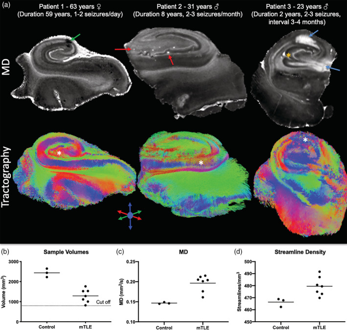
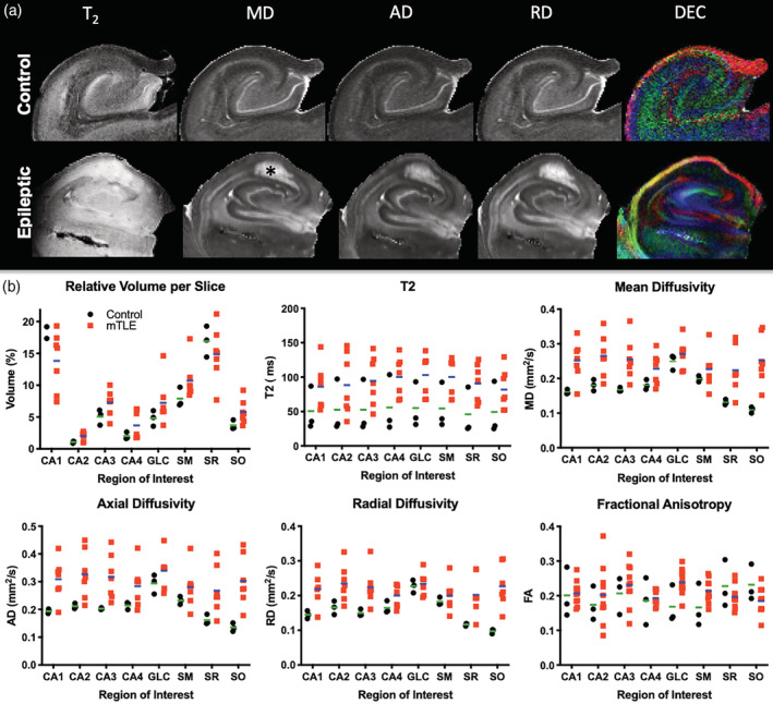
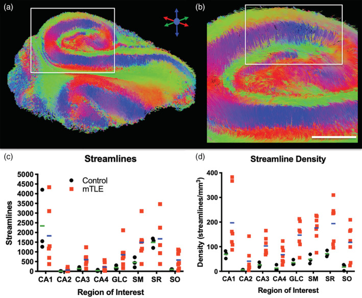
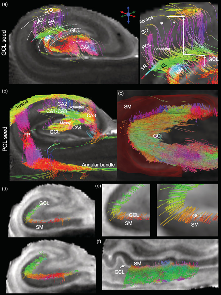
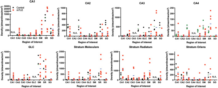
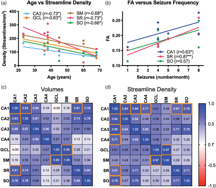
The role of hippocampal connectivity in mesial temporal lobe epilepsy (mTLE) remains poorly understood. The use of ex vivo hippocampal samples excised from patients with mTLE affords mesoscale diffusion magnetic resonance imaging (MRI) to identify individual cell layers, such as the pyramidal (PCL) and granule cell layers (GCL), which are thought to be impacted by seizure activity. Diffusion tensor imaging (DTI) of control (n = 3) and mTLE (n = 7) hippocampi on an 11.7 T MRI scanner allowed us to reveal intra-hippocampal connectivity and evaluate how epilepsy affected mean (MD), axial (AD), and radial diffusivity (RD), as well as fractional anisotropy (FA). Regional measurements indicated a volume loss in the PCL of the cornu ammonis (CA) 1 subfield in mTLE patients compared to controls, which provided anatomical context. Diffusion measurements, as well as streamline density, were generally higher in mTLE patients compared to controls, potentially reflecting differences due to tissue fixation. mTLE measurements were more variable than controls. This variability was associated with disease severity, as indicated by a strong correlation (r = 0.87) between FA in the stratum radiatum and the frequency of seizures in patients. MD and RD of the PCL in subfields CA3 and CA4 also correlated strongly with disease severity. No correlation of MR measures with disease duration was evident. These results reveal the potential of mesoscale diffusion MRI to examine layer-specific diffusion changes and connectivity to determine how these relate to clinical measures. Improving the visualization of intra-hippocampal connectivity will advance the development of novel hypotheses about seizure networks.
Keywords: biomarker; connectivity; diffusion mri; epilepsy; hippocampus; mesoscale; surgical resection; tractography.
© 2020 The Authors. Human Brain Mapping published by Wiley Periodicals LLC.
Conflict of interest statement
The authors have no personal financial or institutional interest in the results described in this article.
Figures
References
-
- Adler, D. H. , Wisse, L. E. M. , Ittyerah, R. , Pluta, J. B. , Ding, S. L. , Xie, L. , … Yushkevich, P. A. (2018). Characterizing the human hippocampus in aging and Alzheimer's disease using a computational atlas derived from ex vivo MRI and histology. Proceedings of the National Academy of Sciences of the United States of America, 115(16), 4252–4257. 10.1073/pnas.1801093115 - DOI - PMC - PubMed
-
- Alizadeh, M. , Kozlowski, L. , Muller, J. , Ashraf, N. , Shahrampour, S. , Mohamed, F. B. , … Sharan, A. (2019). Hemispheric regional based analysis of diffusion tensor imaging and diffusion tensor tractozgraphy in patients with temporal lobe epilepsy and correlation with patient outcomes. Scientific Reports, 9(1), 215 10.1038/s41598-018-36818-x - DOI - PMC - PubMed
-
- Bao, Y. , He, R. , Zeng, Q. , Zhu, P. , Zheng, R. , & Xu, H. (2018). Investigation of microstructural abnormalities in white and gray matter around hippocampus with diffusion tensor imaging (DTI) in temporal lobe epilepsy (TLE). Epilepsy & Behavior, 83, 44–49. 10.1016/j.yebeh.2017.12.002 - DOI - PubMed
-
- Bartsch, T. (2012). The clinical neurobiology of the hippocampus: An integrative view. Oxford, UK: Oxford University Press.
Publication types
MeSH terms
Grants and funding
LinkOut - more resources
Full Text Sources
Miscellaneous
