

Stem cell niche exit in C. elegans via orientation and segregation of daughter cells by a cryptic cell outside the niche
- PMID: 32692313
- PMCID: PMC7467730
- DOI: 10.7554/eLife.56383
Stem cell niche exit in C. elegans via orientation and segregation of daughter cells by a cryptic cell outside the niche
Abstract
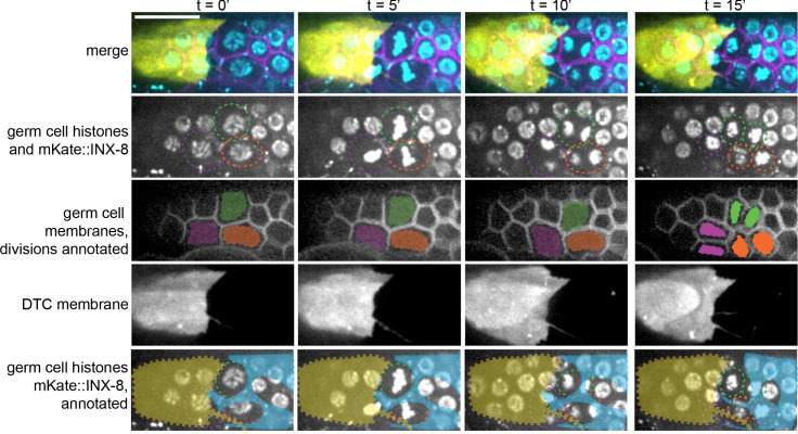
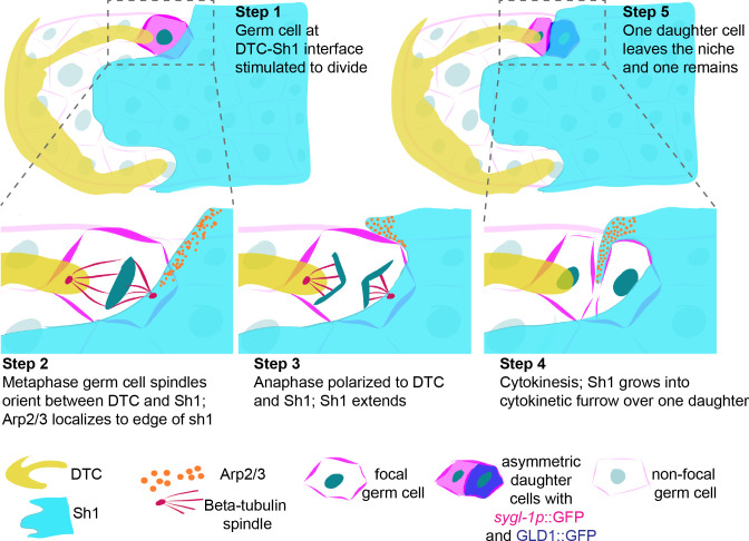
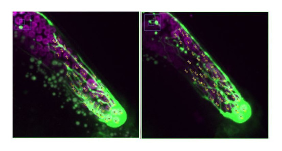
Stem cells reside in and rely upon their niche to maintain stemness but must balance self-renewal with the production of daughters that leave the niche to differentiate. We discovered a mechanism of stem cell niche exit in the canonical C. elegans distal tip cell (DTC) germ stem cell niche mediated by previously unobserved, thin, membranous protrusions of the adjacent somatic gonad cell pair (Sh1). A disproportionate number of germ cell divisions were observed at the DTC-Sh1 interface. Stem-like and differentiating cell fates segregated across this boundary. Spindles polarized, pairs of daughter cells oriented between the DTC and Sh1, and Sh1 grew over the Sh1-facing daughter. Impeding Sh1 growth by RNAi to cofilin and Arp2/3 perturbed the DTC-Sh1 interface, reduced germ cell proliferation, and shifted a differentiation marker. Because Sh1 membrane protrusions eluded detection for decades, it is possible that similar structures actively regulate niche exit in other systems.
Keywords: C. elegans; asymmetric cell division; developmental biology; distal tip cell; germ cell; gonadal sheath; niche exit; regenerative medicine; stem cell niche; stem cells.
Plain language summary
Stem cells have the rare ability to divide and specialize into the many different types of cells necessary for an organism to survive. For instance, germ stem cells can multiply to produce precursor cells that go on to become eggs or sperm needed for reproduction. When a stem cell divides, the daughter cells can either remain ‘naïve’, or start to specialize into a given cell type. In many cases, this decision is strongly influenced by the properties of the environment that surrounds the stem cell. However, in the microscopic worm Caenorhabditis elegans, how the daughters of germ stem cells specialize was thought to be a random process, with nearby cells equally likely to specialize or remain naïve. In this animal, germ stem cells reside in tube-shaped structures called gonads, which are enclosed by a large ‘distal tip’ cell. In addition, cells known as Sh1 surround the gonad. Here, Gordon et al. tracked dividing germ stem cells in the gonads of live worms. This revealed that both the distal tip cell and Sh1 cells have finger-like extensions that form contacts with the germ stem cells. The fate of dividing germ stem cells is shaped by these interactions. If they touch only the distal tip cell, they remain in a naïve state. However, if they contact both the distal tip cell and an Sh1 cell, one daughter of the stem cell becomes an egg precursor – with the daughter closest to the distal tip cell staying naïve. In fact, germ stem cells that are prevented from contacting Sh1 cells divide less often. Many other types of stem cells, for example in human skin, are believed to make the decision to remain naïve or undergo specialization randomly. The results from Gordon et al. could provide a roadmap to discover hidden layers of control in other organisms, some of which may be potentially relevant in health and disease.
© 2020, Gordon et al.
Conflict of interest statement
KG, JZ, XL, CM, DS No competing interests declared
Figures


Comment in
-
More than just a pool.Elife. 2020 Sep 11;9:e61397. doi: 10.7554/eLife.61397. Elife. 2020. PMID: 32915135 Free PMC article.
References
Publication types
MeSH terms
Substances
Grants and funding
LinkOut - more resources
Full Text Sources
Other Literature Sources
Medical
