. 2020 Dec 1;202(11):1540-1550.
doi: 10.1164/rccm.201904-0792OC.

Single-Cell Reconstruction of Human Basal Cell Diversity in Normal and Idiopathic Pulmonary Fibrosis Lungs

Affiliations

Single-Cell Reconstruction of Human Basal Cell Diversity in Normal and Idiopathic Pulmonary Fibrosis Lungs

Gianni Carraro et al. Am J Respir Crit Care Med. .

Abstract

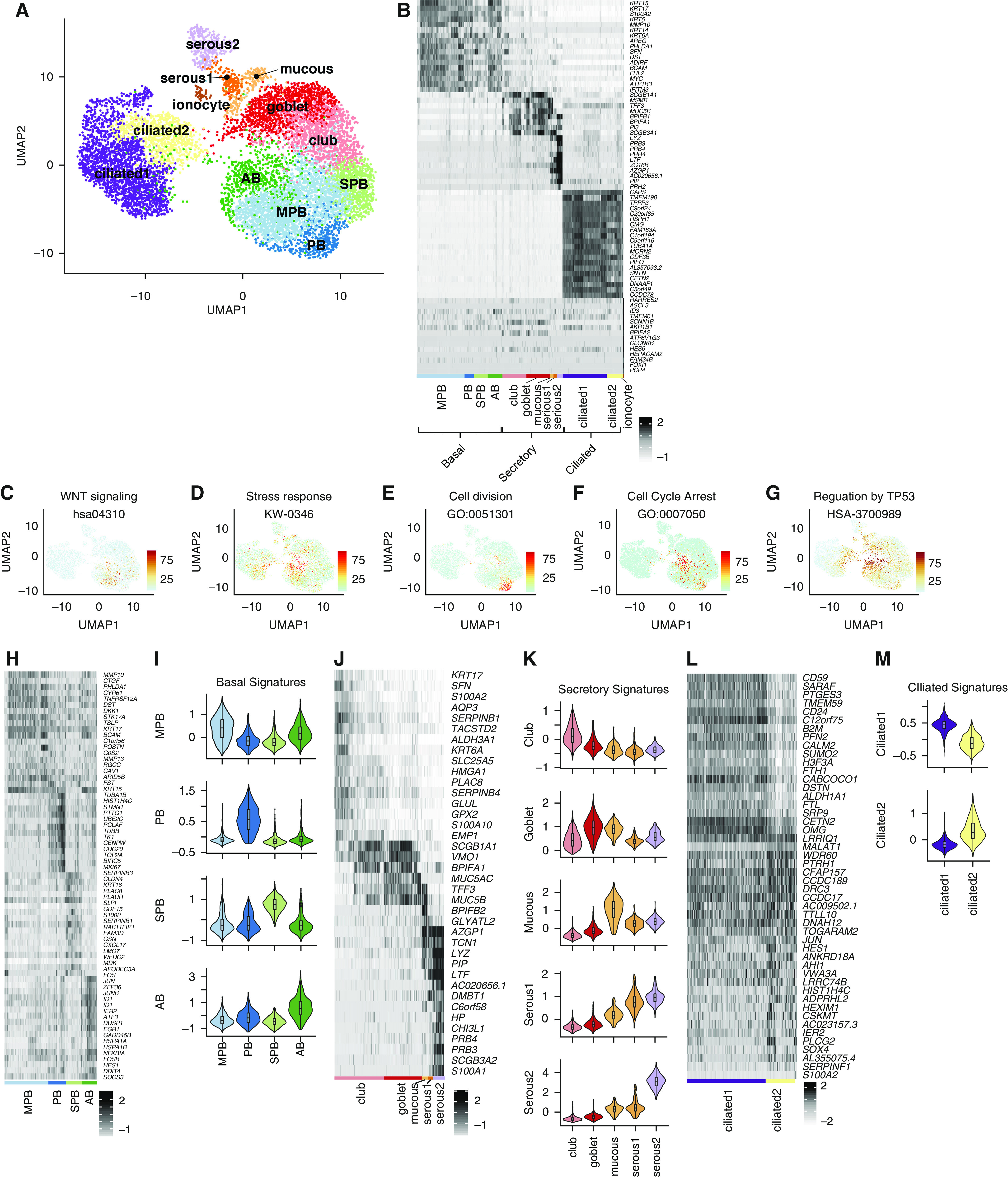
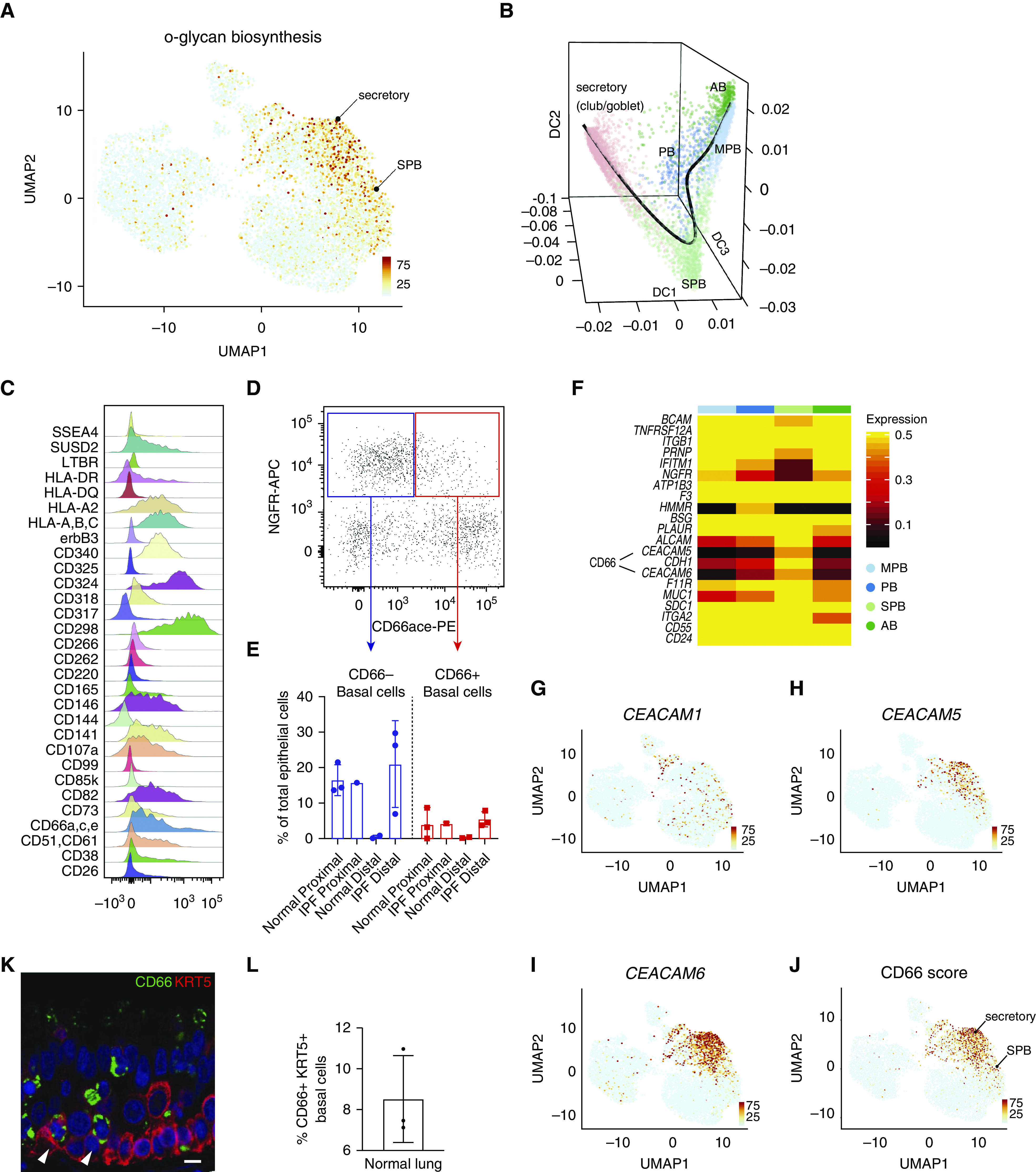
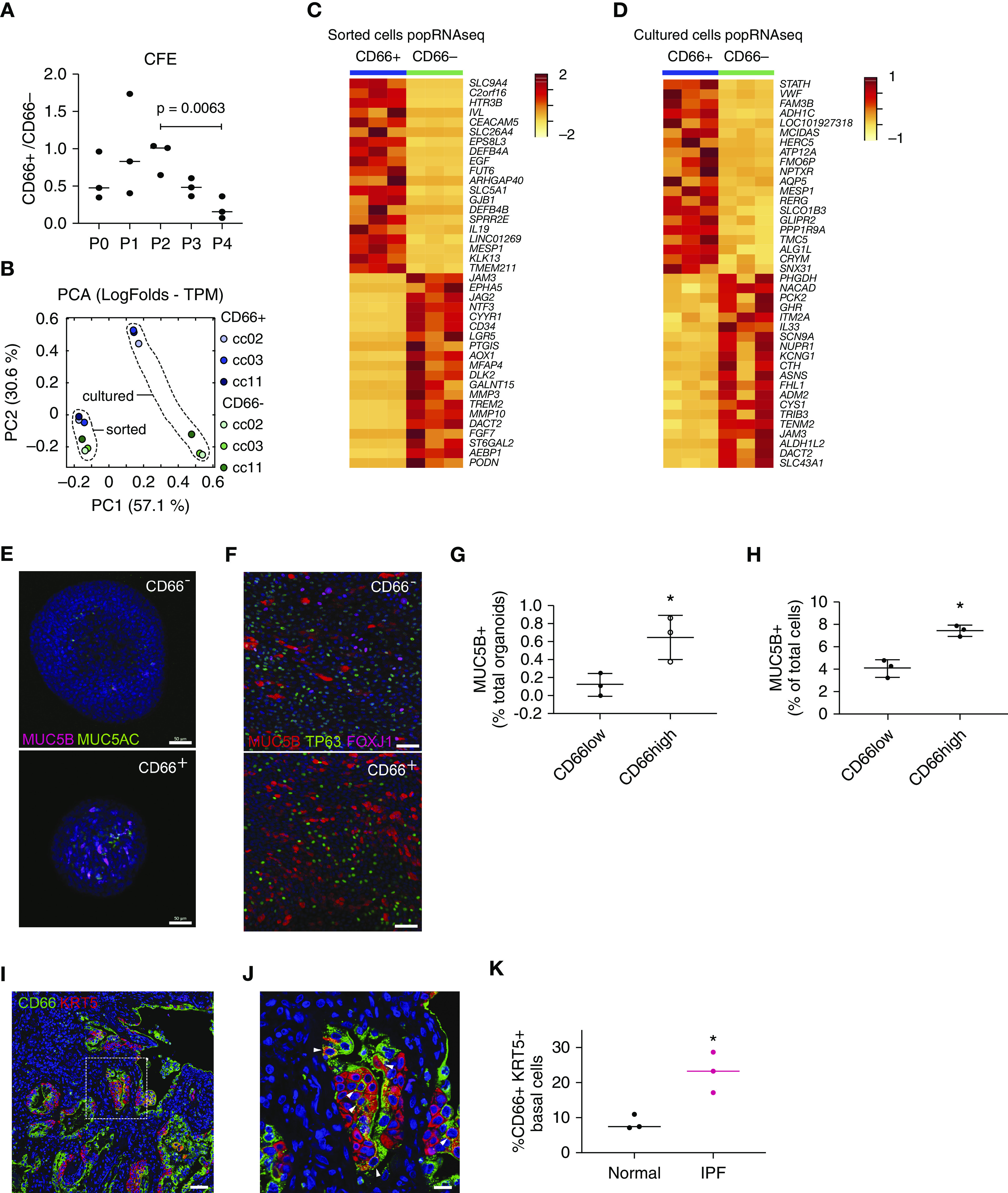
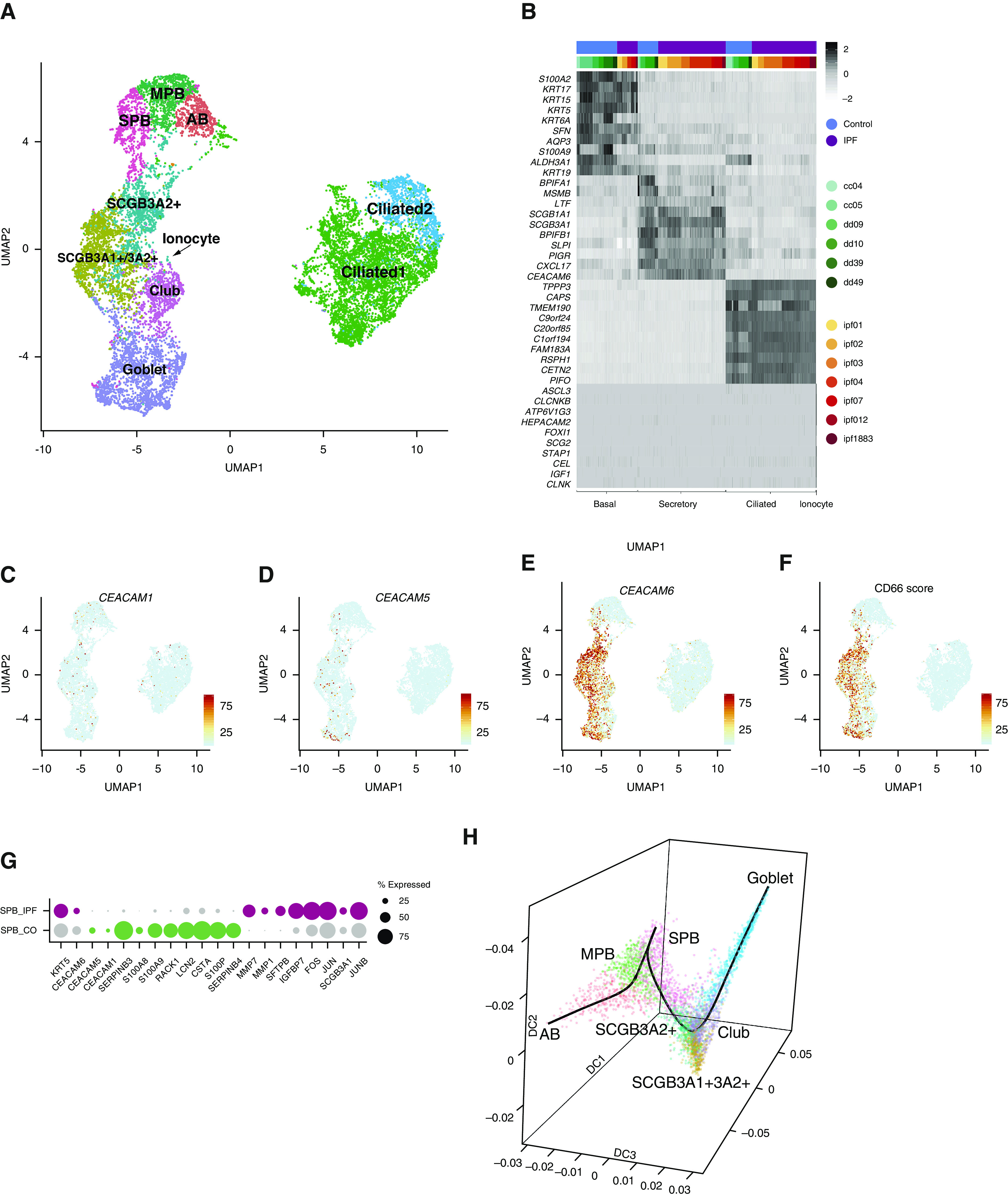
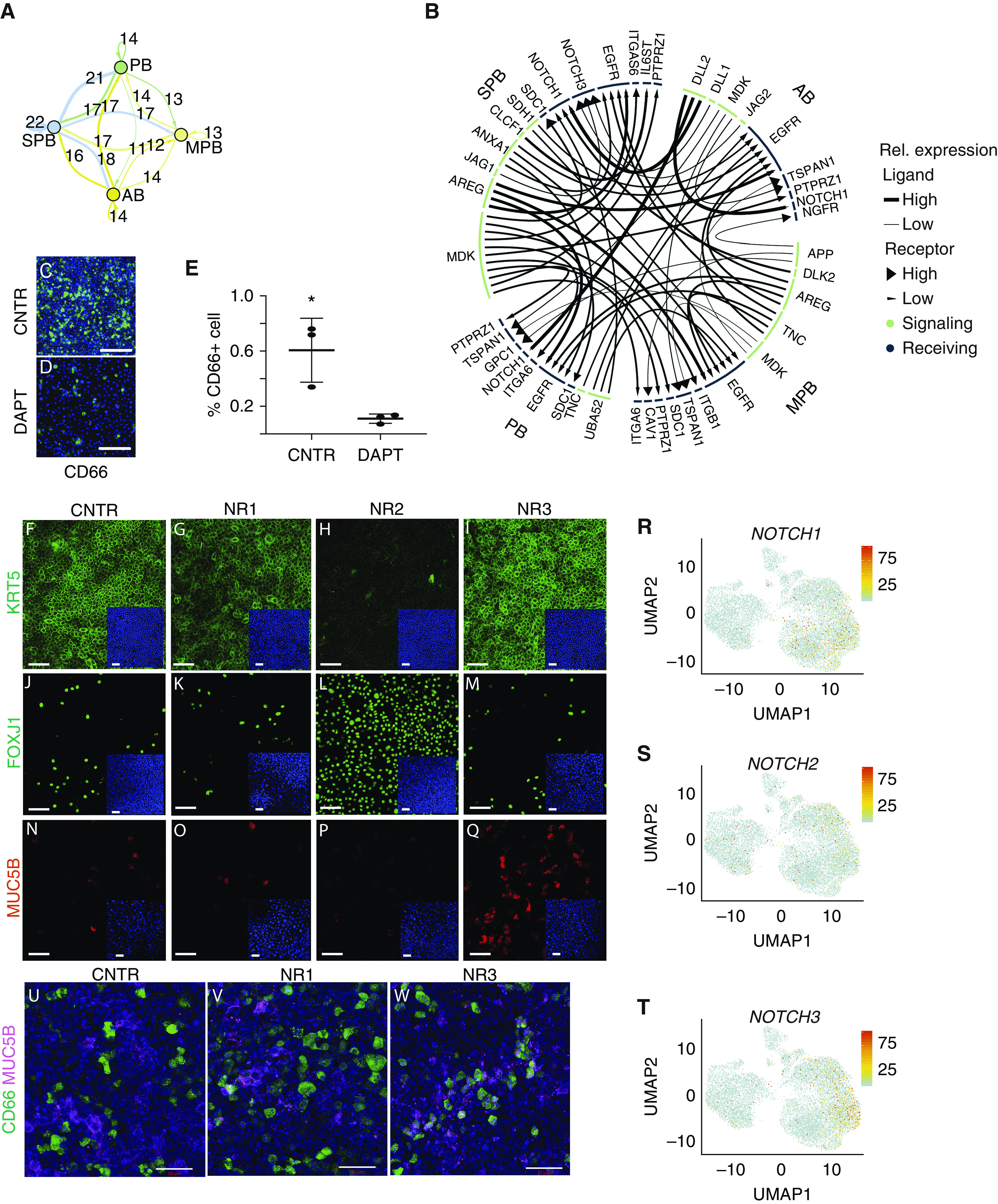
Rationale: Declining lung function in patients with interstitial lung disease is accompanied by epithelial remodeling and progressive scarring of the gas-exchange region. There is a need to better understand the contribution of basal cell hyperplasia and associated mucosecretory dysfunction to the development of idiopathic pulmonary fibrosis (IPF).Objectives: We sought to decipher the transcriptome of freshly isolated epithelial cells from normal and IPF lungs to discern disease-dependent changes within basal stem cells.Methods: Single-cell RNA sequencing was used to map epithelial cell types of the normal and IPF human airways. Organoid and air-liquid interface cultures were used to investigate functional properties of basal cell subtypes.Measurements and Main Results: We found that basal cells included multipotent and secretory primed subsets in control adult lung tissue. Secretory primed basal cells include an overlapping molecular signature with basal cells obtained from the distal lung tissue of IPF lungs. We confirmed that NOTCH2 maintains undifferentiated basal cells and restricts basal-to-ciliated differentiation, and we present evidence that NOTCH3 functions to restrain secretory differentiation.Conclusions: Basal cells are dynamically regulated in disease and are specifically biased toward the expansion of the secretory primed basal cell subset in IPF. Modulation of basal cell plasticity may represent a relevant target for therapeutic intervention in IPF.

Keywords: dynamic contrast enhanced magnetic resonance imaging; prematurity; pulmonary microvascular function; vascular simplification.


Figures

Figure 1.
Figure 1.
Classification of the human airway epithelium. (A) Uniform Manifold Approximation and Projection clustering of single-cell RNA-sequencing data generated for normal human lung airway epithelium. (B) Expression of differentially expressed genes that distinguish major cell types shown by heatmap with z-score values. Cell subtypes are shown within each major cell type. (CG) Scoring derived respectively from Wnt signaling, stress response, cell division, cell-cycle arrest, and TP53-regulation gene signatures visualized by Uniform Manifold Approximation and Projection. (H) Expression of differentially expressed genes within basal cell subclusters shown by heatmap with z-score values. (I) Signatures of basal cell subclusters derived by scoring differentially expressed genes shown by violin plots. (J) Expression of differentially expressed genes within secretory cell subclusters shown by heatmap with z-score values. (K) Signatures of secretory cell subclusters derived by scoring differentially expressed genes shown by violin plots. (L) Expression of differentially expressed genes within ciliated cell subclusters shown by heatmap with z-score values. (M) Signatures of ciliated cell subclusters derived by scoring differentially expressed genes, shown by violin plots. AB = activated basal; MPB = multipotent basal; PB = proliferating basal; SPB = secretory primed basal; UMAP = Uniform Manifold Approximation and Projection.
Figure 2.
Figure 2.
Characterization of secretory primed basal cells (secretory primed basal cluster). (A) Scoring of O-glycan biosynthesis pathway in the normal human airways visualized by Uniform Manifold Approximation and Projection. (B) Lineage reconstruction from basal to secretory cells (black line) was produced with “Slingshot” on the top of a diffusion map generated with “Destiny.” (C) Distribution analysis of CD markers discriminating basal cell subclusters identified by high-content flow cytometry. (D) Discrimination of basal cell subsets by NGFR and CD66 surface reactivity. (E) Percentage of basal subsets over total epithelial cells in proximal and distal compartments of normal and idiopathic pulmonary fibrosis lungs. (F) Differential expression of basal cell–specific CD markers from the single-cell RNA-sequencing data of the four basal cell subclusters. The entire repertoire of human CD molecules available from Human Genome Organization Gene Nomenclature Committee was screened. (GJ) Expression of CEACAM1, CEACAM5, and CEACAM6, and summarized signature score of CD66 in human normal airway epithelium visualized by Uniform Manifold Approximation and Projection. (K) Normal lung cross-section shows immunoreactivity of basal cell subpopulations for CD66 (arrowheads). Scale bar, 15 μm. (L) Quantification of CD66+ basal cells in normal lung, n = 3. AB = activated basal; DC = diffusion component; IPF = idiopathic pulmonary fibrosis; MPB = multipotent basal; NGFR-APC = NGFR allophycocyanin; PB = proliferating basal; PE = phycoerythrin; SPB = secretory primed basal; UMAP = Uniform Manifold Approximation and Projection.
Figure 3.
Figure 3.
Functional properties of secretory primed basal cells. (A) Colony-forming efficiency of CD66+ and CD66 basal cells grown in mesenchyme-free three-dimensional cultures (n = 3; P = 0.0063; ANOVA). (B) Principal component analysis of popRNAseq data from freshly isolated (fluorescence-activated cell-sorting) or cultured CD66+ and CD66 basal cells. (C and D) Transcriptomic differences between freshly isolated and cultured CD66+ and CD66 basal cells visualized by heatmap. Top 20 differentially expressed genes (log2-fold change) with adjusted P value < 0.05 for CD66+ and CD66 basal cells are shown. (E and G) Immunofluorescence detection and quantification of mucins MUC5B and MUC5AC in organoid cultures of CD66+ and CD66 basal cells. Scale bars, 50 μm. (F and H) Immunofluorescence detection of basal (TP63), secretory (MUC5B), and ciliated (FOXJ1) markers for CD66+ and CD66 basal cells grown in air–liquid interface. Scale bars, 50 μm. Quantification of the percentage of MUC5B+ cells is shown. (I and J) Honeycomb regions of idiopathic pulmonary fibrosis lung showing enrichment of CD66+ basal cells. Scale bars, 50 μm for I and 20 μm for J. Arrowheads identify cells that are double positive for KRT5 and CD66. (K) Quantification of difference in proportion of CD66+ basal cells between normal and idiopathic pulmonary fibrosis lung (n = 3; *P < 0.1; Mann-Whitney test ). CFE = colony-forming efficiency; IPF = idiopathic pulmonary fibrosis; PCA = principal component analysis; popRNAseq = population RNA sequencing; TPM = transcript count per million.
Figure 4.
Figure 4.
Analysis of idiopathic pulmonary fibrosis (IPF) datasets. (A) Clustering of human IPF datasets visualized by Uniform Manifold Approximation and Projection. (B) Visualization of conserved genes for major cell categories from integrated data of control and IPF datasets visualized by heatmap with z-score values. (CF) Expression of CEACAM1, CEACAM5, and CEACAM6 and summarized signature score of CD66 in human IPF epithelium visualized by Uniform Manifold Approximation and Projection. (G) Dot plot visualization of top differentially expressed genes for secretory primed basal cells between control and IPF lung. (H) Diffusion map generated with “Destiny” from basal and secretory IPF cells shows a continuous transition of states. The superimposed black line was generated with “Slingshot” and describes the trajectory between cells. AB = activated basal; CO = control; DC = diffusion component; MPB = multipotent basal; SPB = secretory primed basal; UMAP = Uniform Manifold Approximation and Projection.
Figure 5.
Figure 5.
Analysis of Notch receptor specificity in human basal cells. (A) Summary of predicted ligand–receptor interactions for Notch signaling between pairs of basal cell subtypes and for autocrine Notch signaling visualized by network plot. Each arrow is color coded according to basal cell type, and the thickness of the arrow represents the number of ligand–receptor interactions. Arrows that go back to the cell of origin indicate paracrine signaling within the same cell type. Numbers above each arrow indicate how many ligand–receptor interactions were identified. (B) Circus plot showing ligands in green and receptors in blue, with arrows connecting signaling and receiving cells. Each arrow connects signaling and receiving cells, with the tip of the arrow pointing to the receiving cell. The size of the segment underneath each ligand or receptor is proportional to the interactions that were detected. (CE) Air–liquid interface (ALI) coculture of CD66+ and CD66 basal cells showing the effect of DAPT treatment on CD66+ basal cells. Scale bars, 100 μm. *P < 0.1; Mann-Whitney test. (FQ) CD66+ and CD66 basal cell ALI coculture treated with specific Notch-blocking antibody for NOTCH1 (NR1), NOTCH2 (NR2), and NOTCH3 (NR3). Effect on basal cell (KRT5+) maintenance, ciliated cell (FOXJ1+) differentiation at Day 14, and secretory cell (MUC5B+) differentiation at Day 8 is shown by immunoreactivity with cell type–specific antibodies. Scale bars, 50 μm. (RT) Expression of NOTCH1, NOTCH2, and NOTCH3 in normal airway epithelial cells visualized by Uniform Manifold Approximation and Projection. (UW) Coexpression of CD66 and MUC5B in CD66+ and CD66 cells at Day 14 of ALI coculture treated with specific Notch-blocking antibodies. Scale bars, 50 μm. AB = activated basal; CNTR = control; DAPT = N-[N-(3,5-difluorophenacetyl)-l-alanyl]-S-phenylglycine t-butyl ester; MPB = multipotent basal; PB = proliferating basal; Rel. expression = relative expression; SPB = secretory primed basal; UMAP = Uniform Manifold Approximation and Projection.

Comment in

References

    1. Wells JM, Watt FM. Diverse mechanisms for endogenous regeneration and repair in mammalian organs. Nature. 2018;557:322–328. - PubMed
    1. Teixeira VH, Nadarajan P, Graham TA, Pipinikas CP, Brown JM, Falzon M, et al. Stochastic homeostasis in human airway epithelium is achieved by neutral competition of basal cell progenitors. eLife. 2013;2:e00966. - PMC - PubMed
    1. Crystal RG, Randell SH, Engelhardt JF, Voynow J, Sunday ME. Airway epithelial cells: current concepts and challenges. Proc Am Thorac Soc. 2008;5:772–777. - PMC - PubMed
    1. Noble PW, Barkauskas CE, Jiang D. Pulmonary fibrosis: patterns and perpetrators. J Clin Invest. 2012;122:2756–2762. - PMC - PubMed
    1. Seibold MA, Smith RW, Urbanek C, Groshong SD, Cosgrove GP, Brown KK, et al. The idiopathic pulmonary fibrosis honeycomb cyst contains a mucocilary pseudostratified epithelium. PLoS One. 2013;8:e58658. - PMC - PubMed

Publication types

MeSH terms