Review
. 2020 Oct 6:36:85-114.
doi: 10.1146/annurev-cellbio-020520-120627. Epub 2020 Jul 21.

Nuclear Membrane Rupture and Its Consequences

Affiliations
Review

Nuclear Membrane Rupture and Its Consequences

John Maciejowski et al. Annu Rev Cell Dev Biol. .

Abstract

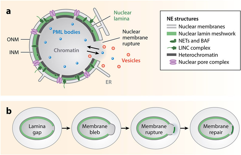
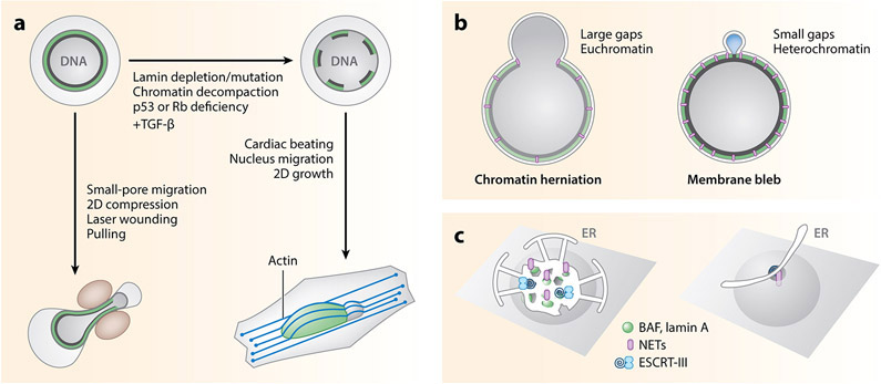
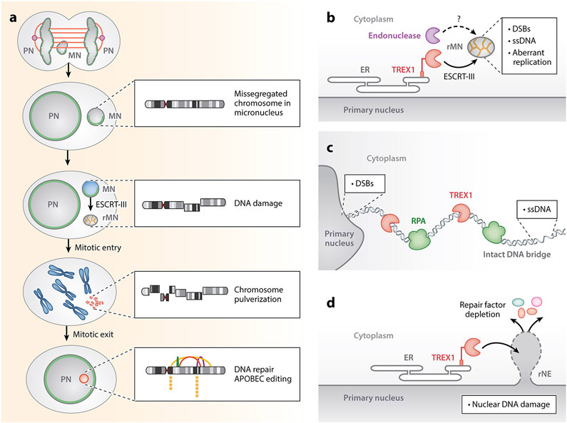
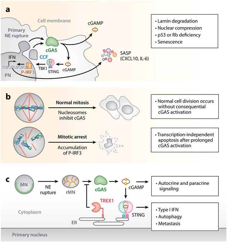
The nuclear envelope is often depicted as a static barrier that regulates access between the nucleus and the cytosol. However, recent research has identified many conditions in cultured cells and in vivo in which nuclear membrane ruptures cause the loss of nuclear compartmentalization. These conditions include some that are commonly associated with human disease, such as migration of cancer cells through small spaces and expression of nuclear lamin disease mutations in both cultured cells and tissues undergoing nuclear migration. Nuclear membrane ruptures are rapidly repaired in the nucleus but persist in nuclear compartments that form around missegregated chromosomes called micronuclei. This review summarizes what is known about the mechanisms of nuclear membrane rupture and repair in both the main nucleus and micronuclei, and highlights recent work connecting the loss of nuclear integrity to genome instability and innate immune signaling. These connections link nuclear membrane rupture to complex chromosome alterations, tumorigenesis, and laminopathy etiologies.

Keywords: ESCRT-III; TREX1; cGAS; chromothripsis; membrane dynamics; nuclear lamina.


Figures

Figure 1
Figure 1
NE structures and membrane rupture. (a) Nucleus undergoing membrane rupture, showing the major NE structures including the membranes, lamina, nuclear pore complexes, chromatin, and peripheral heterochromatin. The nuclear lamina includes the nuclear lamin meshworks, NETs, BAF, and the LINC complex. The nuclear membrane is comprised of the outer and inner nuclear membranes, which are contiguous with each other and the ER. During membrane rupture, a pore is formed in the nuclear membrane that allows the free diffusion of nuclear and cytoplasmic proteins and small organelles, including PML bodies (blue) and vesicles (red) (DeVos et al. 2011, Vargas et al. 2012). (b) Stages of nuclear membrane rupture and repair. Many ruptures begin with the appearance of a gap in the nuclear lamina meshwork, which then becomes the site of membrane blebbing and chromatin herniation followed by membrane rupture when the nucleus is mechanically stressed. A subset of NE proteins (dark green) from the cytoplasm and ER accumulates on the exposed chromatin, along with other cytosolic DNA-binding proteins. These proteins persist at rupture sites even after nuclear compartmentalization is restored in the membrane repair process. This is only one possible order of events. Abbreviations: BAF, barrier-to-autointegration factor; ER, endoplasmic reticulum; INM, inner nuclear membrane; LINC, linker of nucleoskeleton and cytoskeleton; NE, nuclear envelope; NET, NE transmembrane proteins; ONM, outer nuclear membrane; PML, promyelocytic leukemia.
Figure 2
Figure 2
Mechanisms of nuclear membrane rupture and repair. (a) The probability of nuclear membrane rupture depends on the degree of nuclear lamina (green) and peripheral heterochromatin (dark gray) disruption and the degree of mechanical stress. In healthy cells with an intact nuclear lamina, membrane rupture requires a significant amount of force, such as laser-induced rupture or (left) cell migration through a small opening. Disruption of the nuclear lamina by depletion or mutation of lamin proteins, chromatin decompaction, loss of retinoblastoma (Rb) and p53 activity, and activation of EMT genes by TGF-β signaling leads to membrane rupture in less mechanically demanding conditions, such as growth on stiff surfaces where (right) nucleus compression by apical actin bundles drives chromatin herniation and membrane rupture. (b) Nuclear membrane rupture is frequently preceded by or occurs with chromatin herniation and nuclear membrane blebbing. Chromatin herniation, where little to no gap is visible between the membrane and the extruded chromatin, is associated with conditions that cause large lamina gaps and decreased heterochromatin, while membrane blebbing without chromatin is associated with more intact nuclear laminas and significant peripheral heterochromatin. (c) Models of nuclear membrane repair. (Left) Cytosolic BAF binds to exposed chromatin and recruits LEM-domain NETs (purple) embedded in ER sheets and tubules. BAF also recruits lamin A/C and the NET LEMD2, which recruits the ESCRT-III complex. Depletion of BAF, ESCRT-III, or multiple NETs leads to impaired membrane repair. (Right) Alternatively, direct binding of NETs and the ESCRT-III protein Chmp7 to the chromatin and inner nuclear membrane, with or without membrane recruitment, or spreading of the outer nuclear membrane over the membrane gap could recompartmentalize the nucleus in the absence of BAF. Abbreviations: BAF, barrier-to-autointegration factor; Chmp7, charged multivesicular body protein; EMT, epithelial-to-mesenchymal transition; ER, endoplasmic reticulum; ESCRT-III, endosomal sorting complexes required for transport III; LEMD, Lap2, emerin, and Man1 domain; NET, nuclear envelope transmembrane protein; TGF, transforming growth factor.
Figure 3
Figure 3
Sources of DNA damage from nuclear rupture. (a) Micronuclei frequently form around lagging chromosomes during mitosis. Association with midspindle microtubules (orange) may inhibit nuclear lamina (green) and NPC assembly. Insets show the progression of DNA damage and repair in the micronucleated chromosome. Chromosome pulverization is apparent in mitosis, and the chromatin from rMNs can missegregate or reincorporate into the nucleus. DNA repair occurs after nuclear reincorporation of the micronucleated chromatin and is primarily driven by nonhomologous end joining. Reassembly of the damaged micronucleated chromatin frequently gives rise to chromothripsis and other structural aberrations, including translocations, insertions, and deletions. APOBEC3B base editing generates clustered C > T and C > G mutations proximal to chromothripsis-associated breakpoints. (b) Micronucleus membrane rupture is associated with DNA damage. Staining with γH2AX, TUNEL, the bacterial protein Gam, RPA32, and native BrdU indicates the presence of DNA DSBs and ssDNA. The ESCRT-III complex localizes to the rMNs and promotes chromatin compaction. ER invasion into rMNs directs TREX1 to resect micronuclear DNA. (c) DNA bridges accumulate ssDNA as marked by RPA32 prior to DNA bridge breakage. ssDNA on DNA bridges is generated by TREX1 resection. DNA bridges are also associated with evidence of DNA DSBs at the connection site to the primary nucleus. (d) Nuclear rupture induces TREX1-dependent DNA DSBs in primary nuclei. Such DSBs are not restricted to the site of NE breakage and may occur throughout the nucleus. DNA damage is associated with the depletion of DNA repair factors from the nucleus. Abbreviations: APOBEC, apolipoprotein B mRNA-editing enzyme, catalytic polypeptide-like; BrdU, bromodeoxyuridine; DSB, double-strand break; ER, endoplasmic reticulum; ESCRT-III, endosomal sorting complexes required for transport III; MN, micronucleus; NPC, nuclear pore complex; PN, primary nucleus; rMN, ruptured micronucleus; rNE, ruptured nuclear envelope; RPA, replication protein A; ssDNA, single-stranded DNA; TREX1, three-prime repair exonuclease 1; TUNEL, terminal deoxynucleotidyl transferase dUTP nick end labeling.
Figure 4
Figure 4
cGAS activation from nuclear rupture. (a) Primary nuclear rupture enables cGAS access to chromosomal DNA. cGAS localizes to chromatin herniations after primary NE rupture. Lamin depletion during senescence can enable cGAS to access chromosomal DNA via the formation of CCFs that bleb and appear to separate from the PN. cGAS detection of CCFs promotes senescence via the STING-dependent production of SASP factors. cGAS-STING activation in this context may promote immune surveillance. (b) cGAS promotes transcription-independent apoptosis following mitotic NE breakdown. cGAS engages with chromatin but is not activated during an unperturbed mitosis. Prolonged mitotic arrest causes a slow buildup of P-IRF3, which eventually causes transcription-independent apoptosis. (c) Nuclear rupture at micronuclei exposes chromosomal DNA to the cytosol and initiates a cGAS-dependent proinflammatory response. cGAS localizes to micronuclei following MN membrane rupture, where it engages chromatin and initiates a STING-dependent proinflammatory transcriptional response. TREX1 resection of micronuclear DNA limits cGAS activation at micronuclei. cGAS activation at micronuclei can drive metastasis through a noncanonical NF-κB signaling response and is associated with the induction of autophagy during telomere crisis. Abbreviations: AMP, adenosine monophosphate; CCF, cytosolic chromatin fragment; cGAMP, cyclic GMP-AMP; cGAS, cyclic GMP-AMP synthase; CXCL10, C-X-C motif chemokine 10; ER, endoplasmic reticulum; GMP, guanosine monophosphate; IFN, interferon; IL-6, interleukin-6; IRF3, interferon regulatory factor 3; MN, micronucleus; NE, nuclear envelope; NF-κB, nuclear factor κ-light chain–enhancer of activated B cells; P-IRF3, phosphorylated IRF3; PN, primary nucleus; rMN, ruptured micronucleus; SASP, senescence-associated secretory phenotype; STING, stimulator of interferon genes; TBK1, TANK-binding kinase 1; TREX1, three-prime repair exonuclease 1.

References

    1. Ablasser A, Chen ZJ. 2019. cGAS in action: expanding roles in immunity and inflammation. Science 363(6431):eaat8657. - PubMed
    1. Ablasser A, Goldeck M, Cavlar T, Deimling T, Witte G, et al. 2013. cGAS produces a 2′-5′-linked cyclic dinucleotide second messenger that activates STING. Nature 498(7454):380–84 - PMC - PubMed
    1. Anderson DJ, Vargas JD, Hsiao JP, Hetzer MW. 2009. Recruitment of functionally distinct membrane proteins to chromatin mediates nuclear envelope formation in vivo. J. Cell Biol 186(2):183–91 - PMC - PubMed
    1. Bakhoum SF, Cantley LC. 2018. The multifaceted role of chromosomal instability in cancer and its microenvironment. Cell 174(6):1347–60 - PMC - PubMed
    1. Bakhoum SF, Ngo B, Laughney AM, Cavallo J-A, Murphy CJ, et al. 2018. Chromosomal instability drives metastasis through a cytosolic DNA response. Nature 553(7689):467–72 - PMC - PubMed

Publication types

LinkOut - more resources