Review
. 2020 Jul 20;54(2):239-255.
doi: 10.1016/j.devcel.2020.06.029.

Hematopoietic Stem Cell Metabolism during Development and Aging

Affiliations
Review

Hematopoietic Stem Cell Metabolism during Development and Aging

Ayako Nakamura-Ishizu et al. Dev Cell. .

Abstract

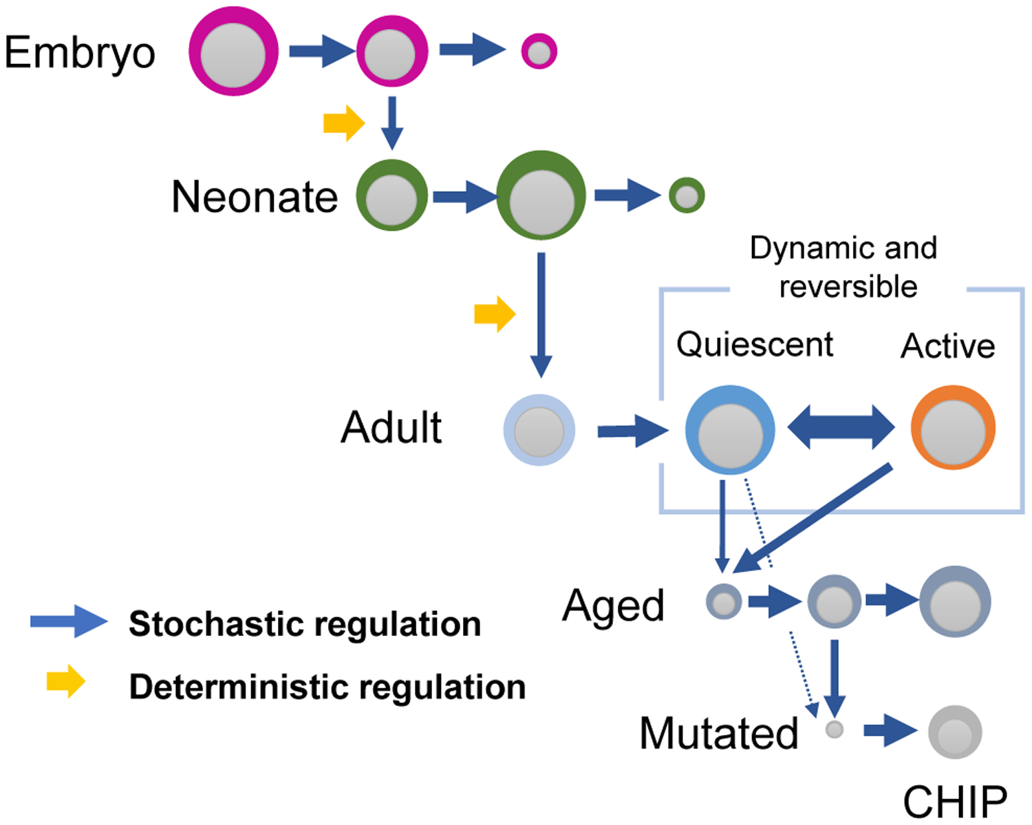
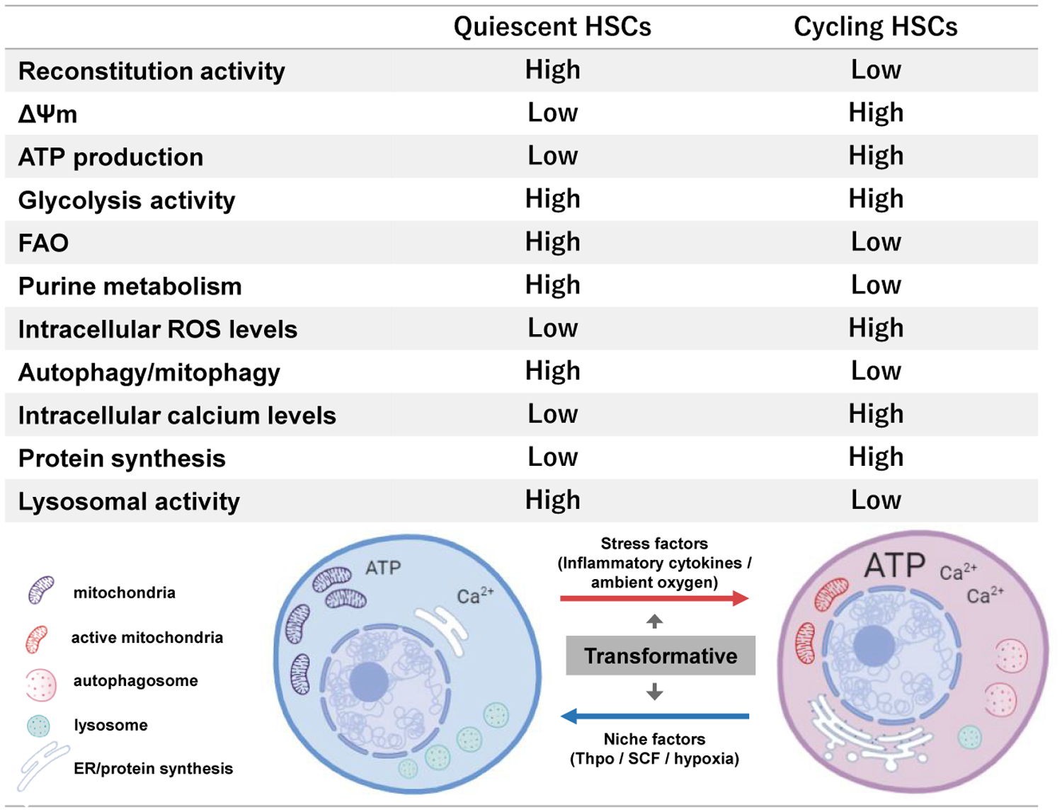
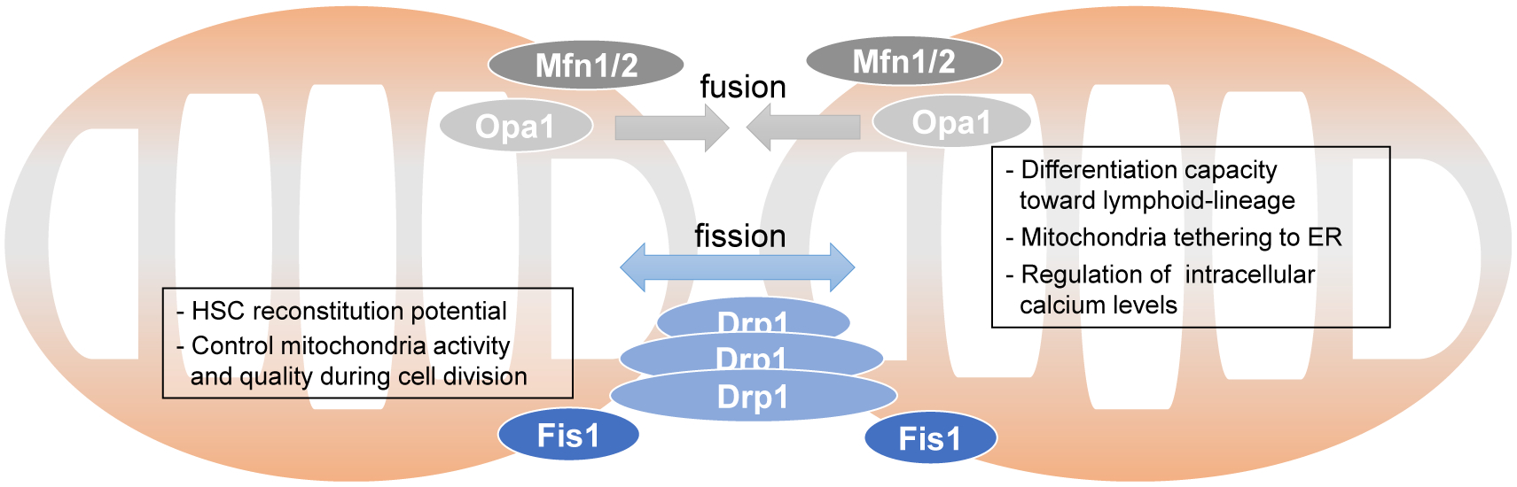
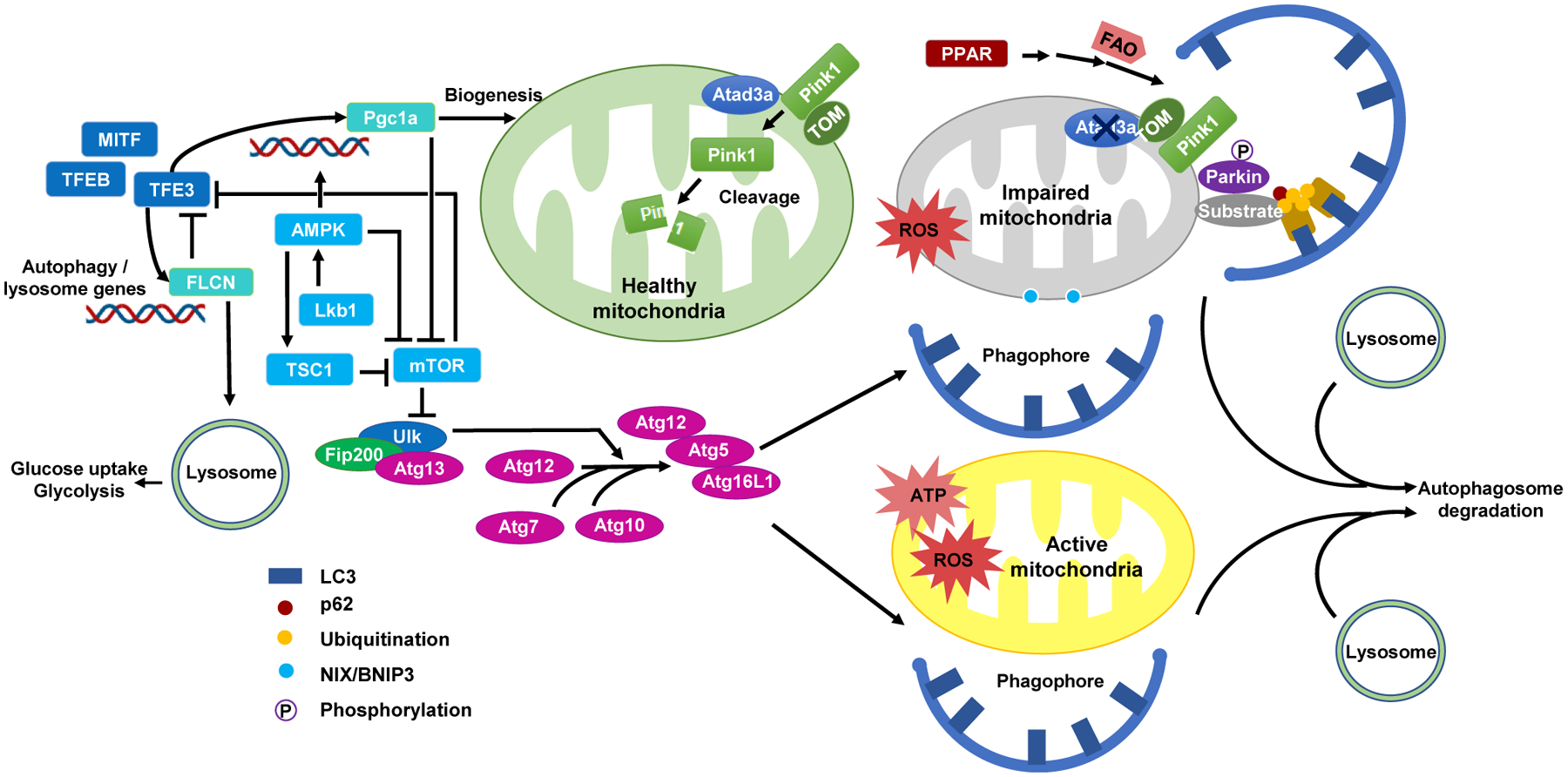
Cellular metabolism in hematopoietic stem cells (HSCs) is an area of intense research interest, but the metabolic requirements of HSCs and their adaptations to their niches during development have remained largely unaddressed. Distinctive from other tissue stem cells, HSCs transition through multiple hematopoietic sites during development. This transition requires drastic metabolic shifts, insinuating the capacity of HSCs to meet the physiological demand of hematopoiesis. In this review, we highlight how mitochondrial metabolism determines HSC fate, and especially focus on the links between mitochondria, endoplasmic reticulum (ER), and lysosomes in HSC metabolism.


Figures

Figure 1.
Figure 1.. Schematic Representation of HSC Dynamics during Development
HSCs of different developmental state (embryonic, neonatal, adult, and aged HSCs) clonally expand through stochastic processes. HSCs give rise to differential clones during development through a deterministic process possibly through the modification of hematopoietic environment. Adult HSCs maintain a quiescent state which may be reversed to an active proliferative state upon stress. Aged HSCs eventually accumulate genetic mutations leading to the expansion of abnormal clones (CHIP).
Figure 2.
Figure 2.. Metabolic Characteristics of Quiescent and Cycling HSCs
Quiescent adult HSCs exhibit high reconstitution potential and differ in organelle (mitochondria, ER, lysosome, and autophagosome) content compared to cycling HSCs. The difference in organelle activity reflects the overall metabolic state (ΔΨm, ATP production, protein synthesis, autophagy, glycolysis, FAO, purine metabolism, ROS levels, and calcium levels).
Figure 3.
Figure 3.. Mitochondria Bioenergetics in HSCs
Mitochondria bioenergetics pathways implicated in HSC metabolism. Other than ATP production through the TCA cycle and electron transport system, HSC mitochondria produce energy through FAO (beta-oxidation). ROS is generated as a byproduct of oxidative phosphorylation and is detoxified by cystolic redox reaction. Mitochondria also home specific pathways for proteostasis and process unfolded proteins through chaperone proteins and proteases. Intracellular calcium levels are regulated by the mitochondria through crosstalk with ER.
Figure 4.
Figure 4.. Mitochondria Dynamics through Fission and Fusion
Mfn1/2 located on the OMM and Opa1 on the IMM stimulate mitochondria fusion. Deletion of Mfn2 affect HSC mitochondria function and stem cell potential. Drp1 and Fis1 stimulate mitochondria fission. Fis1 is implicated to affect HSC mitochondria segregation during division and stem cell functions.
Figure 5.
Figure 5.. Metabolic Crosstalk between Cellular Organelles in HSCs
Mitochondria interact with various cellular organelles. These interactions alter the metabolic and cellular fate of HSCs. The nucleus provides nuclear (nc) DNA encoded proteins, while mitochondria possess mitochondria (mt) DNA for the production of mitochondria proteins. Mitochondria also initiate apoptosis. Mitochondria clearance is regulated through a specific form of autophagy, mitophagy. Mitochondria are firmly associated with ER to which they tether and control intracellular calcium levels.
Figure 6.
Figure 6.. Regulation of Autophagic Clearance of Active and Damaged Mitochondria in HSCs
The removal of active and damaged mitochondria through autophagy maintains HSCs in a low metabolic, quiescent state. Active mitochondria with high ATP production and ROS levels are cleared through macroautophagy. Mitochondria with impaired function, such as deletion of Atad3a, may provoke Pink/Parkin pathway for mitophagy. Phosphorylated Parkin promotes the ubiquitination of mitochondrial substrates, recruits p62, and links mitochondria to LC3 for mitophagy. The presence of Atad3a on healthy mitochondria promotes the degradation of Pink1 via protease cleavage in the mitochondria matrix. Autophagy and mitophagy are regulated through a cascade of reactions involving Atg (Atg5, 7, 10, 12, 13, 16L1), Ulk1, FIP200, and other autophagy-related proteins. Autophagic pathways are interlinked with mitochondria biogenesis (mTOR, AMPK, LKB1, TSC2, and PGC1a) along with lysosomal pathways (FLCN, TFEB, TFE3, and MITF).

References

    1. Agathocleous M, Meacham CE, Burgess RJ, Piskounova E, Zhao Z, Crane GM, Cowin BL, Bruner E, Murphy MM, Chen W, et al. (2017). Ascorbate regulates haematopoietic stem cell function and leukaemogenesis. Nature 549, 476–481. - PMC - PubMed
    1. Amini P, Stojkov D, Felser A, Jackson CB, Courage C, Schaller A, Gelman L, Soriano ME, Nuoffer JM, Scorrano L, et al. (2018). Neutrophil extracellular trap formation requires OPA1-dependent glycolytic ATP production. Nat. Commun 9, 2958. - PMC - PubMed
    1. Ananieva EA, and Wilkinson AC (2018). Branched-chain amino acid metabolism in cancer. Curr. Opin. Clin. Nutr. Metab. Care 21, 64–70. - PMC - PubMed
    1. Ansó E, Weinberg SE, Diebold LP, Thompson BJ, Malinge S, Schumacker PT, Liu X, Zhang Y, Shao Z, Steadman M, et al. (2017). The mitochondrial respiratory chain is essential for haematopoietic stem cell function. Nat. Cell Biol 19, 614–625. - PMC - PubMed
    1. Bai T, Li J, Sinclair A, Imren S, Merriam F, Sun F, O’Kelly MB, Nourigat C, Jain P, Delrow JJ, et al. (2019). Expansion of primitive human hematopoietic stem cells by culture in a zwitterionic hydrogel. Nat. Med 25, 1566–1575. - PubMed

Publication types