. 2020 Jul 21;13(641):eaba9157.
doi: 10.1126/scisignal.aba9157.

Fusobacterium nucleatum host-cell binding and invasion induces IL-8 and CXCL1 secretion that drives colorectal cancer cell migration

Affiliations

Fusobacterium nucleatum host-cell binding and invasion induces IL-8 and CXCL1 secretion that drives colorectal cancer cell migration

Michael A Casasanta et al. Sci Signal. .

Abstract

Fusobacterium nucleatum is implicated in accelerating colorectal cancer (CRC) and is found within metastatic CRC cells in patient biopsies. Here, we found that bacterial invasion of CRC cells and cocultured immune cells induced a differential cytokine secretion that may contribute to CRC metastasis. We used a modified galactose kinase markerless gene deletion approach and found that F. nucleatum invaded cultured HCT116 CRC cells through the bacterial surface adhesin Fap2. In turn, Fap2-dependent invasion induced the secretion of the proinflammatory cytokines IL-8 and CXCL1, which are associated with CRC progression and promoted HCT116 cell migration. Conditioned medium from F. nucleatum-infected HCT116 cells caused naïve cells to migrate, which was blocked by depleting CXCL1 and IL-8 from the conditioned medium. Cytokine secretion from HCT116 cells and cellular migration were attenuated by inhibiting F. nucleatum host-cell binding and entry using galactose sugars, l-arginine, neutralizing membrane protein antibodies, or fap2 deletion. F. nucleatum also induces the mobilization of immune cells in the tumor microenvironment. However, in neutrophils and macrophages, the bacterial-induced secretion of cytokines was Fap2 independent. Thus, our findings show that F. nucleatum both directly and indirectly modulates immune and cancer cell signaling and migration. Because increased IL-8 and CXCL1 production in tumors is associated with increased metastatic potential and cell seeding, poor prognosis, and enhanced recruitment of tumor-associated macrophages and fibroblasts, we propose that inhibition of host-cell binding and invasion, potentially through vaccination or novel galactoside compounds, could be an effective strategy for reducing F. nucleatum-associated CRC metastasis.


Conflict of interest statement

Competing interests: The authors declare that they have no competing interests.

Figures

Fig. 1.
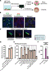
Fig. 1.. F. nucleatum binds to and invades HCT116 cells.
(A) Overview of experiments used to analyze binding and invasion of Fnn and adhesin mutants in HCT116 CRC cells. (B) Cocultures of HCT116 and Fnn (2 hours, MOI 50:1 Fnn:HCT116) stained for intracellular and extracellular Fnn. Fnn was labeled with the fluorescent green membrane–intercalating dye FM 1–43FX to detect intracellular and extracellular bacteria and labeled with a pan-Fusobacterium membrane antisera (Alexa Fluor 594 goat anti-rabbit antibody) for extracellular bacteria. Host-cell nuclei are stained with 4′,6-diamidino-2-phenylindole (DAPI) (blue). Arrows indicate intracellular bacteria. (C) Fnn bacterium that is half intracellular, half extracellular in HCT116 cells. (D) Comparison of HCT116 binding and invasion by wild-type (WT) F. nucleatum 23726 with F. nucleatum 23726 ΔgalKT (Fnn) (n = 3 independent flow cytometry experiments). (E) Binding and invasion analysis of Fnn and adhesin gene deletion strains using flow cytometry (n = 3 independent flow cytometry experiments). (F) Invasion and survival of Fnn and Fnn Δfap2 in HCT116 cells (n = 3 independent antibiotic protection assays). CFU, colony-forming units. In (E) and (F), infection parameters were 50:1 MOI Fnn:HCT116 for 4 hours (tan) and 0.5:1 MOI Fnn:HCT116 for 2 hours (purple). nsP > 0.05, *P < 0.05, and ****P < 0.0001 by unpaired Student’s t test or two-way ANOVA for single or grouped analyses, respectively.
Fig. 2.
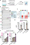
Fig. 2.. F. nucleatum induces cytokine secretion from HCT116 cells.
(A) Schematic of experiments used to analyze Fnn-induced cytokine secretion from HCT116 CRC cells. (B and C) Representative broad cytokine array dot blots [of 36 proteins; (B)] analyzing effect of infection with Fnn and Fnn-adhesin deletion strains. Control spots (B) indicate successful Western blots and provide densitometry controls for quantitation, calculated as fold increase and shown as a heatmap in (C). (D) IL-8 and CXCL1 ELISA to quantitate cytokine secretion from HCT116 cells induced by Fnn and Fnn Δfap. n = 3 independent experiments. (E) IL-8 and CXCL1 ELISA to quantitate and compare cytokine secretion from HCT116 cells induced by F. nucleatum subsp. nucleatum 25586, F. nucleatum subsp. nucleatum 23726, and F. nucleatum subsp. animalis 7_1 (Fna). n = 3 independent experiments. In (D) and (E), infection parameters were 50:1 MOI Fusobacterium: HCT116 for 4 hours (tan) and 50:1 MOI Fusobacterium:HCT116 for 24 hours (purple). ****P < 0.0001 by unpaired Student’s t test or two-way ANOVA for single or grouped analyses, respectively.
Fig. 3.
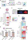
Fig. 3.. Cytokine secretion analysis from mouse neutrophils and macrophages.
(A) Overview of experiments used to analyze Fnn-induced cytokine secretion from immune cells. (B) Heatmap of cytokine array assessing secretion of the indicated cytokines from mouse neutrophils in the presence of E. coli, Fnn, or deletion control (columns as designated by colored dots, legend right). Infection parameters were 50:1 MOI Fnn:neutrophils for 4 hours. (C) Fluorescence microscopy of Fnn interacting with mouse neutrophils. DNA was detected using DAPI, and Fnn was labeled with the fluorescent red membrane–intercalating dye FM 4–64FX. Images are representative of n = 3 experiments. (D) ELISA confirming Fnn induction of CCL3 and CXCL2 and assessing the effect of deletion of fap2 on cytokine secretion in neutrophils. n = 3 independent experiments. (E) Cytokine array assessing the effect of Fnn infection on the secretion of several cytokines from mouse macrophages when compared to that of E. coli and control. Sample conditions designated by colored dots as defined in (B); relative abundance scale as indicated, right. In (B) to (E), infection parameters were 50:1 MOI Fusobacterium:neutrophils/macrophages for 4 hours. nsNot significant (P > 0.05) by unpaired Student’s t test.
Fig. 4.
Fig. 4.. Inhibition of Fnn binding to and invasion of HCT116 cells using small molecules and membrane antibodies.
(A) Fnn invasion is significantly inhibited by galactose-containing sugars and l-arginine (10 mM). n = 3 independent flow cytometry experiments. (B) Effect of sugars and l-arginine (10 mM) on the secretion of IL-8 and CXCL1 from HCT116 cells. n = 3 independent experiments. (C) Fnn invasion in the presence of Fusobacterium membrane antisera (FMAS) versus control rabbit antisera (RAS). n = 3 independent flow cytometry experiments. (D) Effect of FMAS and Fnn Δfap2 on secretion of IL-8 and CXCL1 from HCT116 cells. n = 3 independent experiments. nsP > 0.05, *P < 0.05, ***P < 0.001, and ****P < 0.0001 by unpaired Student’s t test or two-way ANOVA for single or grouped analyses, respectively.
Fig. 5.
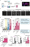
Fig. 5.. HCT116 migration is driven by Fnn-induced secretion of CXCL1 and IL-8.
(A and B) Experimental setup (A) and representative three-dimensional confocal imaging (B) of HCT116 transwell migration experiments. Blue, DAPI-stained DNA; red, CellTracker Red; white arrows, cross-barrier migrated cells. (C) Representative images of migrated HCT116 cells after a 16-hour exposure to the indicated conditioned medium. n = 3 independent experiments. (D) HCT116 cellular migration when cultured in Fnn-conditioned medium containing high CXCL1 and IL-8 concentrations. n = 3 independent experiments. (E) Effect of deletion of fap2 (Fnn Δfap2) and Fusobacterium membrane antisera (FMAS) on bacterial-induced HCT116 cellular migration. n = 3 independent experiments. (F) Effect of the addition of purified IL-8 and CXCL1 to culture medium within the lower transwell chamber on HCT116 cell migration. n = 5 independent experiments. (G) Analysis of CXCL1 and IL-8 levels after concentration, with and without antibody depletion of cytokines and before loading into the bottom chamber of the transwell apparatus. n = 3 independent experiments. (H) HCT116 cellular migration upon depleting CXCL1 and IL-8 from medium versus controls. n = 3 independent experiments. nsP > 0.05, *P < 0.05, **P < 0.01, ***P < 0.001, and ****P < 0.0001 by unpaired Student’s t test or two-way ANOVA for single or grouped analyses, respectively.
Fig. 6.
Fig. 6.. A model of F. nucleatum–induced metastasis through cytokine signaling.
Model of our findings, that Fap2-dependent F. nucleatum invasion into CRC cells induces IL-8 and CXCL1 secretion, and both cytokines have been characterized as key players in cancer metastasis and subsequent downstream cell seeding. HCT116-derived IL-8 and CXCL1 can participate in autocrine signaling back to cancer cells as a metastatic signal, as well as paracrine signaling to recruit neighboring immune cells, which further secrete their own cytokine signatures (CXCL2, CCL3, and TNFα) that alter the tumor microenvironment through metastatic, inflammatory, and immune cell programming. Our data show that Fap2 drives CRC cell docking, which is a key step in initiating the prometastatic cytokine cascade but is not necessary for immune cell signaling.

Comment in

References

    1. van Elsland D, Neefjes J, Bacterial infections and cancer. EMBO Rep. 19, e46632 (2018). - PMC - PubMed
    1. Gagnaire A, Nadel B, Raoult D, Neefjes J, Gorvel J-P, Collateral damage: Insights into bacterial mechanisms that predispose host cells to cancer. Nat. Rev. Microbiol 15, 109–128 (2017). - PubMed
    1. Kaufman HL, Kohlhapp FJ, Zloza A, Oncolytic viruses: A new class of immunotherapy drugs. Nat. Rev. Drug Discov 14, 642–662 (2015). - PMC - PubMed
    1. Brennan CA, Garrett WS , Fusobacterium nucleatum—Symbiont, opportunist and oncobacterium. Nat. Rev. Microbiol 17, 156–166 (2019). - PMC - PubMed
    1. Han YW, Fusobacterium nucleatum: A commensal-turned pathogen. Curr. Opin. Microbiol 23, 141–147 (2015). - PMC - PubMed

Publication types

MeSH terms

LinkOut - more resources