. 2021 Apr;70(4):761-774.
doi: 10.1136/gutjnl-2019-319664. Epub 2020 Jul 21.

Dietary cholesterol drives fatty liver-associated liver cancer by modulating gut microbiota and metabolites

Affiliations

Dietary cholesterol drives fatty liver-associated liver cancer by modulating gut microbiota and metabolites

Xiang Zhang et al. Gut. 2021 Apr.

Abstract

Objective: Non-alcoholic fatty liver disease (NAFLD)-associated hepatocellular carcinoma (HCC) is an increasing healthcare burden worldwide. We examined the role of dietary cholesterol in driving NAFLD-HCC through modulating gut microbiota and its metabolites.

Design: High-fat/high-cholesterol (HFHC), high-fat/low-cholesterol or normal chow diet was fed to C57BL/6 male littermates for 14 months. Cholesterol-lowering drug atorvastatin was administered to HFHC-fed mice. Germ-free mice were transplanted with stools from mice fed different diets to determine the direct role of cholesterol modulated-microbiota in NAFLD-HCC. Gut microbiota was analysed by 16S rRNA sequencing and serum metabolites by liquid chromatography-mass spectrometry (LC-MS) metabolomic analysis. Faecal microbial compositions were examined in 59 hypercholesterolemia patients and 39 healthy controls.

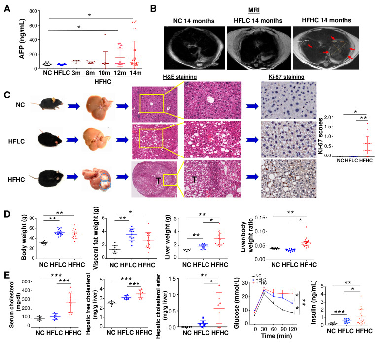
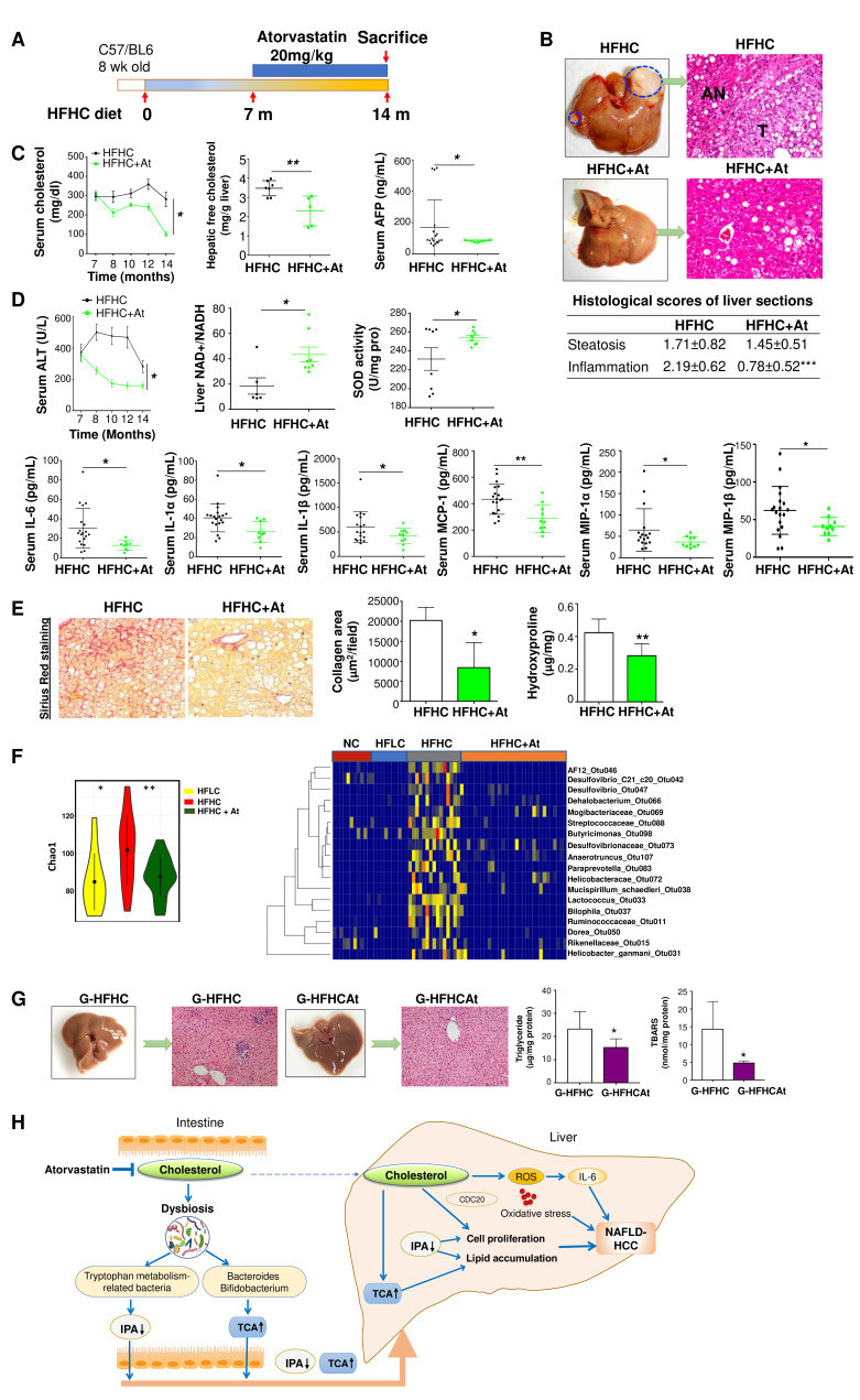
Results: High dietary cholesterol led to the sequential progression of steatosis, steatohepatitis, fibrosis and eventually HCC in mice, concomitant with insulin resistance. Cholesterol-induced NAFLD-HCC formation was associated with gut microbiota dysbiosis. The microbiota composition clustered distinctly along stages of steatosis, steatohepatitis and HCC. Mucispirillum, Desulfovibrio, Anaerotruncus and Desulfovibrionaceae increased sequentially; while Bifidobacterium and Bacteroides were depleted in HFHC-fed mice, which was corroborated in human hypercholesteremia patients. Dietary cholesterol induced gut bacterial metabolites alteration including increased taurocholic acid and decreased 3-indolepropionic acid. Germ-free mice gavaged with stools from mice fed HFHC manifested hepatic lipid accumulation, inflammation and cell proliferation. Moreover, atorvastatin restored cholesterol-induced gut microbiota dysbiosis and completely prevented NAFLD-HCC development.

Conclusions: Dietary cholesterol drives NAFLD-HCC formation by inducing alteration of gut microbiota and metabolites in mice. Cholesterol inhibitory therapy and gut microbiota manipulation may be effective strategies for NAFLD-HCC prevention.

Keywords: dietary factors; fatty liver; intestinal microbiology; nonalcoholic steatohepatitis.


Conflict of interest statement

Competing interests: None declared.

Figures

Figure 1
Figure 1
Cholesterol induces spontaneous HCC formation in C57BL/6 mice. (A) Serum AFP levels in mice fed with HFHC diet for 3, 8, 10, 12 and 14 months as well as mice fed with NC or HFLC diet for 14 months; (B) liver MRI, (C) representative gross morphology, representative microscopic features and immunohistochemistry pictures of Ki67 staining in liver of mice fed with NC, HFLC and HFHC for 14 months, Ki-67 was scored according to the following criteria: 0 (<10% of cells staining), 1 (11%–30% of cells staining), 2 (30%–50% of cells staining) or 3 (>50% of cells staining); (D) body weight, visceral fat, liver weight, liver-to-body weight ratio, (E) serum cholesterol level, hepatic free cholesterol, hepatic cholesterol ester content, glucose tolerance test and fasting insulin levels in mice fed with NC, HFLC and HFHC for 14 months. *p<0.05, **p<0.01, ***p<0.001. AFP, alpha-fetoprotein; NC, normal chow; HFLC, high-fat/low-cholesterol diet; HFHC, high-fat/high-cholesterol diet; H&E, hematoxylin and eosin.
Figure 2
Figure 2
Cholesterol causes NASH and fibrosis in non-HCC liver tissues of mice fed with HFHC diet. (A) Representative H&E staining, histological scores of liver sections; (B) serum ALT and AST levels; (C) serum IL-6, IL-1α and IL-1β protein levels by cytokine profiling assay in mice fed with NC, HFLC and HFHC for 14 months; (D) hepatic proinflammatory cytokines IL-6, IL-1α and IL-1β protein levels by ELISA and Cx3cl1, Mcp1, Cxcl10, Mip1β, Mip1α, Ccl5, Cxcl16, Tnfα mRNA levels by RNA sequencing in mice fed with HFLC and HFHC for 14 months; (E) collagen deposition by Sirius Red staining, α-SMA protein and mRNA levels by immunohistochemistry staining and RT-PCR, respectively; (F) hepatic hydroxyproline content, (G) liver NAD+ to NADH ratio and SOD activity in mice fed with NC, HFLC and HFHC for 14 months. *p<0.05, **p<0.01, ***p<0.001. ALT, alanine aminotransferase; AST, aspartate aminotransferase; α-SMA, alpha-smooth muscle actin; HCC, hepatocellular carcinoma; HFHC, high-fat/high-cholesterol diet; HFLC, high-fat/low-cholesterol diet; H&E, Hematoxylin and eosin; NAD, nicotinamide adenine dinucleotide; NASH, non-alcoholic steatohepatitis; NC, normal chow; RT-PCR, reverse transcription polymerase chain reaction; SOD, superoxide dismutase.
Figure 3
Figure 3
HFHC-fed mice sequentially develop a fatty liver, steatohepatitis, fibrosis and HCC. (A) Schematic illustration of the treatment of C57BL/6 mice with HFHC diet. Mice were sacrificed at 3, 8, 10, and 12 months of age. (B) Representative gross morphology, H&E staining and IHC staining of tumour markers AFP and GP73 in the liver of mice fed with HFLC for 14 months and HFHC for 3, 8, 10, 12 and 14 months; histological scoring of steatosis and inflammation and the quantitation of Sirius Red staining were calculated. (C) Tumour incidence, tumour number and maximum tumour diameter of the largest tumour from mice fed with HFHC at different time points. *p<0.05, **p<0.01, ***p<0.001. AFP, alpha-fetoprotein; HCC, hepatocellular carcinoma; HFHC, high-fat/high-cholesterol diet; HFLC, high-fat/low-cholesterol diet; H&E, hematoxylin and eosin; IHC, immunohistochemistry.
Figure 4
Figure 4
Dietary cholesterol-induced gut microbiota dysbiosis. (A) Principal component ordination analysis (PcoA), Shannon diversity and the richness of gut microbiota between 14 months HFLC and HFHC-fed mice. (B) Heatmap plot of the bacteria in stool of mice fed with HFLC or HFHC for 14 months. (C) PCoA and redundancy analysis of gut microbiota in fed with HFHC for 3 months, 8 months and 14 months, respectively. (D) The microbiota richness measured by chao1 index and sequentially increased bacteria from 3, 8 to 14 months of HFHC feeding. (E) Gut microbiota tryptophan metabolism capacity in HFLC-fed and HFHC-fed mice. (F) Correlation of bacterial abundance with mice phenomes. (G) The association of bacteria by metagenomic sequencing and serum total cholesterol, triglyceride, LDL-cholesterol and HDL cholesterol in 59 human cases of hypercholesterolemia and 39 healthy subjects. *p<0.05, **p<0.01, ***p<0.001. AFP, alpha-fetoprotein; ALT, alanine aminotransferase; AST, aspartate aminotransferase; HDL, high-density lipoprotein; HFHC, high-fat/high-cholesterol diet; HFLC, high-fat/low-cholesterol diet; LDL, low-density lipoprotein; TCHO, total cholesterol; TG, triglyceride.
Figure 5
Figure 5
Dietary cholesterol-induced alteration of metabolic profiles in mice serum. (A) Serum metabolites were significantly different between mice fed with HFLC and HFHC diet by principal component ordination analysis. (B) Pathway analysis of differentially enriched metabolites in mice fed with HFHC diet. (C) Volcano plot of serum metabolomics of mice fed with HFLC and HFHC diet, the outliers of metabolites are indicated. (D) LPS concentration in portal vein blood of mice fed with HFLC and HFHC diet for 3 months and E-cadherin expression in the colon tissues of 14 months HFLC and HFHC diet fed mice. (E) Correlation analysis of the association of the HFHC-altered microbes and metabolites. (F) mRNA levels of Cyp7a1, Cyp8b1, Cyp27a1 and Cyp7b1 in the liver tissues of mice fed with HFLC and HFHC diet. (G) TCA aggravated cholesterol-induced triglyceride accumulation in human LO2 cell line while IPA inhibited cholesterol-induced triglyceride accumulation in NASH–HCC cell lines, HKCI-2 and HKCI-10 by Oil Red O staining. Cholesterol, 200 μg/mL; (H) IPA inhibited cell proliferation in NASH–HCC cell lines. *p<0.05, **p<0.01, ***p<0.001. DMSO, dimethyl sulfoxide; GCA, glycocholic acid; HCC, hepatocellular carcinoma; HFHC, high-fat/high-cholesterol diet; HFLC, high-fat/low-cholesterol diet; IPA, 3-indolepropionic acid; NASH, non-alcoholic steatohepatitis; LPS, lipopolysaccharides; TCA, taurocholic acid; TCDCA, taurochenodeoxycholic acid; TUDCA, tauroursodeoxycholic acid.
Figure 6
Figure 6
High cholesterol-modulated microbiota promotes hepatocyte proliferation in germ-free mouse model. (A) Stools were transplanted from NC-fed, HFLC-fed and HFHC-fed mice (14 months) to germ-free mice (G-NC, G-HFLC and G-HFHC) under NC. Gross morphology, histological examination and Ki-67 staining of livers in stool-gavaged germ-free mice in the G-NC, G-HFLC or GHFHC groups were shown. (B) Liver triglyceride content, lipid peroxidation and liver histology in recipient germ-free mice of the G-NC, G-HFLC or G-HFHC groups at 8, 10 and 14 months. (C) Hepatic IL-6 protein levels and (D) Ki-67 staining of liver sections in the G-NC, G-HFLC or G-HFHC groups at 14 months. (E1) Mouse cancer pathway Finder PCR array in the liver tissues of G-NC, G-HFLC or G-HFHC mice. (E2) CDC20 protein level was validated. (F) Principal component ordination analysis and histogram of the linear discriminant analysis (LDA) scores of gut microbiota among G-NC, G-HFLC or G-HFHC mice. (G) Serum metabolomic analysis of G-HFLC and G-HFHC mice. *p<0.05, **p<0.01, ***p<0.001. HFHC, high-fat/high-cholesterol diet; HFLC, high-fat/low-cholesterol diet; NC, normal chow; PCA, principal component analysis.
Figure 7
Figure 7
Cholesterol inhibition abrogated NAFLD–HCC progression in HFHC-fed mice. (A) Schematic illustration of the atorvastatin treatment of C57BL/6 mice fed with HFHC diet. (B) Representative gross morphology of liver and microscopic features in H&E-stained HFHC-fed mice with or without the treatment of atorvastatin, histological scores of steatosis, inflammation and collagen were calculated. (C) Serum cholesterol levels, hepatic free cholesterol levels, serum AFP levels, (D) serum ALT levels, liver NAD+ to NADH ratio, liver SOD activity, serum IL-6, IL-1α, IL-1β, MCP-1, MIP-1α, MCP-1β protein levels, (E) collagen deposition and hydroxyproline content in HFHC-fed mice with or without the treatment of atorvastatin. (F) Bacterial richness and heatmap plot of the bacteria in stool of mice fed with NC, HFLC and HFHC with or without atorvastatin treatment. (G) Gross morphology, histological examination, triglyceride and lipid peroxidation of livers in HFHCAt stool gavaged germ-free mice (G-HFHCAt). (H) Schematic diagram of the mechanism of cholesterol-induced NAFLD–HCC development. *p<0.05, **p<0.01, ***p<0.001. AFP, alpha-fetoprotein; At, atorvastatin, ALT, alanine aminotransferase; HCC, hepatocellular carcinoma; HFHC, high-fat/high-cholesterol diet; IPA, 3-indolepropionic acid; NAD, nicotinamide adenine dinucleotide; NAFLD, non-alcoholic fatty liver disease; ROS, reactive oxygen species; SOD, superoxide dismutase; TCA, taurocholic acid.

References

    1. Romeo S. Notch and nonalcoholic fatty liver and fibrosis. N Engl J Med 2019;380:681–3. 10.1056/NEJMcibr1815636 - DOI - PubMed
    1. Kanwal F, Kramer JR, Mapakshi S, et al. . Risk of hepatocellular cancer in patients with non-alcoholic fatty liver disease. Gastroenterology 2018;155:1828–37. 10.1053/j.gastro.2018.08.024 - DOI - PMC - PubMed
    1. Wong RJ, Cheung R, Ahmed A. Nonalcoholic steatohepatitis is the most rapidly growing indication for liver transplantation in patients with hepatocellular carcinoma in the U.S. Hepatology 2014;59:2188–95. 10.1002/hep.26986 - DOI - PubMed
    1. Ioannou GN. The role of cholesterol in the pathogenesis of NASH. Trends Endocrinol Metab 2016;27:84–95. 10.1016/j.tem.2015.11.008 - DOI - PubMed
    1. Van Rooyen DM, Larter CZ, Haigh WG, et al. . Hepatic free cholesterol accumulates in obese, diabetic mice and causes nonalcoholic steatohepatitis. Gastroenterology 2011;141:1393–403. 10.1053/j.gastro.2011.06.040 - DOI - PMC - PubMed

Publication types

MeSH terms