. 2020 Jul 21;10(1):12066.
doi: 10.1038/s41598-020-68600-3.

Distinct and stage-specific contributions of TET1 and TET2 to stepwise cytosine oxidation in the transition from naive to primed pluripotency

Affiliations

Distinct and stage-specific contributions of TET1 and TET2 to stepwise cytosine oxidation in the transition from naive to primed pluripotency

Christopher B Mulholland et al. Sci Rep. .

Abstract

Cytosine DNA bases can be methylated by DNA methyltransferases and subsequently oxidized by TET proteins. The resulting 5-hydroxymethylcytosine (5hmC), 5-formylcytosine (5fC), and 5-carboxylcytosine (5caC) are considered demethylation intermediates as well as stable epigenetic marks. To dissect the contributions of these cytosine modifying enzymes, we generated combinations of Tet knockout (KO) embryonic stem cells (ESCs) and systematically measured protein and DNA modification levels at the transition from naive to primed pluripotency. Whereas the increase of genomic 5-methylcytosine (5mC) levels during exit from pluripotency correlated with an upregulation of the de novo DNA methyltransferases DNMT3A and DNMT3B, the subsequent oxidation steps turned out to be far more complex. The strong increase of oxidized cytosine bases (5hmC, 5fC, and 5caC) was accompanied by a drop in TET2 levels, yet the analysis of KO cells suggested that TET2 is responsible for most 5fC formation. The comparison of modified cytosine and enzyme levels in Tet KO cells revealed distinct and differentiation-dependent contributions of TET1 and TET2 to 5hmC and 5fC formation arguing against a processive mechanism of 5mC oxidation. The apparent independent steps of 5hmC and 5fC formation suggest yet to be identified mechanisms regulating TET activity that may constitute another layer of epigenetic regulation.


Conflict of interest statement

The authors declare no competing interests.

Figures
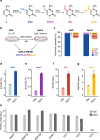
Figure 1
Figure 1
Global increases in cytosine modifications accompany the transition from naive to primed pluripotency. (a) Cytosine modifications depicted with the enzymes responsible for their generation. (b) Schematic overview of experimental design. DNA modifications were measured in murine naive embryonic stem cells (ESC) and epiblast-like stem cells (EpiLC) using UHPLC-MS/MS. (c) Abundance of genomic 5mC, 5hmC, 5fC, and 5caC in wild-type ESCs and EpiLCs shown as the fraction of total modified (mod.) cytosines. Due to their relative scarcity, 5fC and 5caC are depicted with a zoomed-in view. n = 6 (ESCs) and n = 12 (EpiLCs) biological replicates. (dg) Global levels of (d) 5mC, (e) 5hmC, (f) 5fC, and (g) 5caC in wild-type ESCs and EpiLCs as determined by mass spectrometry (UHPLC-MS/MS). DNA modification levels are expressed as a percentage (%) or parts per million (ppm: 1 ppm = 0.0001%) of total cytosine (dC). Error bars indicate mean ± SD calculated from n = 6 (ESCs) and n = 12 (EpiLCs) biological replicates. (h) Protein abundance of DNA modifying enzymes in wild-type ESCs and EpiLCs as determined by LC–MS/MS-based whole proteome profiling. Shown are log2-transformed protein levels. Error bars indicate mean ± SD calculated from n = 3 (ESCs) and n = 3 (EpiLCs) biological replicates. N.D.: no peptides of protein detected.
Figure 2
Figure 2
Quantification of cytosine modifications in Tet1 and Tet2 knockout ESCs and EpiLCs. (af) Global levels of (a, b) 5mC, (c, d) 5hmC, and (e, f) 5fC, in wild-type (WT), Tet1 KO (T1KO), Tet2 KO (T2KO), and Tet1/Tet2 DKO (T12KO) ESCs and EpiLCs as determined by mass spectrometry (UHPLC-MS/MS). DNA modification levels are expressed as a percentage (%) or parts per million (ppm: 1 ppm = 0.0001%) of total cytosine (dC) and shown as the mean ± SD of biological replicates as follows: WT (ESCs: n = 18; EpiLCs: n = 24), T1KO (ESCs: n = 18; EpiLCs: n = 12), T2KO (ESCs: n = 12; EpiLCs: n = 12), and T12KO (ESCs: n = 12; EpiLCs: n = 12). * p < 0.005 to wt as determined using a one-way ANOVA followed by a post-hoc Tukey HSD test. (gh) Correlations between 5hmC and 5fC levels in wt and Tet KO (g) ESCs and (h) EpiLCs. The dashed regression line was generated using the full data set, the solid regression line was generated by excluding Tet2 KO data. Depicted are values from the individual replicates presented in cf. R2: coefficient of determination; r: Pearson correlation coefficient. (i) Box plots of the ratio of 5fC to 5hmC in wt, Tet1 KO and Tet2 KO ESCs and EpiLCs. Unlike the Tet1 KO, Tet2 KO drastically affects the 5fC/5hmC ratio. The median is represented by the central bold line. The lower and upper hinges correspond to the first and third quartiles (the 25th and 75th percentiles). The upper and lower whisker extend from the hinge to the largest and lowest value, respectively, no further than 1.5 * interquartile range (IQR).
Figure 3
Figure 3
Epigenetic changes and distinct contributions of different DNA modifying enzymes during the transition from naive to primed pluripotency. Graphical summary depicting changes in cellular levels of cytosine modifications and their respective DNA modifying enzymes in the transition from naive to primed pluripotency. The relative contributions of TET1 and TET2 to the generation of 5hmC and 5fC as estimated from observations in Tet KO ESCs and EpiLCs are illustrated at the bottom; the number of spheres and tilt of the balance represent the protein abundance of each TET and the contribution of each TET to the levels of the depicted cytosine derivative, respectively. TET1 gains importance in the oxidation of 5mC to 5hmC during differentiation as TET2 abundance decreases. Most remarkably, despite drastic downregulation TET2 remains critical for the formation of 5fC in primed pluripotency.

Similar articles

Cited by

References

    1. Smith ZD, Meissner A. DNA methylation: Roles in mammalian development. Nat. Rev. Genet. 2013;14:204–220. - PubMed
    1. Seisenberger S, et al. Reprogramming DNA methylation in the mammalian life cycle: Building and breaking epigenetic barriers. Philos. Trans. R. Soc. Lond. B Biol Sci. 2013;368:20110330. - PMC - PubMed
    1. Greenberg MVC, Bourc’his D. The diverse roles of DNA methylation in mammalian development and disease. Nat. Rev. Mol. Cell Biol. 2019 doi: 10.1038/s41580-019-0159-6. - DOI - PubMed
    1. Lee HJ, Hore TA, Reik W. Reprogramming the methylome: erasing memory and creating diversity. Cell Stem Cell. 2014;14:710–719. - PMC - PubMed
    1. Monk M, Boubelik M, Lehnert S. Temporal and regional changes in DNA methylation in the embryonic, extraembryonic and germ cell lineages during mouse embryo development. Development. 1987;99:371–382. - PubMed

Publication types

MeSH terms