Review
. 2019 Dec;1(12):1189-1201.
doi: 10.1038/s42255-019-0148-2. Epub 2019 Dec 13.

Integrating the inputs that shape pancreatic islet hormone release

Affiliations
Review

Integrating the inputs that shape pancreatic islet hormone release

Glyn M Noguchi et al. Nat Metab. 2019 Dec.

Abstract

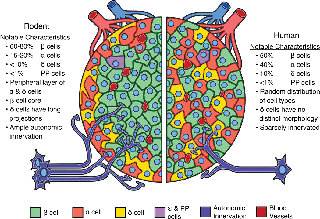
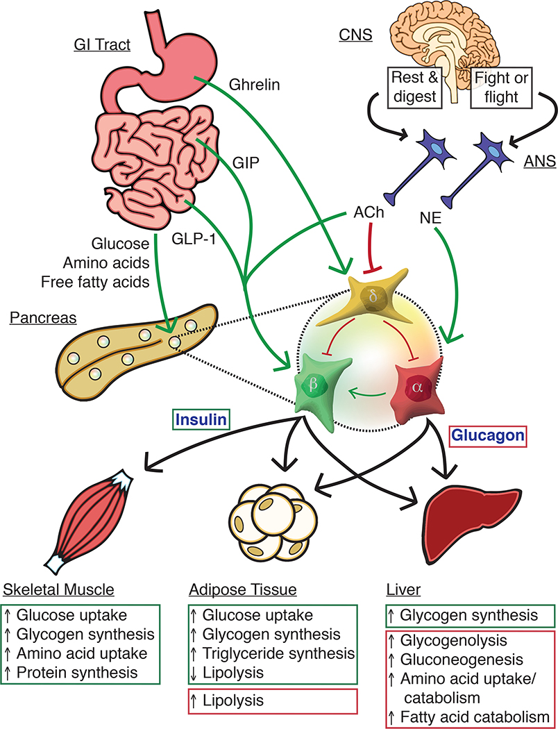
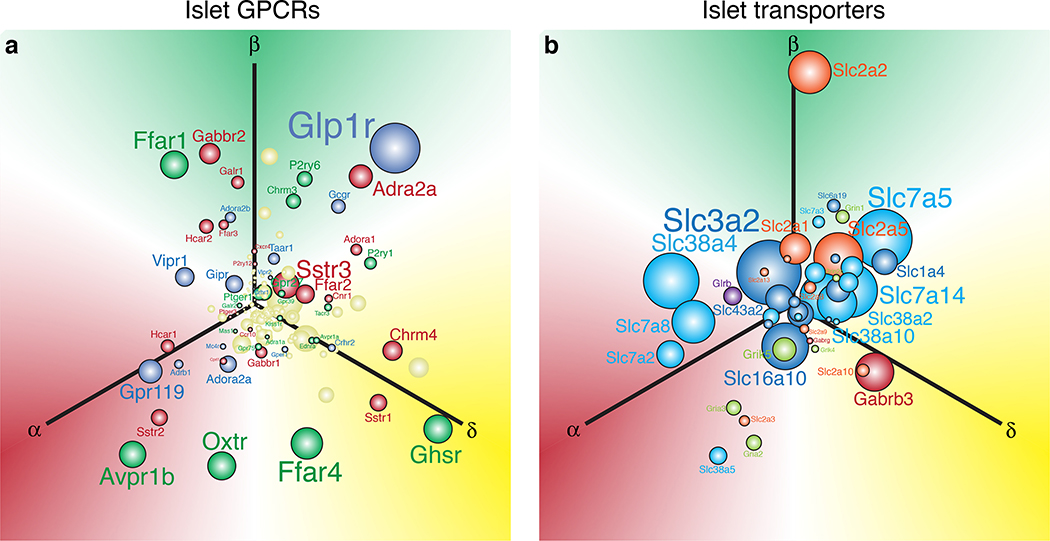
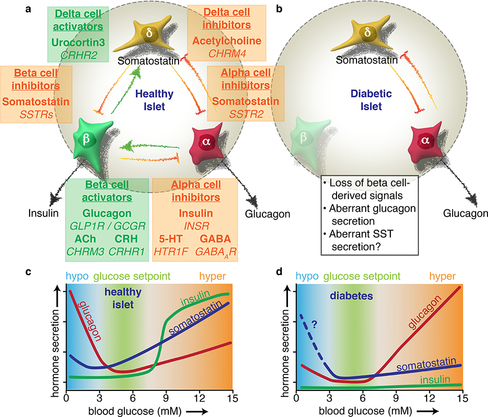
The pancreatic islet is a complex mini organ composed of a variety of endocrine cells and their support cells, which together tightly control blood glucose homeostasis. Changes in glucose concentration are commonly regarded as the chief signal controlling insulin-secreting beta cells, glucagon-secreting alpha cells and somatostatin-secreting delta cells. However, each of these cell types is highly responsive to a multitude of endocrine, paracrine, nutritional and neural inputs, which collectively shape the final endocrine output of the islet. Here, we review the principal inputs for each islet-cell type and the physiological circumstances in which these signals arise, through the prism of the insights generated by the transcriptomes of each of the major endocrine-cell types. A comprehensive integration of the factors that influence blood glucose homeostasis is essential to successfully improve therapeutic strategies for better diabetes management.


Figures

Figure 1.
Figure 1.. Comparative architecture of pancreatic islets of mice and humans.
Pancreatic islets of mice and humans differ in important ways, but also share many features in common. These shared features make mouse islets useful experimental models to study many aspects of human islet biology. The relative proportions of endocrine cell types in mouse (left) and human islets (right) are similar with beta cells (β; green) comprising the majority of the islet cell mass followed by alpha (α; light red) and delta cells (δ; yellow). Other islet endocrine cells such as pancreatic polypeptide and epsilon cells (PP and ε; purple) are more sparse in number. Human islets occur in a wide variety of sizes and conformations that range from highly structured to more random distributions of cells. Mouse islets exhibit a more uniform architecture with alpha and delta cells at the islet periphery surrounding a beta cell core. Islets in both species are vascularised (dark red) and innervated (dark blue) for rapid sensing of changing energy needs, although mouse islets are more densely innervated than humans.
Figure 2.
Figure 2.. Inter-organ signaling from nutrient sensing to islet-mediated metabolic effects.
Nutrition-related signals from the gastrointestinal (GI) tract combine with neuronal input from the autonomic nervous system (ANS) to direct insulin and glucagon secretion from pancreatic islets. Changes in blood glucose levels are sensed by alpha, beta, and delta cells, which respond by restoring blood glucose to homeostatic levels. Alpha cells release glucagon at low glucose to increase hepatic glucose production. During hyperglycemia, beta cells lower blood glucose by releasing insulin to increase glucose storage in the liver, skeletal muscle, and adipose tissue. Insulin release is amplified by the incretin hormones GLP-1 and GIP from the small intestine as well as by glucagon from neighbouring alpha cells. Delta cells secrete somatostatin across a range of glucose levels, but most prominently in response to hyperglycemia. Amino acids and free fatty acids (FFAs) stimulate both alpha and beta cells, and the peripheral effects of both glucagon and insulin result in reduced circulating amino acids and FFAs. The central nervous system (CNS) can augment islet secretion in conditions such as the “rest and digest” state where direct insulin secretion is further facilitate by a suppression of somatostatin secretion by acetylcholine (ACh) associated with parasympathetic innervation. Glucagon secretion is increased during the “fight or flight” response by norepinephrine (NE) released by sympathetic nerves.
Figure 3.
Figure 3.. Visualisation of the abundance and selectivity of GPCR and transporter gene expression in alpha, beta, and delta cells.
We used the natural log of the normalised expression values for a gene (ln[RPKM]) to plot the relative position of that gene along three axes representing alpha, beta, and delta cells. These expression values are derived from transcriptomes of FACS-purified mouse alpha, beta, and delta cells described elsewhere. a) Each of these three individual gene expression values are converted into x and y vectors and then consolidated into a single set of x, y coordinates that represents the overall selectivity of the expression of that gene. The origin represents equal expression (i.e. no enrichment) in each of the three islet cell types, whereas. placement in any direction along one of the axes reflects enrichment in the corresponding cell type. Sphere and font sizes are proportional to abundance of the gene based on the highest RPKM value for that gene in alpha, beta, or delta cells. b) The top 150 most abundant G protein-coupled receptors (GPCR) of the islet cells are color coded in accordance with the predominant signaling cascade associated with each receptor. Blue genes are Gαs-coupled, green are Gαq, red are Gαi, and yellow is ‘unknown’ or ambiguous based on receptor classifications from IUPHAR (International Union of Basic and Clinical Pharmacology). c) Non-GPCR receptors and transporters are colour-coded according to the class of signaling molecules utilised by each receptor/transporter, following IUPHAR classification for solute carriers.
Figure 4.
Figure 4.. Diabetes disrupts the extensive paracrine signaling network of the islet.
Alpha, beta, and delta cells influence each other’s secretion via intra-islet crosstalk. a) Coloured text boxes (green for activating, orange for inactivating) denote the target cell type of paracrine signaling (underlined), the signal molecule involved (bold), and the target receptor gene (italicised). Each box is placed in between the target cell and the source of the signal. Beta cells initiate a negative feedback loop in high glucose whereby they release urocortin 3 to activate delta cells. The resulting somatostatin (SST) release feeds back to mediate insulin release, providing tonic inhibition that establishes the homeostatic glucose setpoint. Beta cells also experience paracrine activation from alpha cells, which release glucagon, acetylcholine (ACh), and corticotropin-releasing hormone (CRH), which all potentiate GSIS. Beta cell-derived products such as insulin, serotonin (5-HT), and GABA – in addition to urocortin 3-induced somatostatin release – all contribute to silence alpha cells during hyperglycemia. b) The onset of diabetes results in a loss of multiple paracrine signals. Due to autoimmune destruction, type 1 diabetic islets effectively lose all beta cell signals. In type 2 diabetes, urocortin 3 is severely downregulated in beta cells, blunting glucose-stimulated somatostatin secretion. c) The physiological impact of paracrine signaling can be visualised with glucose curves for each islet hormone. The homeostatic glucose set point is maintained by glucagon raising blood glucose during hypoglycaemia and insulin lowering glucose during hyperglycaemia. Somatostatin contributes as a fine-tuning mechanism via paracrine inhibition of both alpha and beta cells. d) The absence of beta cell-derived products in diabetes results in inappropriately high glucagon secretion during high glucose, which exacerbates hyperglycaemia. Glucagon counterregulation at low glucose is also impaired, possibly due to aberrant somatostatin secretion, although this is not yet fully understood. Based in part on Ref.

References

    1. Collaboration, N. C. D. R. F. Worldwide trends in diabetes since 1980: a pooled analysis of 751 population-based studies with 4.4 million participants. Lancet 387, 1513–1530, doi:10.1016/S0140-6736(16)00618-8 (2016). - DOI - PMC - PubMed
    1. Unger RH & Cherrington AD Glucagonocentric restructuring of diabetes: a pathophysiologic and therapeutic makeover. J Clin Invest 122, 4–12, doi:10.1172/JCI60016 (2012). - DOI - PMC - PubMed
    1. Cabrera O et al. The unique cytoarchitecture of human pancreatic islets has implications for islet cell function. Proc Natl Acad Sci U S A 103, 2334–2339, doi:10.1073/pnas.0510790103 (2006). - DOI - PMC - PubMed
    1. Steiner DJ, Kim A, Miller K & Hara M Pancreatic islet plasticity: interspecies comparison of islet architecture and composition. Islets 2, 135–145 (2010). - PMC - PubMed
    1. Brissova M et al. Assessment of human pancreatic islet architecture and composition by laser scanning confocal microscopy. J Histochem Cytochem 53, 1087–1097, 10.1369/jhc.5C6684.2005 (2005). - DOI - PubMed

Publication types