. 2020 Jul 21;10(1):12094.
doi: 10.1038/s41598-020-69077-w.

A novel method for assessing the renal biopsy specimens using an activatable fluorescent probe

Affiliations

A novel method for assessing the renal biopsy specimens using an activatable fluorescent probe

Takuji Iyama et al. Sci Rep. .

Abstract

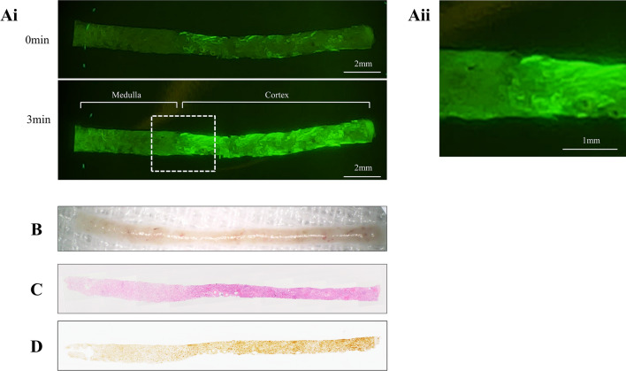
Gamma-glutamyl hydroxymethyl rhodamine green (gGlu-HMRG) is an activatable fluorescent probe that can be activated by γ-glutamyltranspeptidase (GGT). The expression of GGT in the kidney, which is one of the major organs exhibiting enhanced GGT expression, is exclusively localised to the cortex. Here, we aimed to investigate the feasibility of gGlu-HMRG as a probe for the on-site assessment of renal biopsy specimens. gGlu-HMRG fluorescent probe was applied to the renal proximal tubular epithelial cells and cortical collecting duct cells in vitro, mouse kidneys ex vivo, and human biopsy specimens. In addition, the fluorescence intensities in the cortex and the medulla were comparatively evaluated in the biopsy specimens. The fluorescence signal was rapidly detected in the renal proximal tubular epithelial cells, whereas that in the cortical collecting duct cells was not detected. The fluorescence signal was detected in the mouse kidneys ex vivo without markedly affecting the tissue morphology. In the human biopsy specimens, the fluorescence signal in the cortex was significantly distinct from that in the medulla (p < 0.05). Thus, this fluorescent probe can be used to distinctly identify the renal cortex in the biopsy specimens.


Conflict of interest statement

The authors declare no competing interests.

Figures

Figure 1
Figure 1
In vitro experiments with RPTECs and M-1 cells. Phase-contrast and fluorescence images of (A) RPTECs and (B) M-1 cells. gGlu-HMRG was diluted in phosphate-buffered saline and applied at concentrations of 10 or 50 μM. Fluorescence images were captured at 1, 3, 5, and 10 min post-gGlu-HMRG administration. The fluorescence signal was detected only in the RPTECs. gGlu-HMRG; γ-glutamyl hydroxymethyl rhodamine green, RPTEC; renal proximal tubule epithelial cell, M-1; mouse cortical collecting duct.
Figure 2
Figure 2
Ex vivo analysis of mouse kidneys. (A) Fluorescence images of the mouse kidney sections incubated with gGlu-HMRG. (B) The time-course of changes in the fluorescence intensities. The intensity gradually increased and plateaued at 10 min post-gGlu-HMRG administration. (C) Representative images of PAS and PAM staining obtained from paraffin-embedded mouse kidney sections incubated with gGlu-HMRG or PBS. There was no marked change in the two groups. gGlu-HMRG; γ-glutamyl hydroxymethyl rhodamine green, PAS; periodic acid-Schiff, PAM; periodic acid-methenamine silver, PBS; phosphate-buffered saline.
Figure 3
Figure 3
Representative fluorescence images of the human biopsy specimen. (Ai) Fluorescence images captured at 0 and 3 min post-50 μM gGlu-HMRG administration. (Aii) Magnified fluorescence image of the cortico-medullary junction. (B) The image captured from the same specimen using a magnifying lens under white light. Cortico-medullary junction, which was clearly detected in the fluoserscence images, is obscure under magnified lens. (C) PAS staining image of the same specimen. (D) The same specimen was subjected to GGT immunostaining. The region with intense fluorescence intensity corresponds to the cortex and the area with a strong GGT signal. gGlu-HMRG; γ-glutamyl hydroxymethyl rhodamine green, PAS; periodic acid-Schiff, GGT; γ-glutamyltranspeptidase.
Figure 4
Figure 4
Fluorescence intensities in the cortex and medulla. (A) Fluorescence intensities in each 8 parts from 12 patients at 0 and 3 min post-50 μM gGlu-HMRG administration. Each part was divided into the cortex or medulla according to the periodic acid-Schiff staining. Data were analysed by two-way analysis of variance with post hoc Sidak’s test. (B) Fold increase in the intensities after 3 min incubation with gGlu-HMRG. Data were analysed by unpaired t test. Bars indicate average ± standard error of mean (SEM). *P < 0.05. gGlu-HMRG; γ-glutamyl hydroxymethyl rhodamine green.

References

    1. Bandari J, Fuller TW, Turner RM, D’Agostino LA. Renal biopsy for medical renal disease: Indications and contraindications. Can. J. Urol. 2016;23:8121–8126. - PubMed
    1. Corapi KM, Chen JL, Balk EM, Gordon CE. Bleeding complications of native kidney biopsy: A systematic review and meta-analysis. Am. J. Kidney Dis. 2012;60:62–73. doi: 10.1053/j.ajkd.2012.02.330. - DOI - PubMed
    1. Urano Y, et al. Rapid cancer detection by topically spraying a γ-glutamyltranspeptidase-activated fluorescent probe. Sci. Transl. Med. 2011;3:110ra119. doi: 10.1126/scitranslmed.3002823. - DOI - PMC - PubMed
    1. Mitsunaga M, et al. Fluorescence endoscopic detection of murine colitis-associated colon cancer by topically applied enzymatically rapid-activatable probe. Gut. 2013;62:1179–1186. doi: 10.1136/gutjnl-2011-301795. - DOI - PMC - PubMed
    1. Kawakubo K, et al. Feasibility of using an enzymatically activatable fluorescence probe for the rapid evaluation of pancreatic tissue obtained using endoscopic ultrasound-guided fine needle aspiration: A pilot study. Mol. Imaging Biol. 2016;18:463–471. doi: 10.1007/s11307-015-0898-5. - DOI - PubMed

Publication types

LinkOut - more resources