. 2020 Jun;2(6):487-498.
doi: 10.1038/s42255-020-0206-9. Epub 2020 Jun 1.

Metabolic coessentiality mapping identifies C12orf49 as a regulator of SREBP processing and cholesterol metabolism

Affiliations

Metabolic coessentiality mapping identifies C12orf49 as a regulator of SREBP processing and cholesterol metabolism

Erol C Bayraktar et al. Nat Metab. 2020 Jun.

Abstract

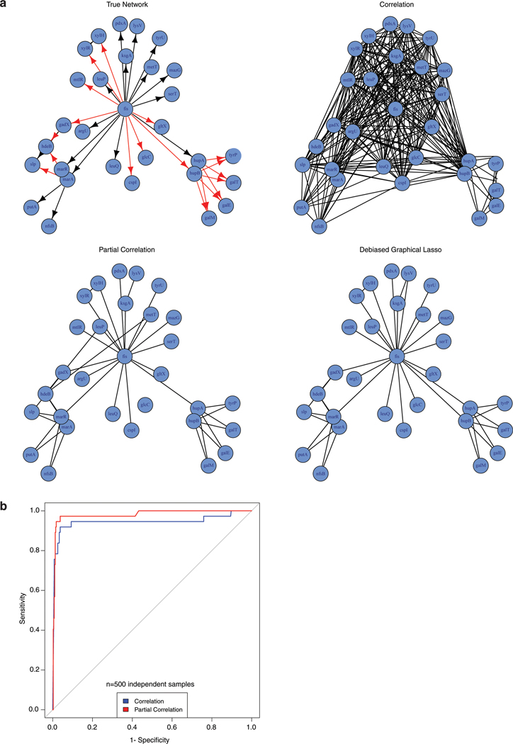
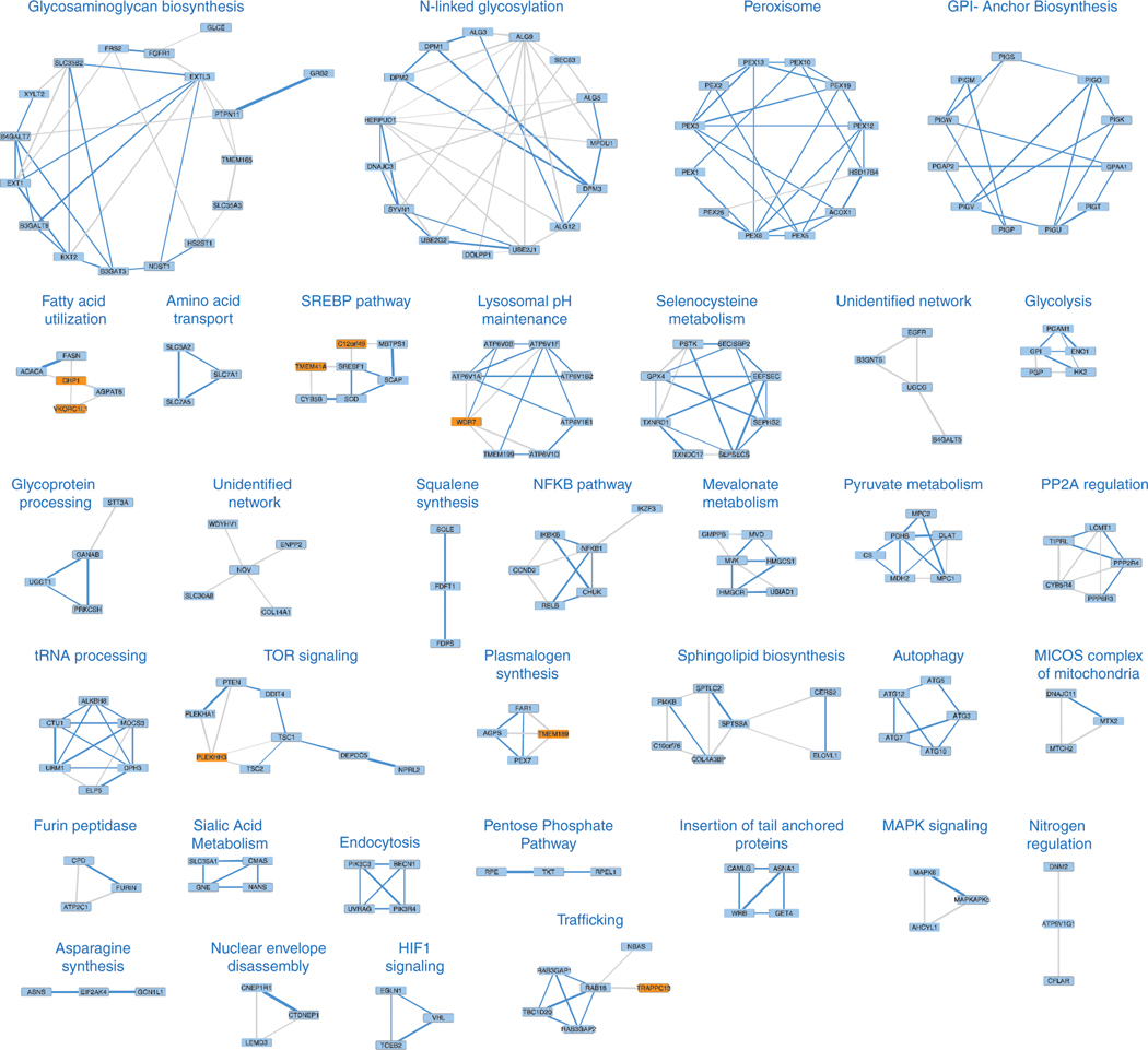
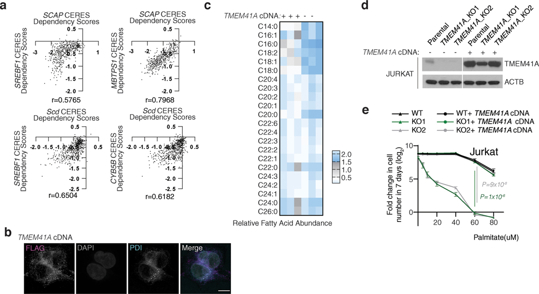
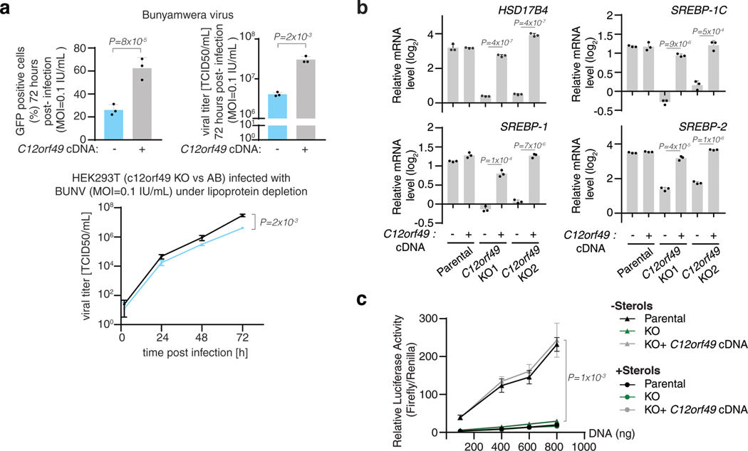
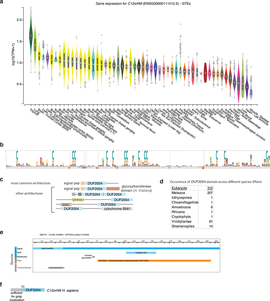
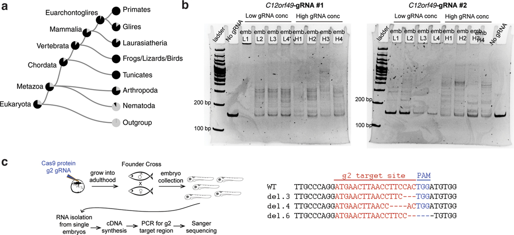
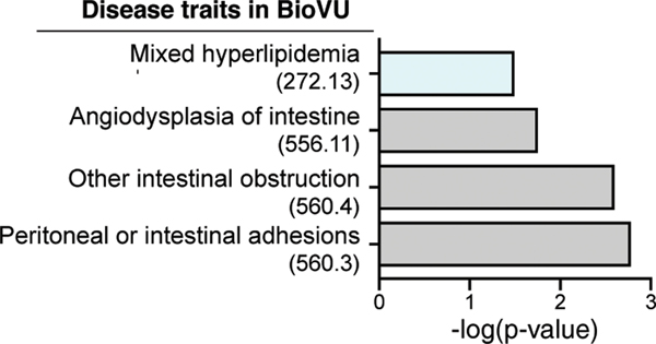
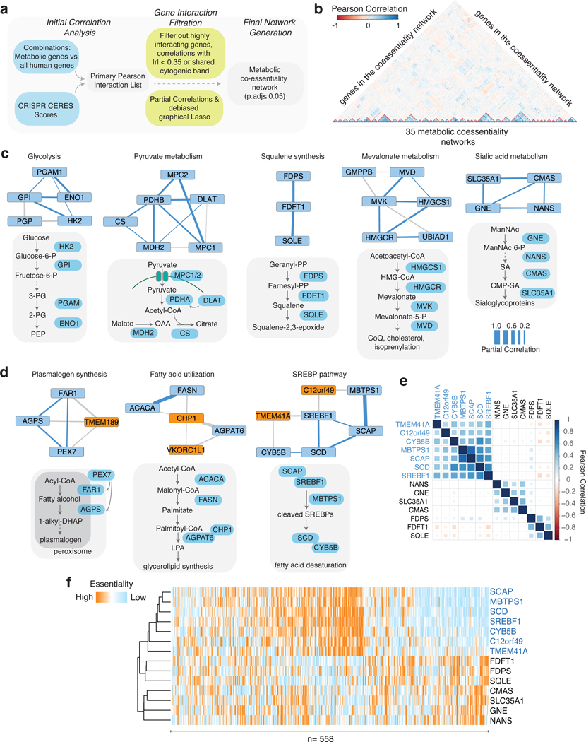
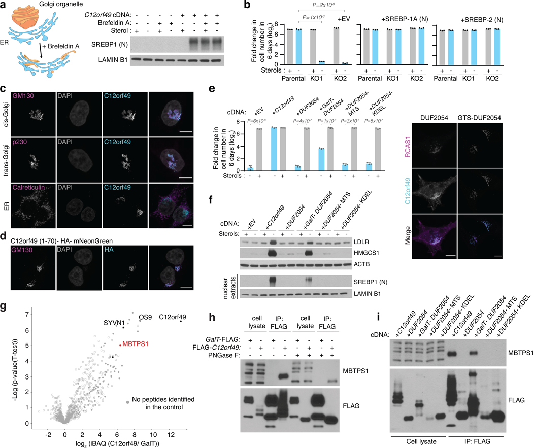
Coessentiality mapping has been useful to systematically cluster genes into biological pathways and identify gene functions1-3. Here, using the debiased sparse partial correlation (DSPC) method3, we construct a functional coessentiality map for cellular metabolic processes across human cancer cell lines. This analysis reveals 35 modules associated with known metabolic pathways and further assigns metabolic functions to unknown genes. In particular, we identify C12orf49 as an essential regulator of cholesterol and fatty acid metabolism in mammalian cells. Mechanistically, C12orf49 localizes to the Golgi, binds membrane-bound transcription factor peptidase, site 1 (MBTPS1, site 1 protease) and is necessary for the cleavage of its substrates, including sterol regulatory element binding protein (SREBP) transcription factors. This function depends on the evolutionarily conserved uncharacterized domain (DUF2054) and promotes cell proliferation under cholesterol depletion. Notably, c12orf49 depletion in zebrafish blocks dietary lipid clearance in vivo, mimicking the phenotype of mbtps1 mutants. Finally, in an electronic health record (EHR)-linked DNA biobank, C12orf49 is associated with hyperlipidaemia through phenome analysis. Altogether, our findings reveal a conserved role for C12orf49 in cholesterol and lipid homeostasis and provide a platform to identify unknown components of other metabolic pathways.


Conflict of interest statement

Competing interests

The authors declare no competing interests.

Figures

Extended Data Fig. 1
Extended Data Fig. 1. Comparative Simulation between partial and Pearson correlation
A. Simulation experiment of a subnetwork from an E. coli network demonstrating the advantage of using partial correlation over Pearson correlation. B. Receiver operating characteristic (ROC) curve based on the simulated data. (n= 500 independent samples)
Extended Data Fig. 2
Extended Data Fig. 2. Metabolic coessentiality modules
35 Metabolic coessentiality modules. Blue line indicates a previously known interaction between the genes. Poorly characterized genes are highlighted as orange.
Extended Data Fig. 3
Extended Data Fig. 3. C12orf49 is necessary for cell growth under sterol depletion
A. Pearson correlation values of the essentiality scores of the indicated genes across different cancer cell lines (n=558). B. Differential sgRNA score for C12orf49 gene of Jurkat cell line in the presence or absence of sterols. C. Fold change in cell number (log2) of U-87 MG or MDA-MB-435 c12orf49_KO cell line following a 6-day growth under lipoprotein depletion in the absence or presence of sterols. (mean ± SD, n=3 biologically independent samples). Statistical significance was determined by two-tailed unpaired t-test. D. Immunoblots of c12orf49 in the indicated knockout cells of HEK293T. Actin was used as the loading control. The experiment was repeated independently twice with similar results. E. (left) Immunoblots of c12orf49 knockout and addback cells in Jurkat cells. Actin was used as the loading control. The experiment was repeated independently twice with similar results. (right) Fold change in cell number (log2) of indicated knockout and rescued addback Jurkat cells following a 6-day growth under lipoprotein depletion in the absence or presence of sterols. (mean ± SD, n=3 biologically independent samples). Statistical significance was determined by two-tailed unpaired t-test. F. Fold change in cell number (log2) of indicated knockout and rescued addback HEK293T cells following a 6-day growth under lipoprotein depletion in the absence or presence of sterols. (mean ± SD, n=3 biologically independent samples). Statistical significance was determined by two-tailed unpaired t-test.
Extended Data Fig. 4
Extended Data Fig. 4. TMEM41A is involved in lipid metabolism
A. Pearson correlation values of the essentiality scores of the indicated genes across different cancer cell lines (n=558). B. Localization of TMEM41A to ER. Wild type HEK293T cells expressing FLAG-TMEM41A cDNA were processed for immunofluorescence analysis using antibodies against FLAG and PDI (ER). White color indicates overlap (Scale bar, 8 μm). The experiment was repeated independently twice with similar results. C. Heatmap showing the relative abundance of indicated lipid species in TMEM41-null Jurkat cells and those expressing sgRNA resistant TMEM41A cDNA. D. Immunoblot of TMEM41A in Jurkat wild type cell line, TMEM41A nulls and those expressing TMEM41A cDNA. Actin was used as the loading control. The experiment was repeated independently twice with similar results. E. Fold change in cell number (log2) of Jurkat wild type cell line, TMEM41A-null cells and those expressing TMEM41A cDNA after a 7-day growth upon treatment of indicated palmitate concentrations (0–80 uM). (mean ± SD, n=3 biologically independent samples). Statistical significance was determined by two-tailed unpaired t-test.
Extended Data Fig. 5
Extended Data Fig. 5. Role of C12orf49 in sterol synthesis and SREBP-mediated transcription
A. (top left) Percentage of Bunyamwera virus-positive cells at 72-hours post-infection (MOI=0.1IU/Ml) in indicated knockout and addback HEK293T cells (mean ± SD, n=3 biologically independent samples). Statistical significance was determined by two-tailed unpaired t-test. (top right) Viral titer measured by TCID50 assays on BHK-21 cells with the harvested supernatant from the Bunyamwera virus infected HEK293T cells of C12orf49 knockouts and addbacks. (mean ± SD, n=3 biologically independent samples) Statistical significance was determined by two-tailed unpaired t-test. (bottom) Growth of the viral titers at different time points in the knockout and addback cells. B. Fold change in mRNA levels (log2) of SREBP target genes in indicated Jurkat cell lines following 8h growth under lipoprotein depletion in the presence and absence of sterols (mean ± SD, n=3). C. Relative luminescence activity (Luciferase/Renilla) in the indicated HEK293 cell lines following transfection with firefly luciferase under SRE promoter and Renilla luciferase for normalization of transfection following 24h growth under lipoprotein depletion in the presence and absence of sterols (mean ± SD, n=3 biologically independent samples). Statistical significance was determined by two-tailed unpaired t-test.
Extended Data Fig. 6
Extended Data Fig. 6. C12orf49 gene expression in various tissues
A. Gene expression analysis across different tissues for C12orf49. Box plots are shown as median and 25th and 75th percentiles; points are displayed as outliers if they are above or below 1.5 times the interquartile range (Source: GTEx Portal). B. DUF2054 profile hidden Markov Model (HMM) logo from Pfam shows 14 conserved cysteines, 3 of which are CC-dimers. C. Different architectures of DUF2054 in different species. (Source: Pfam) D. Occurrence of DUF2054 domain across different species. E. Predicted N-glycosylation site (UniProtKB) and transmembrane domains (predicted with TMHMM v.2.0) for C12orf49. F. Scheme for different functional domains of C12orf49.
Extended Data Fig. 7
Extended Data Fig. 7. The impact of C12orf49 loss on the cleavage of MBTPS1 targets
A. Immunoblot analysis of OS9 in the C12orf49 immunoprecipitates of the HEK293T cell line expressing the indicated cDNAs. The experiment was repeated independently twice with similar results. B. Immunoblot analysis of cleavage of other site-1 protease targets, GNPTAB, CREB3L2 and CREB4 at 24-hours following transfection in the C12orf49-knockout and addback HEK293T cells. Actin was used as loading control. The experiment was repeated independently twice with similar results. C. Localization of SCAP-GFP in c12orf49 null HEK293T cells expressing control or C12orf49 cDNA under lipoprotein depletion in the presence or absence of sterols (Scale bar, 8 μm). The experiment was repeated independently twice with similar results.
Extended Data Fig. 8
Extended Data Fig. 8. Conservation of C12orf49 function in metazoa and zebrafish
A. Phylogenetic tree of the C12orf49 genes across species (Source: TreeFam). B. DNA gel showing the cutting efficiencies of c12orf49 sgRNAs used in the zebrafish experiments. Upper bands (smears) represent DNA heteroduplexes caused by CRISPR-Cas9 mutations; lower band is unedited DNA. This assay was repeated twice with similar results. C. Strategy to evaluate the effect of CRISPR-Cas9-generated c12orf49 mutations at transcript level. c12orf49-g2 founder F0 fish were crossed and F1 progeny was individually analyzed. Briefly, RNA was isolated from individual larvae, then cDNA was synthesized. Using exon-specific primers g2 target site was PCR amplified and sequenced. Various mutations detected from transcripts are shown.
Extended Data Fig. 9
Extended Data Fig. 9. GReX analysis identifies C12orf49 association with mixed hyperlipidemia
Disease traits associated with reduced c12orf49 GReX in BioVU biobank. Phecodes are indicated in parentheses. Traits are categorized into systems (y-axis), and significance is displayed on x-axis. Significance is tested by logistic regression analysis (two-sided), n = 25,000. Multiple testing adjustment is done using Bonferroni correction.
Figure 1,
Figure 1,. Genetic coessentiality analysis assigns metabolic functions to uncharacterized genes
A. Scheme of the computational steps to generate the metabolic coessentiality network. B. Heatmap depicting the partial correlation values of the essentialities of genes in the metabolic coessentiality networks. C. Correlated essentialities of the genes encoding members of glycolysis, pyruvate metabolism, squalene synthesis, mevalonate and sialic acid metabolism. The thickness of the lines indicates the level of partial correlation. D. Genetic coessentiality analysis assigns metabolic functions to uncharacterized genes. Orange and blue boxes show genes with unknown and known functions, respectively. The thickness of the lines is indicative of partial correlation. E. Pearson correlation values of the essentiality scores of genes in indicated metabolic networks. F. Unbiased clustering of fitness variation of indicated genes across 558 human cancer cell lines.
Figure 2,
Figure 2,. C12orf49 is necessary for cholesterol synthesis and SREBP-induced gene expression in human cells
A. Schematic for the focused CRISPR-Cas9 based genetic screen. B. Differential sgRNA scores for the indicated genes. Blue bars indicate genes that are significantly and differentially essential under lipoprotein depletion. Boxes represent the median, and the first and third quartiles, and the whiskers represent the minimum and maximum of all data points. n=8 independent sgRNAs targeting each gene except for previously validated sgRNAs for ACSL3 (n=3) and ACSL4 (n=4). C. Immunoblot of C12orf49 in the indicated cancer cell lines (left). Actin was used as the loading control. Fold change in cell number (log2) of Jurkat wild type and C12orf49_KO cells following 6-day growth under lipoprotein depletion with the indicated treatments (mean ± SD, n=3 biologically independent samples) (middle). Representative images of indicated cell lines under lipoprotein depletion at the end of the experiment (right). D. Fold change in cell number (log2) of HEK293T wild type and C12orf49_KO cells following 6-day growth under lipoprotein depletion with the indicated treatments (mean ± SD, n=3 biologically independent samples). E. Mass isotopologue analysis of cholesterol in Jurkat wild type and C12orf49_KO cells in the absence and presence of sterols after 48 hours of incubation with 13C-acetate (mean ± SD, n=3 biologically independent samples). F. Fold change in mRNA levels (log2) of SREBP target genes in indicated Jurkat cell lines following 8h growth under lipoprotein depletion in the presence and absence of sterols (mean ± SD, n=3 biologically independent samples). G. Immunoblots of SREBP target proteins in indicated Jurkat cell lines following 24h growth under lipoprotein depletion in the presence and absence of sterols. Actin was used as the loading control. H. Immunoblots of mature SREBP1 and SREBP2 in indicated Jurkat cell lines following 24h growth under lipoprotein depletion in the presence and absence of sterols. Lamin B1 was used as the loading control. I. Localization of SREBP1 in C12orf49-null HEK293T cells expressing control or C12orf49 cDNA under lipoprotein depletion in the presence or absence of sterols (Scale bar, 8 μm). The experiments were repeated independently at least twice with similar results. Statistical significance was determined by two-tailed unpaired t-test.
Figure 3,
Figure 3,. C12orf49 is a Golgi localized protein and binds S1P to regulate cholesterol metabolism
A. Scheme depicting the action of Brefeldin A which disassembles the Golgi compartments and redistributes them to the ER (left). Immunoblots of mature SREBP1 and SREBP2 in indicated Jurkat cells in the presence and absence of sterols or Brefeldin A (1 ug/ml) for 6 hours in the lipoprotein depleted serum (right). Lamin B1 was used as the loading control. B. Fold change in cell number (log2) of Jurkat wild type and C12orf49_KO cells overexpressing a control or mature SREBP cDNA following 7-day growth under lipoprotein depleted serum in the absence or presence of sterols (mean ± SD, n=3 biologically independent samples). C. Localization of C12orf49 to the Golgi. Wild type HEK293T cells expressing C12orf49 cDNA were processed for immunofluorescence analysis using antibodies against c12orf49, calreticulin (ER), p230 (trans-Golgi) and GM130 (cis-Golgi). White color indicates overlap. (Scale bar, 8 μm). D. N-terminal region of C12orf49 is sufficient for Golgi localization. Wild type HEK293T cells expressing C12orf49(1–70)- HA-mNeonGreen cDNA were processed for immunofluorescence analysis using antibodies against HA and GM130 (Golgi). White color indicates overlap. (Scale bar, 8 μm) E. Fold change in cell number (log2) of Jurkat C12orf49_KO cells overexpressing indicated cDNAs following 6-day growth under lipoprotein depletion serum with indicated sterol concentrations (mean ± SD, n=3 biologically independent samples) (left). Immunofluorescence analysis of overexpressed DUF2054 domain alone or tagged with the Golgi targeting sequence of B3GALT1 (amino acids 1–61) in HEK293T cells (right). White indicates overlap (Scale bar, 8 μm). F. Immunoblots of SREBP1 and several SREBP target proteins of Jurkat C12orf49_KO cell lines expressing the indicated cDNAs following 24h growth under lipoprotein depletion in the presence and absence of sterols. Actin and Lamin B1 were used as the loading controls for whole cell and nuclear extracts, respectively. G. iBAQ based mass spectrometric analysis identified proteins immunoprecipitated from HEK293T cells expressing FLAG-C12orf49 (n=6 biologically independent samples) or GalT-FLAG cDNA (n=2 biologically independent samples). Log2 transformed fold differences are indicated on x-axis. Selected proteins are marked to show proteins of particular interest. Filled circles indicates that a protein was not detectable in the control samples. For visualization, an unpaired two-tailed t-test was performed. H. Immunoblot analysis of C12orf49 interaction partners. Glycosylated MBTPS1 co-immunoprecipitated with c12orf49. GalT- FLAG was used as a near-neighbor control immunoprecipitation. I. Immunoblot analysis of c12orf49 immunoprecipitates in the HEK293T C12orf49_KO cells expressing the indicated cDNAs. DUF2054 was localized to mitochondria, ER or Golgi using specified targeted sequences. The experiments were repeated independently at least twice with similar results. Statistical significance was determined by two-tailed unpaired t-test.
Figure 4,
Figure 4,. C12orf49 function is conserved and essential for organismal lipid homeostasis
A. Phylogenetic tree of C12orf49 in organisms. B. Fold change in cell number (log2) of Jurkat C12orf49_KO cells overexpressing indicated C12orf49 cDNAs of different organisms following a 6-day growth under lipoprotein depletion in the presence or absence of sterols (mean ± SD, n=3 biologically independent samples). Statistical significance was determined by two-tailed unpaired t-test. C. Immunoblots of SREBP1 (nuclear) and SREBP target proteins of Jurkat c12orf49_KO cell lines expressing the indicated cDNAs following 24h growth under lipoprotein depletion in the presence and absence of sterols. Actin and Lamin B1 were used as the loading controls for whole cell and nuclear extracts, respectively. The experiment was repeated independently twice with similar results. D. Schematic showing genomic locus of zebrafish c12orf49, g1 and g2 guide RNA target sites are marked by arrows. E. Experimental strategy for feeding and dietary clearance assay. F. Lipid absorption defects are marked by Oil Red O staining (full gut) in mutant larvae. Quantification shows similar defects in c12orf49 g1/g2 (trans-heterozygous germline mutant) and mbtps1hi1487/hi1487 germline mutants, as well as c12orf49-gRNA injected larvae (c12orf49 g1and c12orf49 g2). Number of larvae with represented phenotype is indicated on corresponding images. Gut is demarcated by dashed lines. G. CRISPR-Cas9 generated mutations detected in c12orf49 g1 and c12orf49 g2 injected larvae. del: deletion, ins: insertion, sub: substitution. Number of base pair changes are indicated. Dashes indicate deletions, insertions are shown in green, substitutions in small-case letters. H. Flow chart describing disease association study using PrediXcan method in BioVU biobank. Significance is tested by logistic regression analysis (two-sided), n = 25,000. Multiple testing adjustment is done using Bonferroni correction. GTEx: Genotype-Tissue Expression, EHR: electronic health record.

References

    1. Wang T et al. Gene Essentiality Profiling Reveals Gene Networks and Synthetic Lethal Interactions with Oncogenic Ras. Cell 168, 890–903 e815, doi:10.1016/j.cell.2017.01.013 (2017). - DOI - PMC - PubMed
    1. Pan J et al. Interrogation of Mammalian Protein Complex Structure, Function, and Membership Using Genome-Scale Fitness Screens. Cell Syst 6, 555–568 e557, doi:10.1016/j.cels.2018.04.011 (2018). - DOI - PMC - PubMed
    1. Wainberg M et al. A genome-wide almanac of co-essential modules assigns function to uncharacterized genes. bioRxiv, 827071, doi:10.1101/827071 (2019). - DOI - PMC - PubMed
    1. Kanehisa M, Furumichi M, Tanabe M, Sato Y & Morishima K KEGG: new perspectives on genomes, pathways, diseases and drugs. Nucleic Acids Res 45, D353–D361, doi:10.1093/nar/gkw1092 (2017). - DOI - PMC - PubMed
    1. Rozman J et al. Identification of genetic elements in metabolism by high-throughput mouse phenotyping. Nat Commun 9, 288, doi:10.1038/s41467-017-01995-2 (2018). - DOI - PMC - PubMed

Publication types

Substances