. 2020 Mar;2(3):278-289.
doi: 10.1038/s42255-020-0184-y. Epub 2020 Mar 16.

Meteorin-like facilitates skeletal muscle repair through a Stat3/IGF-1 mechanism

Affiliations

Meteorin-like facilitates skeletal muscle repair through a Stat3/IGF-1 mechanism

Gurpreet S Baht et al. Nat Metab. 2020 Mar.

Erratum in

Abstract

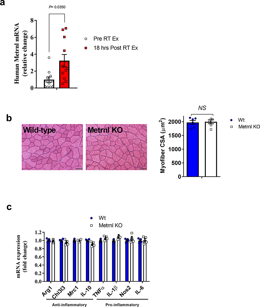
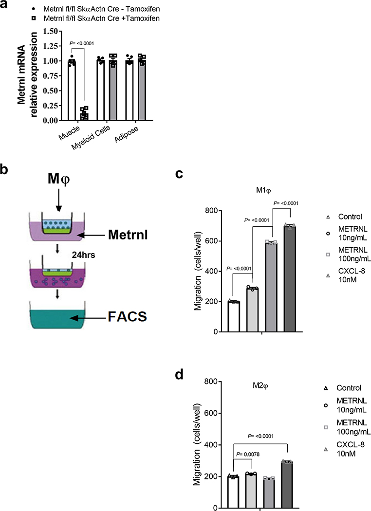
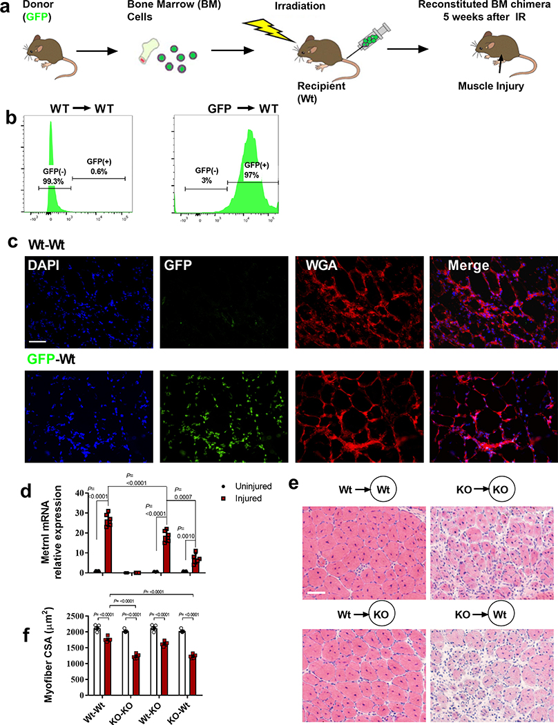
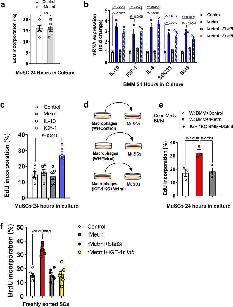
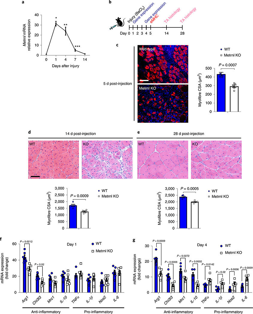
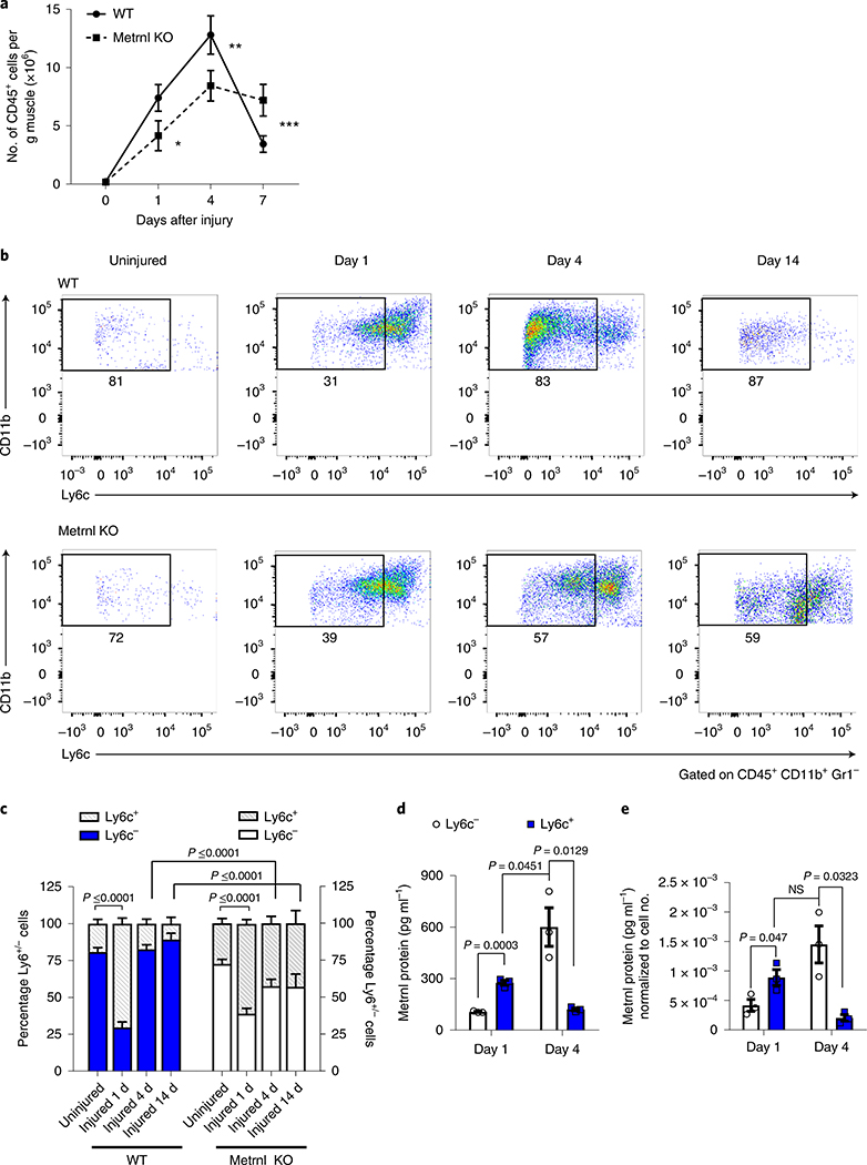
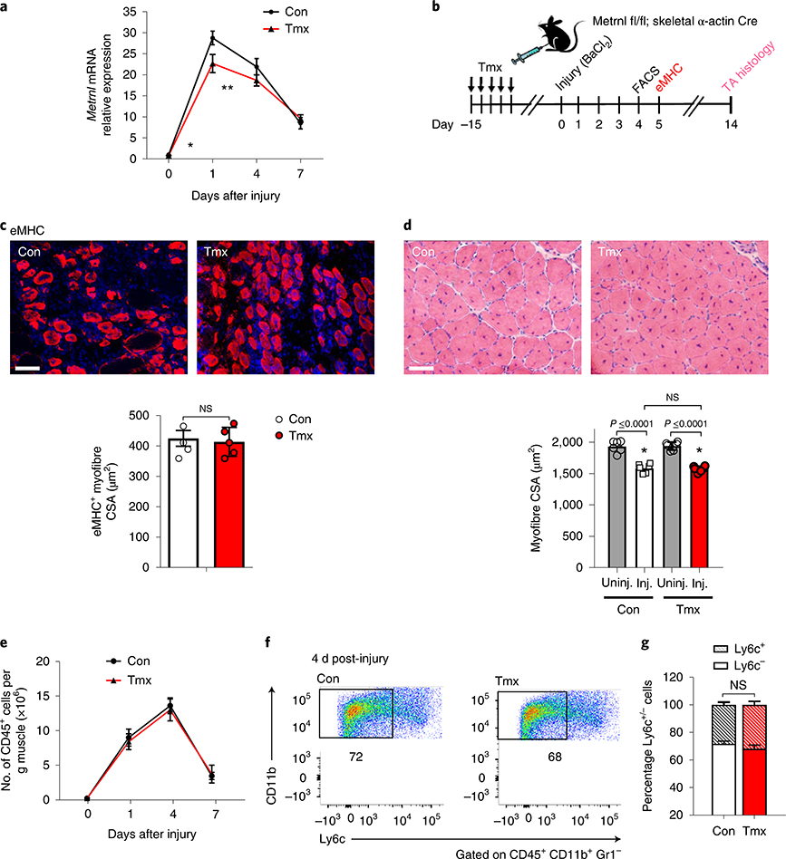
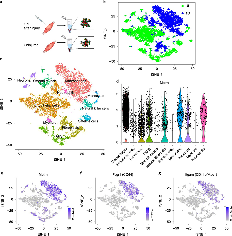
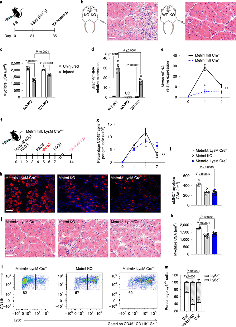
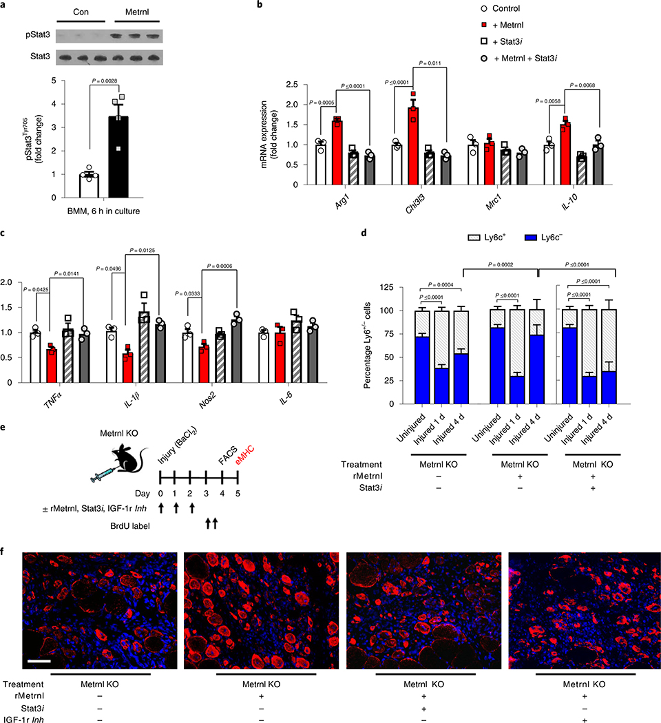
The immune system plays a multifunctional role throughout the regenerative process, regulating both pro-/anti-inflammatory phases and progenitor cell function. In the present study, we identify the myokine/cytokine Meteorin-like (Metrnl) as a critical regulator of muscle regeneration. Mice genetically lacking Metrnl have impaired muscle regeneration associated with a reduction in immune cell infiltration and an inability to transition towards an anti-inflammatory phenotype. Isochronic parabiosis, joining wild-type and whole-body Metrnl knock-out (KO) mice, returns Metrnl expression in the injured muscle and improves muscle repair, providing supportive evidence for Metrnl secretion from infiltrating immune cells. Macrophage-specific Metrnl KO mice are also deficient in muscle repair. During muscle regeneration, Metrnl works, in part, through Stat3 activation in macrophages, resulting in differentiation to an anti-inflammatory phenotype. With regard to myogenesis, Metrnl induces macrophage-dependent insulin-like growth factor 1 production, which has a direct effect on primary muscle satellite cell proliferation. Perturbations in this pathway inhibit efficacy of Metrnl in the regenerative process. Together, these studies identify Metrnl as an important regulator of muscle regeneration and a potential therapeutic target to enhance tissue repair.


Conflict of interest statement

Competing interests

The authors declare no competing interests.

Additional information

Extended data is available for this paper at https://doi.org/10.1038/s42255-020-0184-y.

Supplementary information is available for this paper at https://doi.org/10.1038/s42255-020-0184-y.

Peer review information Primary Handling Editor: Elena Bellafante.

Reprints and permissions information is available at www.nature.com/reprints.

Publisher’s note Springer Nature remains neutral with regard to jurisdictional claims in published maps and institutional affiliations.

Figures

Extended Data Fig. 1 |
Extended Data Fig. 1 |. Metrnl mRNA expression with human muscle damage and CSA analysis of uninjured Metrnl Ko muscle.
a, Human muscle Metrnl mRNA expression before and after unaccustomed resistance training (N=11/group). b, left Myofiber cross-sections from uninjured wild-type and Metrnl KO mice and right corresponding myofiber cross-sectional area (N=6/group). c, Whole muscle mRNA expression of anti-inflammatory and pro-inflammatory macrophage makers in uninjured muscle (N=4 for both groups). Scale bar is 50μm; Two-tailed, unpaired Student’s t-test (ac). Data are presented as mean ± SE.
Extended Data Fig. 2 |
Extended Data Fig. 2 |. Skeletal muscle specific deletion of Metrnl and the chemotaxis ability of Metrnl.
a, Metrnl mRNA across tissues with or without tamoxifen treatment. Samples were harvested two weeks after tamoxifen IP injections (N=5/group). b, Migration assays using LPS and IL-4-treated macrophages. c, FACS analysis of migrating M1 or (d) M2 differentiated macrophages to recombinant Metrnl protein (N=3/group). Two-tailed, unpaired Student’s t-test (a) and ANOVA with Tukey’s post hoc comparison (c, d). Data are presented as mean ± SE.
Extended Data Fig. 3 |
Extended Data Fig. 3 |. Hematopoietic-derived Metrnl expression is necessary and sufficient for successful muscle regeneration.
a, Experimental design for the bone marrow transplant. Wild-type or Metrnl KO mice served as donor mice to wild-type or Metrnl KO recipient mice. Mice received BaCL2 injection five weeks after irradiation and recovered for 14 days before harvest. b, left GFP+ cells in circulating blood from a wild-type donor into a wild-type recipient 5 weeks after transplant. right GFP+ cells in circulating blood from a GFP donor into a wild-type recipient 5 weeks after transplant. N=5 biologically independent samples. c, GFP+ cells infiltrating muscle 1 day after injury in the respective transplant groups. N=5 biologically independent samples. Scale bar 100 μm. d, Metrnl mRNA expression. (N=5/group). e, Representative images of the respective transplantation groups 14 days after injury. N=5 biologically independent samples. f, Quantification of myofiber cross sectional area of the respective transplantation groups. (N=5/group). Scale bar 100 μm. ANOVA with Tukey’s post hoc comparison (d, f). Data are presented as mean ± SE.
Extended Data Fig. 4 |
Extended Data Fig. 4 |. Metrnl signals directly to macrophages through Stat3 and indirectly to satellite cells through IGF-1.
a, EdU incorporation in MuSCs treated with recombinant Metrnl or control 24 h in culture (N=6/group). b, untreated/M0 BMM mRNA expression of IL-10, IGF-1 and IL-6 with or without recombinant Metrnl and/or Stat inhibitors, N=3/group. c, EdU incorporation in MuSCs treated with recombinant Metrnl, IL-10 and IGF-1 for 24 h in culture. (N=6/group). d, Experimental design of BMM-conditional media treatment to MuSCs. e, EdU incorporation in media-treated MuSCs in culture for 24 h. (N=3/group). f, BrdU incorporation in satellite cells 4 days after injury with or without recombinant Metrnl or Stat3 inhibitor. (N=6/group) Two-tailed, unpaired Student’s t-test (a). One-way ANOVA with Tukey’s post hoc comparison (bf). Data are presented as mean ± SE.
Fig. 1 |
Fig. 1 |. Metrnl is necessary for successful muscle regeneration.
a, Metrnl mRNA expression in the TA throughout 14 d of recovery from BaCl2 injury (n = 5 per group). Asterisks indicate the means that are significantly different from controls: *P = 0.0002, **P = 0.0045, ***P = 0.0211. b, Experimental design showing the time scale of BaCl2 injection and tissue analysis at 5 d (eMHC), 14 d (H&E stain) and 28 d (H&E stain) post-injection. c, The eMHC staining (left) and quantification of eMHC+ fibres’ cross-sectional area (right) in WT and Metrnl KO TA muscle (n = 5 per group). CSA, cross-sectional area. Scale bar, 100 μm. d, TA muscle H&E staining (upper) at 14 d after injury and related myofibre cross-sectional area (lower) (n = 5 per group). Scale bar, 100 μm. e, TA muscle H&E staining (upper) and related myofibre cross-sectional area (lower) 28 d after injury (n = 5 per group). f,g, Whole-muscle mRNA expression of anti-inflammatory and pro-inflammatory macrophage makers at 1 d post-injury (f) and 4 d post-injury (g) (n = 6 for both groups). Two-tailed, unpaired, Student’s t-test (a,cg). Data are presented as mean ± s.e.m.
Fig. 2 |
Fig. 2 |. Metrnl deletion alters immune cell migration and phenotype in injured muscle.
a, Total counts of CD45+ cells in muscle throughout 7 d of recovery (n = 5 per group). Asterisks indicate the means that are significantly different from controls: *P = 0.0029, **P = 0.0016, ***P = 0.006. b, Immune inflammatory profile showing CD45+, CD11b+, Gr1 cells stained with Ly6c to show pro- and anti-inflammatory status (n = 5 biologically independent samples). c, Quantification of percentage Ly6c+/− cells in uninjured and 1, 4 and 14 d post-injury, wild-type and Metrnl KO muscle (n = 4 or 5 per group). d, Metrnl protein expression in medium from cultured Ly6c+/− cells sorted 1 and 4 d after injury (n = 3 per group). e, Metrnl protein expression in the medium normalized to cell number (n = 3 per group). NS, not significant. Two-tailed, unpaired, Student’s t-test (a); one-way ANOVA with Tukey’s post hoc comparison (c); and multiple, two-tailed, unpaired, Student’s t-tests (d,e). Data are presented as mean ± s.e.m.
Fig. 3 |
Fig. 3 |. Myofibre-specific Metrnl mRNA expression is not required for successful muscle regeneration.
a, Metrnl mRNA expression through 7 d of recovery in Metrnl fl/fl × skeletal α-actin Cre mice treated with control or tamoxifen (n = 6 per group). Asterisks indicate the means that are significantly different from controls: *P = 0.0004, **P = 0.0475. Con, control; Tmx, tamoxifen. b, Experimental design to induce Cre recombinase in an inducible skeletal muscle Cre line, followed by BaCl2-induced muscle injury and subsequent cellular and histological measurements. c, The eMHC staining 5 d after injury in the TA muscle from Metrnl fl/fl × skeletal α-actin Cre mice (n = 5 per group). CSA, cross-sectional area. d, TA myofibre cross-sections stained for H&E after 14 d of recovery from BaCl2 injury (n = 6 per group). Inj., injured; Uninj., uninjured. Scale bars, 100 μm. e, Quantification of TA muscle CD45+ immune cells throughout 7 d of recovery from injury (n = 5 per group). f, Ly6c staining after 4 d of recovery (n = 5 biologically independent samples). g, Quantification of percentage of Ly6c+ cells within the CD45+, CD11b+, Gr1 population (n = 5 per group). Multiple, two-tailed, unpaired, Student’s t-test (a,e); two-tailed, unpaired, Student’s t-test (c,g); ANOVA with Tukey’s post hoc comparison (d). Data are presented as mean ± s.e.m.
Fig. 4 |
Fig. 4 |. Metrnl gene expression after injury is identified in macrophage populations.
a, Single-cell RNA-seq analysis of uninjured and injured muscle (1 d after injury). b, Batch plot showing cell populations present in uninjured (green) and injured (blue) groups (n = 1 per group). c, The t-distributed stochastic neighbour embedding (tSNE) plots showing various cell populations in the uninjured and injured muscle (n = 1 per group). d, Violin plot of Metrnl expression throughout the identified cell clusters. Each dot represents individual cells and their relative gene expression. FAPS, fibro-/adipogenic progenitors. eg, Gene expression plots of Metrnl (e), CD64 (f) and CD11b (g) (n = 1 per group).
Fig. 5 |
Fig. 5 |. Macrophage-mediated Metrnl expression is necessary for successful muscle regeneration.
a, Experimental design of parabiosis, BaCl2-induced muscle injury and the time course of recovery. b, TA cross-sections of injured Metrnl KO mice anastomosed to Metrnl KO or WT donor mice (n = 6 biologically independent samples). Muscles were harvested 14 d after injury. c, Myofibre cross-sectional area from uninjured and injured Metrnl KO muscle during anastomosis with WT or Metrnl KO mice (n = 6 per group). d, Metrnl mRNA expression in uninjured and injured host muscle. Pairs include WT–WT, KO–KO and WT–KO (n = 5 per group). e, Whole-muscle Metrnl mRNA expression through 4 d of recovery from injury in Metrnl fl/fl × LysM Cre+/− mice (n = 5 per group). Asterisks indicate the means that are significantly different from controls: *P = 0.0025, **P = 0.0449. f, Experimental design of BaCl2-induced muscle injury and the time course of assays used to assess regeneration in the Metrnl fl/fl × LysM Cre mice. g, Quantification of CD45+ cells in the TA muscle throughout a 7-d recovery period (n = 5 per group). Asterisks indicate the means that are significantly different from controls: *P = 0.0022, **P = 0.0009. h, The eMHC staining after 5 d of recovery in Metrnl KO and Metrnl fl/fl × LysM Cre+/− mice (n = 5 biologically independent samples). i, Quantification of eMHC+ myofibre cross-sectional area 5 d after injury (n = 5 per group). j, Myofibre cross-sections stained with H&E 14 d after injury in Metrnl KO and Metrnl fl/fl × LysM Cre+/− (n = 6 biologically independent samples). k, Quantification of TA muscle cross-sectional area (CSA) (n = 6 per group). l, LysM staining in the TA muscle after 4 d of recovery from injury in Metrnl KO and Metrnl fl/fl × LysM Cre+/− mice (n = 5 biologically independent samples). m, Quantification of percentage LysM+/− populations (n = 5 per group). Scale bars, 100 μm. Two-way ANOVA with Tukey’s post hoc comparison (c,d); multiple, two-tailed, unpaired, Student’s t-tests (e,g); one-way ANOVA with Tukey’s post hoc comparison (i,k,m). Data are presented as mean ± s.e.m.
Fig. 6 |
Fig. 6 |. Metrnl signals directly to macrophages through Stat3 and indirectly to satellite cells through IGF-1.
a, Stat3 phosphorylation in BMMs with or without treatment (6 h) of recombinant Metrnl (100 ng ml−1) (n = 4 per group). Pro-inflammatory macrophages were treated with Metrnl and/or a Stat3 inhibitor for 24 h. b,c, Macrophage mRNA expression of anti-inflammatory (b) and pro-inflammatory (c) genes after treatment (n = 3 per group). d, Quantification of percentage Ly6c+/− populations in the TA muscle 1 and 4 d after injury with or without recombinant Metrnl (rMetrnl) and/or a Stat3 inhibitor (Stat3i) (n = 5 per group). e, Experimental design of f and Supplementary Fig. 6f. TA muscle of Metrnl KO mice was treated with a combination of recombinant Metrnl, Stat3 inhibitor and IGF-1 receptor inhibitor during and after injury, and assessed for satellite cell proliferation and muscle regeneration. f, The eMHC staining 5 d after injury with or without recombinant Metrnl or Stat3 inhibitor (n = 6 per group). Scale bar, 100 μm. Two-tailed, unpaired, Student’s t-test (a) and one-way ANOVA with Tukey’s post hoc comparison (b,c,d). Data are presented as mean ± s.e.m.

References

    1. Heredia JE et al. Type 2 innate signals stimulate fibro/adipogenic progenitors to facilitate muscle regeneration. Cell 153, 376–388 (2013). - PMC - PubMed
    1. Du H et al. Macrophage-released ADAMTS1 promotes muscle stem cell activation. Nat. Commun 8, 669 (2017). - PMC - PubMed
    1. Mounier R & Chazaud B PPARgamma transcription factor controls in anti-inflammatory macrophages the expression of GDF3 that stimulates myogenic cell fusion during skeletal muscle regeneration. Med. Sci 33, 466–469 (2017). - PubMed
    1. Tonkin J et al. Monocyte/Macrophage-derived IGF-1 orchestrates murine skeletal muscle regeneration and modulates autocrine polarization. Mol. Ther 23, 1189–1200 (2015). - PMC - PubMed
    1. Deng B et al. IL-10 triggers changes in macrophage phenotype that promote muscle growth and regeneration. J. Immunol 189, 3669–3680 (2012). - PMC - PubMed

Publication types

MeSH terms

Substances