. 2020 Jun 30:11:1481.
doi: 10.3389/fmicb.2020.01481. eCollection 2020.

Identification and Functional Analysis of BmNPV-Interacting Proteins From Bombyx mori (Lepidoptera) Larval Midgut Based on Subcellular Protein Levels

Affiliations

Identification and Functional Analysis of BmNPV-Interacting Proteins From Bombyx mori (Lepidoptera) Larval Midgut Based on Subcellular Protein Levels

Shang-Zhi Zhang et al. Front Microbiol. .

Abstract

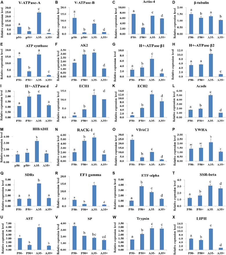
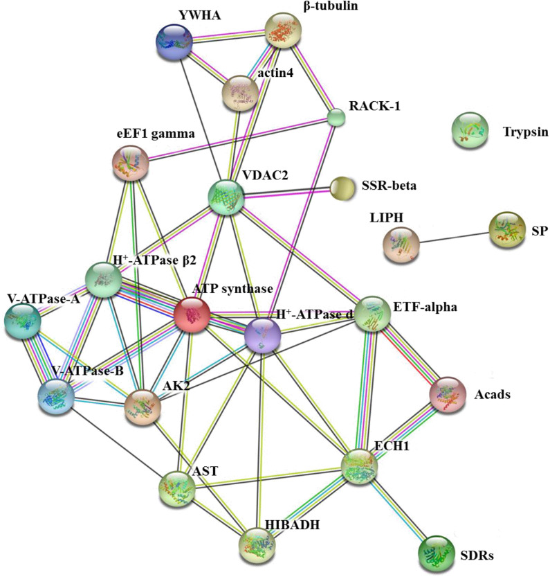
Bombyx mori nucleopolyhedrovirus (BmNPV) is a major pathogen causing severe economic loss. However, the molecular mechanism of silkworm resistance to BmNPV and the interactions of this virus with the host during infection remain largely unclear. To explore the virus-binding proteins of silkworms, the midgut subcellular component proteins that may interact with BmNPV were analyzed in vitro based on one- and two-dimensional electrophoresis and far-western blotting combined with mass spectrometry (MS). A total of 24 proteins were determined to be specifically bound to budded viruses (BVs) in two subcellular fractions (mitochondria and microsomes). These proteins were involved in viral transportation, energy metabolism, apoptosis and viral propagation, and they responded to BmNPV infection with different expression profiles in different resistant strains. In particular, almost all the identified proteins were downregulated in the A35 strain following BmNPV infection. Interestingly, there were no virus-binding proteins identified in the cytosolic fraction of the silkworm midgut. Two candidate proteins, RACK1 and VDAC2, interacted with BVs, as determined with far-western blotting and reverse far-western blotting. We speculated that the proteins interacting with the virus could either enhance or inhibit the infection of the virus. The data provide comprehensive useful information for further research on the interaction of the host with BmNPV.

Keywords: BmNPV; BmNPV-interacting proteins; Bombyx mori (B. mori); far-western blotting; subcellular proteins; two-dimensional electrophoresis.


Figures

FIGURE 1
FIGURE 1
P50 silkworm strains BmNPV free (P50-) and infected (P50+). The DNA of the silkworm midgut was used as a template for baculovirus major protein gp64 gene PCR amplification. BmNPV DNA was used as a positive control (NPV). M, DNA molecular weight marker.
FIGURE 2
FIGURE 2
Identification of virus-binding of the mitochondrial proteins fraction of the P50 midgut on SDS-PAGE. (A) Separation of Bombyx mori midgut mitochondrial (MC) and BmNPV budded viruses (BVs) proteins by SDS-PAGE. (B) Virus overlay binding experiment. Mitochondrial protein blots, overlaid with (MC + BV) and without BVs (MC-BV) were incubated with antibodies against the AcMNPV gp64 protein to detect the BVs. BmNPV Budded virus was used as a positive control (BV) to gp64 protein. Arrows a, b, and c in (A,B) refer to the bands detected in the stained gel and the PVDF membrane. M, protein molecular weight marker.
FIGURE 3
FIGURE 3
Identification of virus-binding of the mitochondrial proteins fraction of the P50 midgut of the P50 midgut on 2-DE. (A) Bombyx mori midgut mitochondrial proteins submitted to 2-DE. Mitochondrial proteins blot were overlaid (B) or not (C; negative control) with BV, and antibodies against the AcMNPV gp64 protein were used to detect the BVs. Arrows 1–8 in (A,B) refer to the spots detected on corresponding proteins in the stained gel and the PVDF membrane. M, protein molecular weight marker.
FIGURE 4
FIGURE 4
Identification of virus-binding in the microsomal proteins fraction of the P50 midgut on SDS-PAGE. (A) Separation of Bombyx mori midgut microsomes (MS) and BmNPV budded viruses (BVs) proteins by SDS-PAGE. (B) Virus overlay binding experiment. Microsomes protein blots, overlaid with (MS + BV) and without BVs (MS-BV) were incubated with antibodies against the AcMNPV gp64 protein to detect the BVs. BmNPV Budded virus was used as a positive control (BV) to gp64 protein. Arrows d and e in (A,B) refer to the bands detected in the stained gel and the PVDF membrane. M, protein molecular weight marker.
FIGURE 5
FIGURE 5
Identification of virus-binding in the microsomal proteins fraction of the P50 midgut on 2-DE. (A) Bombyx mori midgut microsomal proteins submitted to 2-DE. Microsomal proteins blot were overlaid (B) or not (C; negative control) with BV, and antibodies against the AcMNPV gp64 protein were used to detect the BVs. Arrows 9–13 in (A,B) refer to the spots detected on corresponding proteins in the stained gel and the PVDF membrane. M, protein molecular weight marker.
FIGURE 6
FIGURE 6
Identification of virus-binding in the cytosol proteins fraction of the P50 midgut on SDS-PAGE. (A) Separation of cytosolic proteins of the BmNPV and Bombyx mori midgut samples by SDS-PAGE. (B) Virus overlay binding experiment. BmNPV and cytosolic protein samples blot were overlaid with BV (CS + BV), and a blot with CS without overlaid with BVs (CS-BV) are detected by antibodies against AcMNPV gp64 protein to detect the BVs. M, protein molecular weight marker.
FIGURE 7
FIGURE 7
Analysis of the interaction between BVs and recombinant proteins by far western blot. Purified recombinant proteins (RACK1 and VDAC2) were overlaid with BVs, and antibodies against AcMNPV gp64 protein were used to detect the BVs. BmNPV was used as a positive control. The negative overlay control consisted of binding buffer without BVs before incubation with monoclonal antibodies against AcMNPV gp64. The plus and minus signs on the top indicate membranes incubated with or without BVs, respectively. M, protein molecular weight marker.
FIGURE 8
FIGURE 8
Analysis of the interaction between recombinant proteins and BVs by far western blot. The BVs were overlaid with purified recombinant proteins (RACK1 and VDAC2), and antibodies against His-tagged proteins were used to detect the purified recombinant proteins. M, protein molecular weight marker; NC, negative control, BmNPV incubated with PBS.
FIGURE 9
FIGURE 9
RT-qPCR analysis of the expression profiles of the BmNPV interacting proteins in different resistant silkworm midguts. (A–X) represents the transcription level of 24 BmNPVinteracting proteins in different resistant silkworm strains following BmNPV infection. The data were normalized using BmGAPDH and presented as the means ± standard error of the means from three independent experiments. The relative expression levels were calculated using the 2–ΔΔCt method. The statistical analysis was conducted using SPSS software. Significant differences are indicated by letters (P < 0.05).
FIGURE 10
FIGURE 10
The network of 24 BmNPV-interacting proteins was mapped based on the STRING website information using a database of information on another well-studied insect, D. melanogaster. VDAC2, voltage-dependent anion-selective channel isoform X2; SSR-beta, signal sequence receptor beta subunit precursor; AK2, adenylate kinase 2; actin4, actin-4; eEF1 gamma, elongation factor 1 gamma; AST, aspartate aminotransferase; HIBADH, 3-hydroxyisobutyrate dehydrogenase isoform X1; RACK1, receptor for activated protein kinase C RACK isoform 1; LIPH, lipase member H-A-like; Acads, short-chain specific acyl-CoA dehydrogenase; ECH1, enoyl-coa hydratase precursor 1; H + -ATPase β2, H + transporting ATP synthase beta subunit isoform 2; V-ATPase-B, vacuolar ATP synthase subunit B; β-tubulin, beta-tubulin; YWHA, tyrosine 3-monooxygenase/tryptophan 5-monooxygenase activation protein epsilon polypeptide; ETF-alpha, electron transfer flavoprotein subunit alpha; SDRs, short-chain dehydrogenase/reductase; V-ATPase-A, vacuolar ATP synthase catalytic subunit A; SP, serine protease precursor; Trypsin, trypsin, alkaline C-like; H+-ATPase d, H+transporting ATP synthase subunit.

Similar articles

Cited by

References

    1. Abu-Hamad S., Sivan S., Shoshan-Barmatz V. (2006). The expression level of the voltage-dependent anion channel controls life and death of the cell. Proc. Natl. Acad. Sci. U.S.A. 103 5787–5792. 10.1073/pnas.0600103103 - DOI - PMC - PubMed
    1. Achilonu I., Siganunu T. P., Dirr H. W. (2014). Purification and characterisation of recombinant human eukaryotic elongation factor 1 gamma. Protein Expr. Purif. 99 70–77. 10.1016/j.pep.2014.04.003 - DOI - PubMed
    1. Agnihotri G., Liu H. W. (2003). Enoyl-CoA hydratase: reaction, mechanism, and inhibition. Bioorg. Med. Chem. 11 9–20. 10.1016/s0968-0896(02)00333-4 - DOI - PubMed
    1. Baker M. E., Yan L., Pear M. R. (2000). Three-dimensional model of human TIP30, a coactivator for HIV-1 Tat-activated transcription, and CC3, a protein associated with metastasis suppression. Cell. Mol. Life Sci. 57 851–858. 10.1007/s000180050047 - DOI - PMC - PubMed
    1. Barabasi A. L., Gulbahce N., Loscalzo J. (2011). Network medicine: a network-based approach to human disease. Nat. Rev. Genet. 12 56–68. 10.1038/nrg2918 - DOI - PMC - PubMed

LinkOut - more resources