Review
. 2020 May 28;7(Pt 4):630-638.
doi: 10.1107/S205225252000562X. eCollection 2020 Jul 1.

The data universe of structural biology

Affiliations
Review

The data universe of structural biology

Helen M Berman et al. IUCrJ. .

Abstract

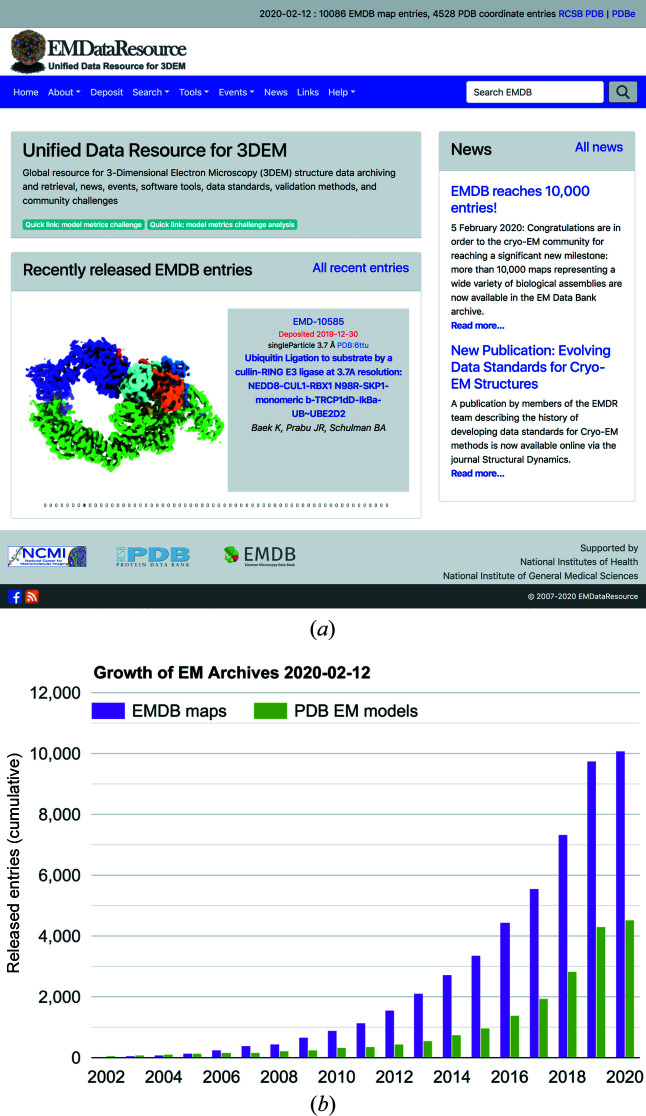
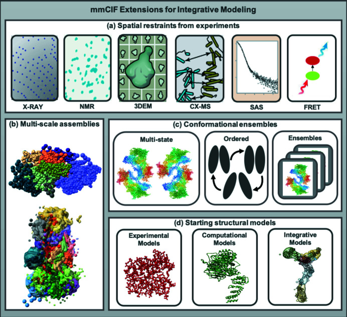
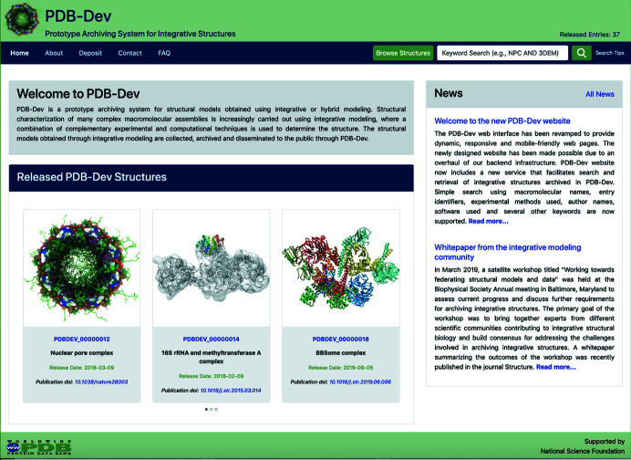
The Protein Data Bank (PDB) has grown from a small data resource for crystallographers to a worldwide resource serving structural biology. The history of the growth of the PDB and the role that the community has played in developing standards and policies are described. This article also illustrates how other biophysics communities are collaborating with the worldwide PDB to create a network of interoperating data resources. This network will expand the capabilities of structural biology and enable the determination and archiving of increasingly complex structures.

Keywords: Protein Data Bank; X-ray crystallography; data resources; data standards; structural biology.


Figures

Figure 1
Figure 1
Formats for representation of atomic coordinates. (a) PDB format. All data items are in fixed-sized fields and definitions are implicit. (b) mmCIF format. The names of the data items as defined in the mmCIF dictionary are listed first using a loop directive. The values of the data items then follow in a tabular form. This representation enables mmCIF to be flexible, self-consistent and software compatible.
Figure 2
Figure 2
Key elements of the wwPDB validation report for X-ray structures are shown for PDB entry 6pzd, a recent crystal structure of Influenza A neuraminidase, determined at 1.12 Å (Zhu et al., 2019 ▸). (a) Graphical display of key metrics (‘sliders’). For each metric, two percentile ranks are calculated: an absolute rank with respect to the entire PDB archive and a relative rank with respect to structures determined at similar resolution. Slider markers in the blue region on the right are indicative of a high-quality structure. Lower-quality structures have the markers in the red region on the left. (b) Residue property plot: residues are color-coded green if no issues are detected, yellow if there are outliers for one criterion (e.g. unusual bond lengths), orange if there are outliers for two criteria (e.g. unusual bond lengths and clashes) and red for three or more criteria. A horizontal stack bar plot presents the fraction of residues with each color code. Unmodeled regions of the chain, if present, are represented by a gray segment. The upper red bar indicates the fraction of residues with poor fit to the electron density.
Figure 3
Figure 3
Cumulative holdings of the PDB at the end of each decade for each of the three major structure determination methods, X-ray crystallography, NMR and 3DEM, respectively. 3DEM methods include structures determined by electron microscopy (single-particle, helical, subtomogram averaging and tomography) and electron crystallography.
Figure 4
Figure 4
Total annual downloads of PDB archive files. Plotted values represent the sum of annual downloads from all of the wwPDB partner ftp and websites. Data Source: https://www.wwpdb.org/stats/download.
Figure 5
Figure 5
(a) Recent screenshot of the EMDataResource website (https://www.emdataresource.org). The website is updated weekly to highlight all newly released EMDB maps. (b) Cumulative number of 3DEM maps available in the EMDB and coordinate models available in the PDB by year; 2020 statistics are through February 2. Source: https://www.emdataresource.org/statistics.html.
Figure 6
Figure 6
The IHM dictionary provides definitions for (a) spatial restraints from experimental methods such as X-ray diffraction, NMR, 3DEM, CX-MS, SAS and FRET; (b) multi-scale assemblies consisting of both atomic coordinates and coarse-grained representations; (c) ensembles representing multiple conformational states or ensembles related by time or other criteria such as events in a sequential pathway; and (d) starting structural models used in integrative modeling.
Figure 7
Figure 7
Screenshot of the current PDB-Dev website (https://pdb-dev.wwpdb.org). PDB-Dev currently consists of over 40 integrative structures including several that are on hold for publication. The structures archived in PDB-Dev vary in complexity from simple atomic structures in a single conformational state to complex coarse-grained assemblies in multiple conformational states. The data model underlying PDB-Dev supports the representation of these complex structures as well as the diverse set of spatial restraints used in building them.

References

    1. Adams, P. D., Afonine, P. V., Baskaran, K., Berman, H. M., Berrisford, J., Bricogne, G., Brown, D. G., Burley, S. K., Chen, M., Feng, Z., Flensburg, C., Gutmanas, A., Hoch, J. C., Ikegawa, Y., Kengaku, Y., Krissinel, E., Kurisu, G., Liang, Y., Liebschner, D., Mak, L., Markley, J. L., Moriarty, N. W., Murshudov, G. N., Noble, M., Peisach, E., Persikova, I., Poon, B. K., Sobolev, O. V., Ulrich, E. L., Velankar, S., Vonrhein, C., Westbrook, J., Wojdyr, M., Yokochi, M. & Young, J. Y. (2019). Acta Cryst. D75, 451–454. - PMC - PubMed
    1. Barinaga, M. (1989). Science, 245, 1179–1181. - PubMed
    1. Berman, H. M., Adams, P. D., Bonvin, A. A., Burley, S. K., Carragher, B., Chiu, W., DiMaio, F., Ferrin, T. E., Gabanyi, M. J., Goddard, T. D., Griffin, P. R., Haas, J., Hanke, C. A., Hoch, J. C., Hummer, G., Kurisu, G., Lawson, C. L., Leitner, A., Markley, J. L., Meiler, J., Montelione, G. T., Phillips, G. N. Jr, Prisner, T., Rappsilber, J., Schriemer, D. C., Schwede, T., Seidel, C. A. M., Strutzenberg, T. S., Svergun, D. I., Tajkhorshid, E., Trewhella, J., Vallat, B., Velankar, S., Vuister, G. W., Webb, B., Westbrook, J. D., White, K. L. & Sali, A. (2019). Structure, 27, 1745–1759.
    1. Berman, H. M., Henrick, K. & Nakamura, H. (2003). Nat. Struct. Biol. 10, 980. - PubMed
    1. Berman, H. M., Kleywegt, G. J., Nakamura, H., Markley, J. L. & Burley, S. K. (2010). Nature, 463, 425. - PMC - PubMed