

MiR-21 nanocapsules promote early bone repair of osteoporotic fractures by stimulating the osteogenic differentiation of bone marrow mesenchymal stem cells
- PMID: 32695607
- PMCID: PMC7349941
- DOI: 10.1016/j.jot.2020.04.007
MiR-21 nanocapsules promote early bone repair of osteoporotic fractures by stimulating the osteogenic differentiation of bone marrow mesenchymal stem cells
Abstract
Objective: The healing of osteoporotic fractures in the elderly patients is a difficult clinical problem. Currently, based on the internal fixation of fractures, the available drug treatments mainly focus on either inhibiting osteoclast function, such as bisphosphonate, calcitonin, oestrogen or promoting osteogenesis, such as parathyroid hormones. However, the availability of current antiosteoporotic drugs in promoting osteoporotic fracture healing is limited. The objective of the present study was to investigate the ability of the MiR-21/nanocapsule to enhance the early bone repair of osteoporotic fractures.
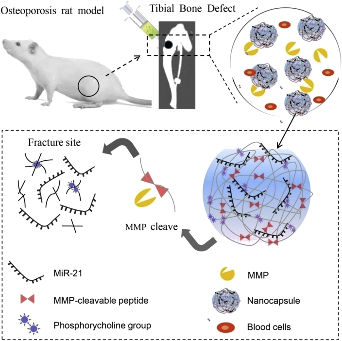
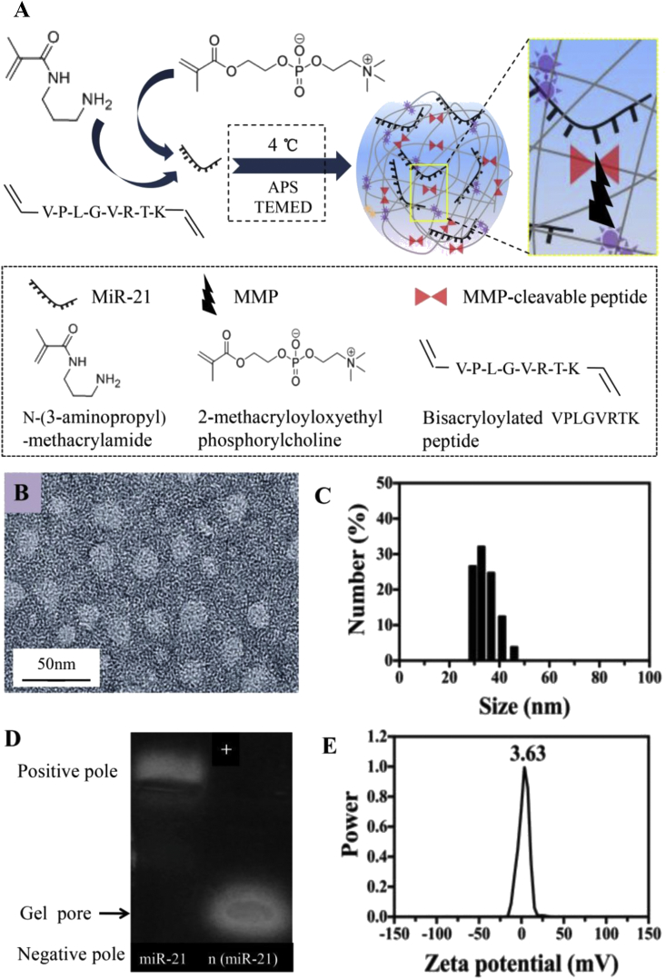
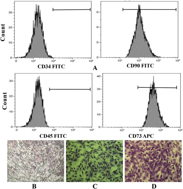
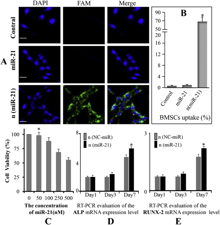
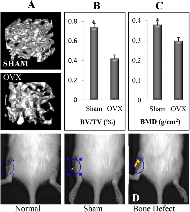
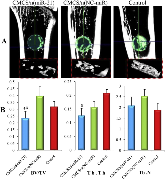
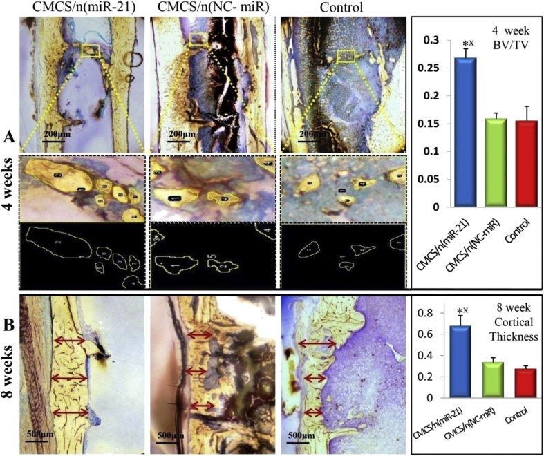
Methods: Based on the presence of matrix metalloproteinases that are overexpressed at the fracture site, we designed the matrix metalloproteinase-sensitive nanocapsules which were formed by in situ free radical polymerisation on the surface of MiR-21 with 2-(methacryloyloxy) ethyl phosphorylcholine and the bisacryloylated VPLGVRTK peptide. The MiR-21/nanocapsule [n (miR-21)] and O-carboxymethyl chitosan (CMCS) were mixed until they formed a gel-like material [CMCS/n (miR-21)] with good fluidity and injectability. Thirty elderly Sprague Dawley (SD) rats (female, 14-month-old, 380 ± 10 g) were subjected to bilateral removal of the ovaries (ovariectomised). All rats were subjected to bilateral bone defects (2 mm diameter) of the proximal tibia and randomly divided into three groups (groups A, B, and C): separately injected with CMCS/n (miR-21), CMCS/n (NC-miR), and saline. Micro-computed tomography (CT) imaging was performed to evaluate newly formed bone volume and connectivity. Nondecalcified histology and toluidine blue staining were performed to measure the effects of CMCS/n (miR-21) on bone repair. In vitro, the effect of n (miR-21) on osteogenic differentiation to bone marrow mesenchymal stem cells (BMSCs) which derived from the ovariectomised rat model was observed.
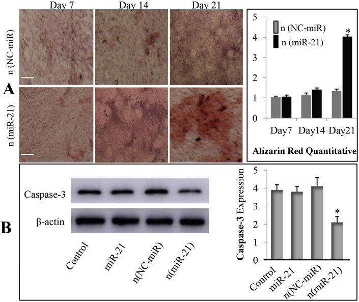
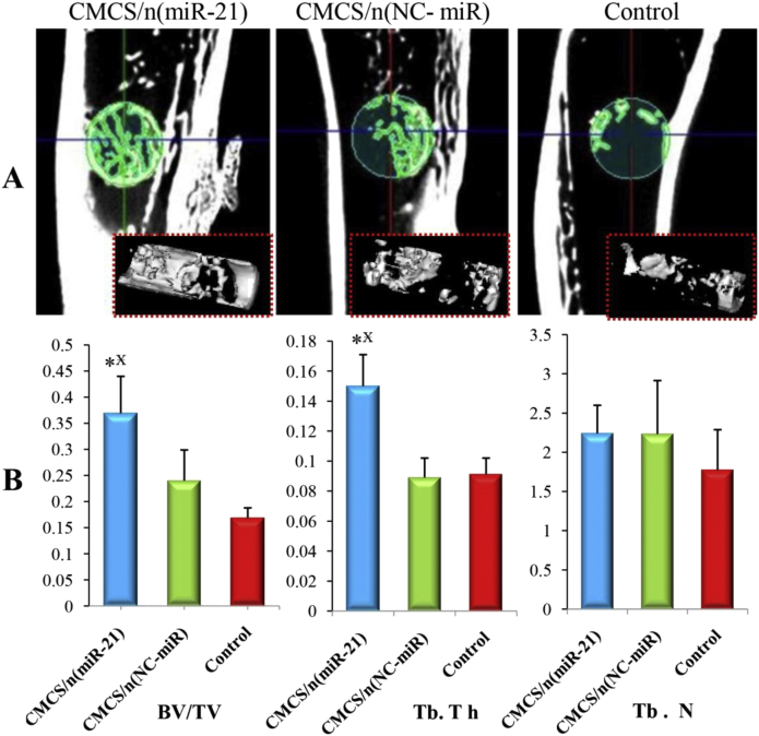
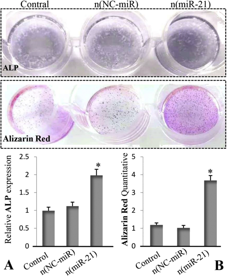
Results: The morphology of n (miR-21) was a regular spherical nanocapsule with a uniform small size (25-35 nm). The results confirmed that n (miR-21) could be efficiently phagocytosed by BMSCs and released in the cytoplasm to promote osteogenesis. The expression level of alkaline phosphatase and Runt-related transcription factor 2 mRNA in the n (miR-21) group was higher than that in the n (NC-miR) group. Animal experiments proved that CMCS/n (miR-21) produced better bone repair compared with the CMCS/n (NC-miR) group in the early stages of fracture healing at 4 weeks. In the late stage of fracture healing (8 weeks), micro-CT quantitative analysis showed that the new bone trabeculae in the CMCS/n (miR-21) group has decreased compared with the CMCS/n (NC-miR) group. In the CMCS/n (miR-21) group, the new cancellous bone had been absorbed, and the process of bone healing was almost completed. In contrast, the new bone in the CMCS/n (NC-miR) and the control groups was still in the healing process.
Conclusion: The cytological tests confirmed that n (miR-21) can promote osteogenic differentiation of BMSCs derived from the osteoporosis rat model. Furthermore, the results of animal tests demonstrated that local injection of CMCS/n (miR-21) promoted the early healing of osteoporotic bone defects. Consequently CMCS/n (miR-21) promoted the bone repair process to enter the moulding phase earlier.
The translational potential of this article: CMCS/n (miR-21) can be widely applied to elderly patients with osteoporotic fractures. This method can help patients with osteoporotic fractures recover earlier and avoid serious complications. It provides a potential approach for the clinical treatment of osteoporotic fractures in the elderly.
Keywords: Bone repair; MicroRNA-21; Nanocapsules; Osteoporotic fractures.
© 2020 Published by Elsevier (Singapore) Pte Ltd on behalf of Chinese Speaking Orthopaedic Society.
Figures
References
-
- Sims N.A., Walsh N.C. Intercellular cross-talk among bone cells: new factors and pathways. Curr Osteoporos Rep. 2012;10:109–117. - PubMed
-
- Matsuo K., Irie N. Osteoclast-osteoblast communication. Arch Biochem Biophys. 2012;473:201–209. - PubMed
-
- Collison J. Osteoporosis: teriparatide preferable for fracture prevention. Nat Rev Rheumatol. 2018;14:4. - PubMed
-
- Simpson A.H., Mills L., Noble B. The role of growth factors and related agents in accelerating fracture healing. J Bone Jt Surg Br Vol. 2006;88:701–705. - PubMed
-
- Lieberman J.R., Daluiski A., Einhorn T.A. The role of growth factors in the repair of bone. J Bone Jt Surg Am Vol. 2002;84:1032–1044. - PubMed
LinkOut - more resources
Full Text Sources
