. 2020 Jun 30:8:426.
doi: 10.3389/fcell.2020.00426. eCollection 2020.

The Impact of Di-2-Ethylhexyl Phthalate on Sperm Fertility

Affiliations

The Impact of Di-2-Ethylhexyl Phthalate on Sperm Fertility

Liliya Gabelev Khasin et al. Front Cell Dev Biol. .

Abstract

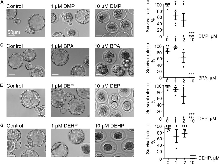
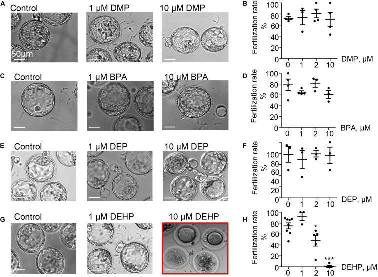
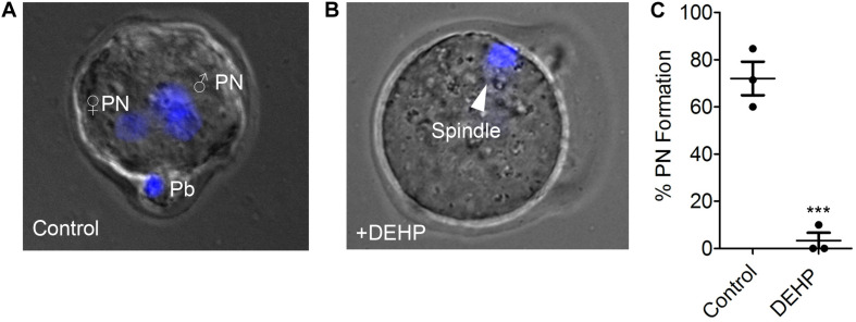
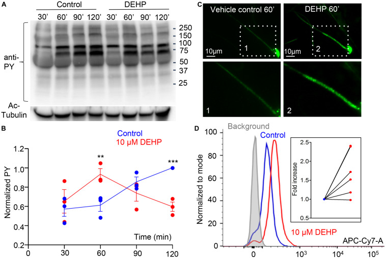
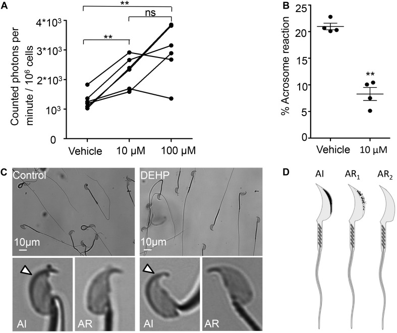
A growing number of studies point to reduced fertility upon chronic exposure to endocrine-disrupting chemicals (EDCs) such as phthalates and plasticizers. These toxins are ubiquitous and are often found in food and beverage containers, medical devices, as well as in common household and personal care items. Animal studies with EDCs, such as phthalates and bisphenol A have shown a dose-dependent decrease in fertility and embryo toxicity upon chronic exposure. However, limited research has been conducted on the acute effects of these EDCs on male fertility. Here we used a murine model to test the acute effects of four ubiquitous environmental toxins: bisphenol A (BPA), di-2-ethylhexyl phthalate (DEHP), diethyl phthalate (DEP), and dimethyl phthalate (DMP) on sperm fertilizing ability and pre-implantation embryo development. The most potent of these toxins, di-2-ethylhexyl phthalate (DEHP), was further evaluated for its effect on sperm ion channel activity, capacitation status, acrosome reaction and generation of reactive oxygen species (ROS). DEHP demonstrated a profound hazardous effect on sperm fertility by producing an altered capacitation profile, impairing the acrosome reaction, and, interestingly, also increasing ROS production. These results indicate that in addition to its known chronic impact on reproductive potential, DEHP also imposes acute and profound damage to spermatozoa, and thus, represents a significant risk to male fertility.

Keywords: acrosome reaction; capacitation; di-2-ethylhexyl phthalate (DEHP); embryo development; endocrine-disrupting chemicals (EDC); phthalates; reactive oxygen species (ROS); spermatozoa.


Figures

FIGURE 1
FIGURE 1
Murine embryo development is impacted by DMP, BPA, DEP and DEHP. In vitro embryo development on day 5 post fertilization. Panels (A,C,E,G) are representative images of blastocysts previously exposed at the zygote stage to 0, 1, or 10 μM of the indicated EDC for 20 h. The subsequent embryo culture was done in the absences of the indicated EDC. (A) Shown are representative images of DMP-exposed embryos. (B) The survival rate of DMP exposed zygotes was calculated based on the percentage of embryos that have reached the morula or blastocyst stage. (C) Representative images of embryos previously exposed to BPA. (D) The survival rate of BPA exposed zygotes was calculated as in (B). (E) Representative images of embryos previously exposed to DEP. (F) The survival rate of DEP exposed zygotes was calculated as in (B). (G) Representative images of embryos previously exposed to DEHP. (H) The survival rate of DEHP- exposed zygotes was calculated as in (B). Data are means ± S.E.M. Asterisk indicates a statistical difference between control embryos and embryos exposed to EDCs. *P ≤ 0.05, **P < 0.01, ***P < 0.001. Scale bars for all images are 50 μm.
FIGURE 2
FIGURE 2
Fertilization rate of murine eggs exposed to EDCs treated spermatozoa. Panels (A,C,E,G) are representative images of blastocysts obtained after murine eggs were introduced to sperm previously exposed to 0, 1, or 10 μM of the indicated EDC. Subsequent embryo culturing was done in the absences of EDCs, and the images were taken on day 5 post insemination. (A) Shown are representative images of blastocysts obtained after murine eggs were introduced to sperm previously exposed to DMP. (B) Percentage of eggs that were fertilized by DMP- treated sperm and were able to reach morula or blastocyst stage. (C) Representative images of embryos obtained after IVF with BPA-treated spermatozoa. (D) The percentage of eggs fertilized by BPA-treated sperm was calculated as in (B). (E) Representative images of embryos obtained after IVF with DEP-treated sperm. (F) The percentage of eggs fertilized by DEP-treated sperm was calculated as in (B). (G) Representative images of embryos obtained after IVF with DEHP-treated sperm. (H) The percentage of eggs fertilized by DEHP-treated sperm was calculated as in (B). Data are means ± S.E.M. Asterisk indicates a statistical difference between control embryos and embryos exposed to EDCs. *P < 0.05, **P < 0.01, ***P < 0.001. Scale bars for all images are 50 μm.
FIGURE 3
FIGURE 3
Pronuclei formation after in vitro fertilization by DEHP-treated sperm. Pronuclear formation was assessed 9 h after IVF; genomic DNA staining was done with DAPI. (A) Representative image of a successfully fertilized egg with two pronuclei (PN) and a polar body (Pb). The egg was inseminated with sperm treated with vehicle control solution. (B) Shown are representative images of unfertilized eggs following an IVF with sperm that was treated with 10 μM DEHP. The arrowhead indicates the position of the second metaphase spindle. (C) Percentage of fertilized eggs with PN detected. Each data point represents the mean of one of the three independent experiments ± S.E.M. *** indicates statistical significance (P < 0.005). The total number of eggs: 25 (control), 36 (+10 μM DEHP).
FIGURE 4
FIGURE 4
Capacitation-associated tyrosine phosphorylation of murine sperm is altered in the presence of DEHP. (A) Representative western blot image shows the time course of protein tyrosine phosphorylation under capacitating conditions in the presence or absence of 10 μM DEHP. DEHP was added to the media immediately before the start of the capacitation process. Sperm lysates were obtained at the indicated times (30, 60, 90, and 120 min) and subjected to SDS-PAGE immunoblotting. Tyrosine phosphorylation was detected with a monoclonal phospho-tyrosine (PY) antibody. Acetylated-tubulin (Ac-Tubulin) was used as a loading control. (B) Levels of relative tyrosine phosphorylation obtained as total densities extracted from (A) and normalized to the densities of the loading control. Each data point represents the mean of one of the three independent experiments. (C) Immunofluorescent localization of tyrosine phosphorylated proteins as visualized by PY antibody. Increased phosphorylation detected after 60 min of capacitation in the mid-piece region of spermatozoa in DEHP- treatment group (right panels) as compared to control untreated spermatozoa (left panels). Lower panels represent insets from the corresponding region of interests indicated on the upper panels by dashed rectangular. (D) A representative flow cytometry data showing an increase in global tyrosine phosphorylation in 10 μM DEHP- treated spermatozoa (red) at 60 min of capacitation compared to the vehicle control (blue). Tyrosine phosphoproteins were detected using a CF 647 dye conjugated to an anti-PY antibody. Inset: fold increase in mean fluorescent intensity normalized to mode as detected by the flow cytometer compared to control conditions. Data are means ± S.E.M. ** indicates statistical significance (P < 0.01) between control spermatozoa and spermatozoa exposed to 10 μM DEHP. *** indicates statistical significance (P < 0.001).
FIGURE 5
FIGURE 5
DEHP increases ROS production and decreases spontaneous acrosome reaction in capacitated sperm cells. (A) Luminol-dependent chemiluminescence assay showed increased rates of ROS production in sperm treated with 10 and 100 μM DEHP in comparison to the vehicle control (0 μM DEHP). Connected dots represent spermatozoa extracted from the same mouse (a total of six mice were used for this experiment). Each sample was divided into three aliquots and subjected to different conditions. A minimum of 1.35*106 sperm cells/mL was used per condition. (B) Percentage of spontaneously acrosome-reacted spermatozoa subjected to 10 μM DEHP or vehicle control (0.1% ethanol) during capacitation. Each data point represents the mean of one of four independent experiments. A minimum of 500 cells was evaluated per experiment. (C) Acrosome-reacted (AR) and acrosome-intact (AI) spermatozoa in control and 10 μM DEHP. Arrowheads point at intact acrosome caps. “AI” cells have bright blue staining on the dorsal region of the acrosome. “AR” cells have patchy or absent staining. (D) Diagram showing acrosome-reacted (two distinct patterns are indicated as AR1 or partially reacted, and AR2 as fully reacted) and acrosome-intact (AI) spermatozoa. Images were produced using Biorender.com. Data are means ± S.E.M. ** indicates statistical significance (P < 0.01) between control spermatozoa and spermatozoa exposed to 10 μM DEHP.

References

    1. Agarwal A., Cocuzza M., Abdelrazik H., Sharma R. K. (2008). Oxidative Stress Measurement in Patients with Male or Female Factor Infertility. Trivandrum: Transworld Research Network.
    1. Aitken R. J., Clarkson J. S. (1987). Cellular basis of defective sperm function and its association with the genesis of reactive oxygen species by human spermatozoa. J. Reprod. Fertil. 81 459–469. 10.1530/jrf.0.0810459 - DOI - PubMed
    1. Aitken R. J., Paterson M., Fisher H., Buckingham D. W., Van Duin M. (1995). Redox regulation of tyrosine phosphorylation in human spermatozoa and its role in the control of human sperm function. J. Cell Sci. 108 2017–2025. - PubMed
    1. Aitken R. J., Wingate J. K., De Iuliis G. N., Koppers A. J., Mclaughlin E. A. (2006). Cis-unsaturated fatty acids stimulate reactive oxygen species generation and lipid peroxidation in human spermatozoa. J Clin Endocrinol Metab 91 4154–4163. 10.1210/jc.2006-1309 - DOI - PubMed
    1. Alvarez J. G., Storey B. T. (1982). Spontaneous lipid peroxidation in rabbit epididymal spermatozoa: its effect on sperm motility. Biol. Reprod. 27 1102–1108. 10.1095/biolreprod27.5.1102 - DOI - PubMed