Review
. 2020 Jun 15;2(6):e0154.
doi: 10.1097/CCE.0000000000000154. eCollection 2020 Jun.

Review of Viral Testing (Polymerase Chain Reaction) and Antibody/Serology Testing for Severe Acute Respiratory Syndrome-Coronavirus-2 for the Intensivist

Affiliations
Review

Review of Viral Testing (Polymerase Chain Reaction) and Antibody/Serology Testing for Severe Acute Respiratory Syndrome-Coronavirus-2 for the Intensivist

Michael P Motley et al. Crit Care Explor. .

Abstract

Objective: As the severe acute respiratory syndrome-coronavirus-2 pandemic develops, assays to detect the virus and infection caused by it are needed for diagnosis and management. To describe to clinicians how each assay is performed, what each assay detects, and the benefits and limitations of each assay.

Data sources: Published literature and internet.

Study selection: As well done, relevant and recent as possible.

Data extraction: Sources were read to extract data from them.

Data synthesis: Was synthesized by all coauthors.

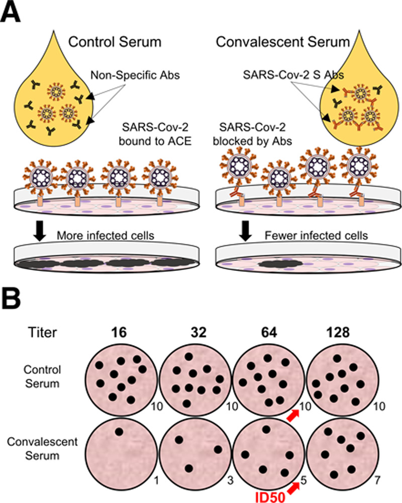
Conclusions: Available assays test for current or previous severe acute respiratory syndrome-coronavirus-2 infection. Nucleic acid assays such as quantitative, or real-time, polymerase chain reaction and loop-mediated isothermal amplification are ideal for acute diagnosis with polymerase chain reaction testing remaining the "gold standard" to diagnose acute infection by severe acute respiratory syndrome-coronavirus-2, specifically the presence of viral RNA. Assays that detect serum antibodies can theoretically diagnose both acute and remote infection but require time for the patient to develop immunity and may detect nonspecific antibodies. Antibody assays that quantitatively measure neutralizing antibodies are needed to test efficacy of convalescent plasma therapy but are more specialized.

Keywords: coronavirus-2; molecular diagnostic techniques; neutralizing antibodies; real-time polymerase chain reaction; serologic tests; severe acute respiratory syndrome.


Figures

Figure 1.
Figure 1.
Severe acute respiratory syndrome-coronavirus-2 (SARS-Cov-2) structure and quantitative, or real-time, polymerase chain reaction (qPCR) overview. A, Rough structure of the SARS-CoV-2 virion (Left) and spike protein (Right), with relevant structural proteins and subunits labeled. B, Outline of qPCR. (Top) The general workflow of qPCR, from isolation of the virion, extraction of the RNA (red), reverse-transcription into DNA (blue), and amplification of DNA regions into amplicons (green). (Middle) Simulation of a qPCR reaction with a negative control, low viral sample, and high viral sample, demonstrating that as cycle number increases, differences between viral copy numbers are exaggerated exponentially. (Bottom) Example of a plotted qPCR graph, with the high viral load sample (teal) reaching the Ct threshold at replication cycle 17 (Ct = 17), the low viral load (pink) reaching Ct at cycle 32 (Ct = 32), and the control sample (black) failing to reach threshold.
Figure 2.
Figure 2.
Examples of enzyme-linked immunosorbent assay (ELISA) and lateral flow assays (LFAs) for serum antibody detection against spike protein. A, An indirect ELISA detecting immunoglobulin (Ig) G antibodies (Ab) against anti-severe acute respiratory syndrome-coronavirus-2 (SARS-Cov-2) S protein (red). (Top) Rows of wells coated with SARS-Cov-2 S protein (the antigen) are filled with various dilutions of serum containing anti-S antibodies (red). After washing, the wells are incubated with IgG-specific detection antibody (green) that is linked to an enzyme (black), which bind anti-S antibodies, forming a “sandwich”. Finally, a developing solution is added, and wells with reactive IgG antibodies accumulate color (gold). Using a different detection antibody, this assay can also be used to detect IgM antibodies (not pictured). (Bottom) The intensity in color after the final step of ELISA is read and plotted in a semilog graph. Strongly reactive sera (Row A) demonstrate maximum signal at lower titers, while weakly reactive sera (Row B) show less signal with fewer dilutions. The dotted lines indicate the test titer value according to an arbitrary threshold above the background signal (Row C). B, A simple single-channel LFA, such as that developed by Cellex, that uses antigen as the detection molecule. Serum is added to the left Loading Zone, and all antibodies in the serum, either reactive to the S antigen (red) or not (black), move right to left by capillary action. Reactive Abs pick up detector-labeled (purple/red) S protein (which forms top of the “sandwich”) as they travel to the test lines. IgG antibodies bind to the IgG test-line, which contains immobilized anti-human-IgG antibodies (green). IgM antibodies bind to the IgM test line, which contains immobilized anti-IgM antibodies (blue). When anti-S antibodies complexed with detector-labeled S-protein bind to the correct appropriate test line, the “sandwich” is completed, and the detector causes a color change at that line. Control detection antibodies that join the serum antibodies during loading travel with the serum and bind species-specific secondary antibodies at the far end of the strip, causing a color change that indicates the assay is finished and results can be read. Other assays examining one isotype per channel may swap the top and bottom components of the “sandwich”, such that the detector is bound to free isotype-specific antibodies, while the antigen is immobilized at the test lines.
Figure 3.
Figure 3.
Images of lateral flow assays (LFA, Top) and rapid immunochromatographic test (below). (A) A single channel LFA, as used by Cellex. (B) A multichannel immunochromatographic test produced by ChemBio. Plates for three separate individuals are shown. The test on the left shows only control bands for immunoglobulin (Ig) M (top) and IgG (bottom), that is, a “nonreactive” or negative test for antibodies. The test in the middle shows a weak band for IgG to the left of the control band. The test on the right shows a weak band for IgM (top) and a strong band for IgG (bottom). COVID-19 = coronavirus disease 2019.
Figure 4.
Figure 4.
Example of a plaque-reduction neutralization titer assay for detection of severe acute respiratory syndrome-coronavirus-2 (SARS-Cov-2) neutralizing antibodies (Ab). A, Non-reactive serum from a control patient, or reactive serum from a convalescent patient is mixed with live virus and plated on monolayers of healthy cells expressing angiotensin-converting enzyme 2 (ACE2). Neutralizing antibodies against SARS-Cov-2 (red) bind the virus and prevent attachment to ACE2 (orange squares), leading to fewer infected cells (black plaques). B, Dilutions of serum are screened for reductions in the number of infected cell groups (plaques). Red arrows indicate the titer at which the patient’s convalescent serum reduces the number of plaques by 50% relative to the control (ID50).

References

    1. Zhu N, Zhang D, Wang W, et al. ; China Novel Coronavirus Investigating and Research Team China Novel Coronavirus Investigating and Research Team. A novel coronavirus from patients with Pneumonia in China, 2019. N Engl J Med 2020; 382:727–733 - PMC - PubMed
    1. Zhou P, Yang XL, Wang XG, et al. A pneumonia outbreak associated with a new coronavirus of probable bat origin. Nature 2020; 579:270–273 - PMC - PubMed
    1. Fehr AR, Perlman S. Coronaviruses: an overview of their replication and pathogenesis. Methods Mol Biol 2015; 1282:1–23 - PMC - PubMed
    1. Ou X, Liu Y, Lei X, et al. Characterization of spike glycoprotein of SARS-CoV-2 on virus entry and its immune cross-reactivity with SARS-CoV. Nat Commun 2020; 11:1620. - PMC - PubMed
    1. Okba NMA, Muller MA, Li W, et al. Severe acute respiratory syndrome coronavirus 2-specific antibody responses in coronavirus disease 2019 patients. Emerg Infect Dis 2020 Apr 8. [online ahead of print]. - PMC - PubMed