. 2020 Aug 28;40(8):BSR20200453.
doi: 10.1042/BSR20200453.

Ionizing radiation induces epithelial-mesenchymal transition in human bronchial epithelial cells

Affiliations

Ionizing radiation induces epithelial-mesenchymal transition in human bronchial epithelial cells

Bo Tang et al. Biosci Rep. .

Abstract

Objective: The present study aimed to analyze the mechanism by which long-term occupational exposure of workers to low-dose ionizing irradiation induces epithelial-mesenchymal transition (EMT) of the human bronchial epithelial cells using transcriptome profiling.

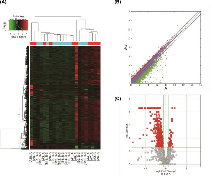
Methods: RNA-seq transcriptomics was used to determine gene expression in blood samples from radiation-exposed workers followed by bioinformatics analysis. Normal bronchial epithelial cells (16HBE) were irradiated for different durations and subjected to immunofluorescence, Western blotting, scratch healing, and adhesion assays to detect the progression of EMT and its underlying molecular mechanisms.

Results: Transcriptomics revealed that exposure to ionizing radiation led to changes in the expression of genes related to EMT, immune response, and migration. At increased cumulative doses, ionizing radiation-induced significant EMT, as evidenced by a gradual decrease in the expression of E-cadherin, increased vimentin, elevated migration ability, and decreased adhesion capability of 16HBE cells. The expression of fibronectin 1 (FN1) showed a gradual increase with the progression of EMT, and may be involved in EMT.

Conclusion: Ionizing radiation induces EMT. FN1 may be involved in the progression of EMT and could serve as a potential biomarker for this process.

Keywords: 16HBE; EMT; FN1; RNA-seq; ionizing radiation.


Conflict of interest statement

The authors declare that there are no competing interests associated with the manuscript.

Figures

Figure 1
Figure 1. Differentially expressed mRNAs in workers with or without radiation exposure
(A) Cluster analysis of differentially expressed mRNA based on FPKM values via heatmap2. In the picture, A is the control group and B is the exposed group. (B) Scatter plot based on the expression of the two groups. Red dots: up-regulated genes, green dots: down-regulated genes, purple dots: no significant change. Fold-change was 2.0. (C) Volcano plot based on fold-change and P-value. The red rectangle represents the differentially expressed mRNA, P≤0.05, fold-change ≥ 2.0.
Figure 2
Figure 2. GO analysis of differentially expressed mRNA in workers with or without radiation exposure
(A) The top 10 GO terms with the largest number of genes. (B) The ratio of the number of differentially expressed genes in the top 10 enriched GO terms with high fold enrichment to the total number of differentially expressed genes. The left column pertains to upregulated mRNAs, the right column pertains to down-regulated mRNAs, P≤0.05.
Figure 3
Figure 3. KEGG analysis of differentially expressed mRNA in workers with or without radiation exposure
(A) The top 10 pathways with high enrichment scores associated with the up-regulated mRNA. (B) The top 10 pathways with high enrichment scores associated with the down-regulated mRNA.
Figure 4
Figure 4. Validation of RNA-seq by RT-PCR
*P<0.05 vs control.
Figure 5
Figure 5. Changes in VIM, E-cad, migration, and proliferation of 16HBE cells following irradiation
Images of E-cad (A) and VIM (B) expression at different cumulative doses of radiation observed under a confocal microscope. (C and D) Wound healing assay was performed to determine the cell migration capability. (E and F) Cell migration and activity were determined by adhesion and CCK-8 assays, respectively; *P<0.05 vs 0 Gy.
Figure 6
Figure 6. Expression of FN1 in 16HBE cells following different cumulative doses of radiation
*P<0.05 vs 0vGy.

References

    1. Hamra G.B., Richardson D.B., Cardis E., Daniels R.D., Gillies M., O'Hagan J.A. et al. . (2016) Cohort Profile: The International Nuclear Workers Study (INWORKS). Int. J. Epidemiol. 45, 693–699 10.1093/ije/dyv122 - DOI - PMC - PubMed
    1. Abbott A. (2015) Researchers pin down risks of low-dose radiation. Nature 523, 17–18 10.1038/523017a - DOI - PubMed
    1. Keil A.P., Richardson D.B. and Troester M.A. (2015) Healthy worker survivor bias in the Colorado Plateau uranium miners cohort. Am. J. Epidemiol. 181, 762–770 10.1093/aje/kwu348 - DOI - PMC - PubMed
    1. Kamiya K., Ozasa K., Akiba S., Niwa O., Kodama K., Takamura N. et al. . (2015) Long-term effects of radiation exposure on health. Lancet 386, 469–478 10.1016/S0140-6736(15)61167-9 - DOI - PubMed
    1. Hall J., Jeggo P.A., West C., Gomolka M., Quintens R., Badie C. et al. . (2017) Ionizing radiation biomarkers in epidemiological studies - An update. Mutat. Res. 771, 59–84 10.1016/j.mrrev.2017.01.001 - DOI - PubMed

Publication types

MeSH terms