. 2020 Sep;29(9):1945-1963.
doi: 10.1002/pro.3915.

Cumulative deamidations of the major lens protein γS-crystallin increase its aggregation during unfolding and oxidation

Affiliations

Cumulative deamidations of the major lens protein γS-crystallin increase its aggregation during unfolding and oxidation

Calvin J Vetter et al. Protein Sci. 2020 Sep.

Abstract

Age-related lens cataract is the major cause of blindness worldwide. The mechanisms whereby crystallins, the predominant lens proteins, assemble into large aggregates that scatter light within the lens, and cause cataract, are poorly understood. Due to the lack of protein turnover in the lens, crystallins are long-lived. A major crystallin, γS, is heavily modified by deamidation, in particular at surface-exposed N14, N76, and N143 to introduce negative charges. In this present study, deamidated γS was mimicked by mutation with aspartate at these sites and the effect on biophysical properties of γS was assessed via dynamic light scattering, chemical and thermal denaturation, hydrogen-deuterium exchange, and susceptibility to disulfide cross-linking. Compared with wild type γS, a small population of each deamidated mutant aggregated rapidly into large, light-scattering species that contributed significantly to the total scattering. Under partially denaturing conditions in guanidine hydrochloride or elevated temperature, deamidation led to more rapid unfolding and aggregation and increased susceptibility to oxidation. The triple mutant was further destabilized, suggesting that the effects of deamidation were cumulative. Molecular dynamics simulations predicted that deamidation augments the conformational dynamics of γS. We suggest that these perturbations disrupt the native disulfide arrangement of γS and promote the formation of disulfide-linked aggregates. The lens-specific chaperone αA-crystallin was poor at preventing the aggregation of the triple mutant. It is concluded that surface deamidations cause minimal structural disruption individually, but cumulatively they progressively destabilize γS-crystallin leading to unfolding and aggregation, as occurs in aged and cataractous lenses.

Keywords: cataracts; crystallins; deamidation; dynamic and static light scattering; hydrogen-deuterium exchange; mass spectrometry; oxidation; protein aggregation; protein unfolding.


Conflict of interest statement

The authors have no conflict of interests to declare.

Figures

FIGURE 1
FIGURE 1
Solution structure of human γS showing the location of Asn residues 14, 76, and 143. Deamidation was mimicked at Asn residues 14 (teal), 76 (firebrick red), and 143 (green). Asparagine residues 14 and 76 are located on the surface of the N‐terminal domain (N‐td) and residue 143 is on the surface of the C‐terminal domain (C‐td). Residues of interest are shown in spheres. Gray area represents solvent accessibility, green arrows represent β‐sheets and red ribbons represent α‐helices as generated with PyMol (Schrodinger, Inc. New York, NY). (PDB: 2M3T)
FIGURE 2
FIGURE 2
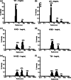
DLS measurements of the R h distributions of γS and deamidated mutants under native conditions. The intensity of DLS (% Intensity) derived from regularization analysis was plotted as a function of R h with R h values binned to 1–10 nm (peak 1), 10–100 nm (peak 2), and 100–1,000 nm (peak 3). (a and b) The % Intensity of WT species at 1 mg/ml and 5 mg/ml respectively. (c–f) The % Intensity of N14D, N76D, N143D and TM γS at 1 mg/ml. Data were collected at 25°C. Data are representative of three independent experiments
FIGURE 3
FIGURE 3
Molecular weight of γS and triply deamidated mutant (TM) under native conditions. Multi‐angle static light scattering (MALS) of WT and TM γS was measured in‐line with size exclusion chromatography (SEC‐MALS). The right axis is relative Rayleigh LS of WT (black line) and TM (magenta line). The left axis is the weight average molecular weight (noted by a blue arrow). (a) Analysis of freshly prepared WT and TM γS. Monomer peaks eluted at 14.5 ml and high Mw peaks eluted at 7.5 ml. The Mw for the monomer peaks was 20.5 × 103 (± 0.2) g/mol (blue arrow) compared with the predicted Mw of 20.9 × 103. Inset: SDS‐PAGE analysis of chromatography fractions. Lane 1 contains molecular weight markers. Lanes 2 and 7 contain 2.5 μg of unfractionated WT γS‐crystallin and TM proteins, respectively. Lanes 3–6 contain fractions of WT γS, 12–15 ml. Lanes 8–10 contain fractions of TM γS, 12–15 ml. (b) Analysis of incubated WT and TM γS monomers. The monomer peaks isolated in panel A were incubated at 37°C for 6 days and then reanalyzed by SEC‐MALS. Monomer peaks were detected with Mw of 20.6 × 103 (±0.2) g/mol (right blue arrow) and dimer peaks eluted at 13.5 ml with a Mw of 46.8 × 103 (± 1.2) g/mol (left blue arrow). The high Mw peak for the TM in panel B was estimated to range from 3–9 × 106 g/mol (off scale)
FIGURE 4
FIGURE 4
Mass spectra of WT and TM γS during chemical unfolding. Mass spectra of the +23 charge state of WT and TM γS following partial unfolding in 2.75 M GuHCl. Three major peaks were detected after 16 min in GuHCl with mass increases of 36–43 Da (peak 1), 83–85 Da (peak 2), and 120–130 Da (peak 3) over the peak of the unlabeled proteins at the position of the arrow (spectra not shown). (a) The spectrum of WT γS incubated in the absence of GuHCl is overlaid with that incubated in GuHCl for 16 min. (b) The spectrum of TM γS incubated in the absence of GuHCl is overlaid with that incubated in GuHCl for 16 min. In both panels a and c, there is a decrease in Peak 1 (folded species) and an increase in Peak 2 (partially unfolded intermediate) and Peak 3 (nearly fully unfolded species). Spectra are averaged from three independent experiments
FIGURE 5
FIGURE 5
Relative abundance of γS intermediates during chemical unfolding. Percentage under the peak for each species of γS as shown in Figure 5, calculated using raw mass spectra from pulse labeled protein in D2O containing buffer. Relative abundance of peaks 1, 2, and 3 for (a–c) N76D and TM compared with WT, and (d–f) N14D and N143D compared with WT. Significant differences from WT γS determined using Student's t‐test, p‐values ≤.01 and ≤.05 are denoted by ** and *, respectively (n = 3)
FIGURE 6
FIGURE 6
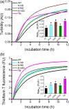
Time‐course of aggregation of γS and its deamidation mimics monitored by turbidity and ThT binding. WT and deamidated γS at 5 mg/ml and in 2.0 M GuHCl were incubated at 37°C. (a) Changes in turbidity at 405 nm. (b) Changes in thioflavin T fluorescence at 440/485 nm. AU and FU represent absorbance and fluorescence units, respectively. Aggregation curves shown are average measurements (n ≥ 2) representative of three independent experiments. Insets show maximum aggregation rate (Ratet50) derived from absorbance and fluorescence curves fitted with Boltzmann and Hill sigmoidal functions, respectively (n ≥ 4, error bars = SEM). Symbols (i.e., formula image, formula image, and formula image) denote groups of ratet50 values that are significantly different from each other (p‐value ≤.05)
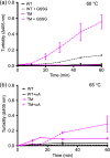
FIGURE 7
FIGURE 7
Effect of deamidation on thermal‐induced aggregation of WT and TM γS monitored by turbidity and its modulation by either oxidation or the molecular chaperone αA‐crystallin (αA). (a). Changes in turbidity during thermal‐induced denaturation of WT and TM γS in the presence of 6 mM GSSG and at 60°C (n = 3, error bars = SEM). (b) Changes in turbidity during thermal‐induced denaturation of WT and TM γS in the presence and absence of αA. WT and TM γS incubated at 65°C with and without αA at a 2:1 ratio of γS:αA (n = 4, error bars = SEM)
FIGURE 8
FIGURE 8
Molecular dynamics simulations of γS. (a) The population distribution of conformations of WT (gray), N14D (teal), N76D (dark red), and N143D (green) γS. (b) The root‐mean‐square‐deviation (RMSD) in the structural distances during simulation. The solid black lines represent the general trend of the RMSD in the structure. (c) Molecular dynamics simulations showing a potential salt bridge between D76 and R78 (black arrow). Hydrogen bonds are also shown (red arrows)
FIGURE 9
FIGURE 9
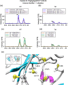
Mass spectra of a potential disulfide bond in peptide 20–35 of WT and TM γS. (a and b) Extracted ion chromatograms of the precursor, M + 1, and M + 2 peaks for the γS peptide 20–35 containing one internal disulfide bond and one alkylation in its cysteines 22, 24, and 26 for WT and TM γS, respectively. (c and d) The tracings for the coincident y6, 7, and 9 fragment ions for the peptide generated during the parallel reaction monitoring scans during the LC/MS run for WT and TM γS, respectively. (e) Distances between the proximal C22, C26, and C82 in the solution monomer as generated with http://www.rbvi.ucsf.edu/chimera/. (PDB 2M3T)
FIGURE 10
FIGURE 10
Potential γS aggregation pathways due to cataract‐associated deamidations. Our data support a model where native γS monomer has an internal disulfide bond that “locks” the N‐terminal domain (N‐td) into a conformation that is “unlocked” by increased conformational dynamics due to deamidation (Step 1). The deamidated monomer favors a “disulfide‐unlocked” state (blue arrow between the two Greek Key motifs in the N‐td) that is predisposed to oxidation at surface cysteines (Step 2, dotted line), leading to more aggregation‐prone crosslinked‐dimers (Step 3). A small amount of the deamidated monomer can also form large light scattering aggregates directly (thin arrow). Created with BioRender.com

References

    1. Delaye M, Tardieu A. Short‐range order of crystallin proteins accounts for eye lens transparency. Nature. 1983;302:415–417. - PubMed
    1. Truscott RJ, Friedrich MG. The etiology of human age‐related cataract. Proteins don't last forever. Biochim Biophys Acta. 2016;1860:192–198. - PMC - PubMed
    1. Congdon N, Vingerling JR, Klein BE, et al. Prevalence of cataract and pseudophakia/aphakia among adults in the United States. Arch Ophthalmol. 2004;122:487–494. - PubMed
    1. Ji F, Jung J, Gronenborn AM. Structural and biochemical characterization of the childhood cataract‐associated R76S mutant of human γD‐crystallin. Biochemistry. 2012;51:2588–2596. - PMC - PubMed
    1. Ji F, Jung J, Koharudin LM, Gronenborn AM. The human W42R gammaD‐crystallin mutant structure provides a link between congenital and age‐related cataracts. J Biol Chem. 2013;288:99–109. - PMC - PubMed

Publication types