. 2020 Jul 22;16(7):e1008920.
doi: 10.1371/journal.pgen.1008920. eCollection 2020 Jul.

Developmental loss of neurofibromin across distributed neuronal circuits drives excessive grooming in Drosophila

Affiliations

Developmental loss of neurofibromin across distributed neuronal circuits drives excessive grooming in Drosophila

Lanikea B King et al. PLoS Genet. .

Abstract

Neurofibromatosis type 1 is a monogenetic disorder that predisposes individuals to tumor formation and cognitive and behavioral symptoms. The neuronal circuitry and developmental events underlying these neurological symptoms are unknown. To better understand how mutations of the underlying gene (NF1) drive behavioral alterations, we have examined grooming in the Drosophila neurofibromatosis 1 model. Mutations of the fly NF1 ortholog drive excessive grooming, and increased grooming was observed in adults when Nf1 was knocked down during development. Furthermore, intact Nf1 Ras GAP-related domain signaling was required to maintain normal grooming. The requirement for Nf1 was distributed across neuronal circuits, which were additive when targeted in parallel, rather than mapping to discrete microcircuits. Overall, these data suggest that broadly-distributed alterations in neuronal function during development, requiring intact Ras signaling, drive key Nf1-mediated behavioral alterations. Thus, global developmental alterations in brain circuits/systems function may contribute to behavioral phenotypes in neurofibromatosis type 1.


Conflict of interest statement

The authors have declared that no competing interests exist.

Figures

Fig 1
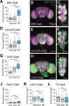
Fig 1. Neurons in the brain and ventral nervous system that modulate grooming are sensitive to loss of Nf1.
(A) An intact fly grooming its anterior (prothoracic) legs. (B) An intact fly grooming its posterior (metathoracic) legs. (C) Grooming duration in intact Nf1P1 vs. wCS10 control flies at 0, 5, and 10 min after introduction to the chamber. n = 18; **p < 0.01, ***p < 0.001 (Wilcoxon rank-sum test). (D) Grooming movement directed toward the anterior body (missing head) in a decapitated fly. (E) A headless fly grooming its hind (metathoracic) legs. (F) Grooming duration in decapitated flies. n = 25–26; *p < 0.05 (Wilcoxon rank-sum test). (G) Grooming duration in flies with Nf1 knockdown in tsh-Gal4+ neurons. n = 16; p < 0.001 (Kruskal-Wallis); ***p < 0.001 (Dunn/Sidak). (H) Maximum-intensity projection showing tsh>UAS-mCD8::GFP (green) and bruchpilot (brp; magenta) in the central brain. Scale bar = 100 μm. (I) VNS expression of tsh and brp, as in panel H. (J) Grooming duration with pan-neuronal knockdown (KD) of Nf1 via the R57C10-Gal4, with or without the tsh-Gal80 repressor. n = 40–42; p < 0.001 (Kruskal-Wallis); *p < 0.05 (Dunn/Sidak). (K) Grooming duration from the same data set in panel J, graphed by body part groomed. Individual data points are graphed (zero values are not plotted), with the mean shown as a line. Stars represent significance re: UAS control. (L) Maximum-intensity projection of anti-GFP (green) and anti-brp (magenta) immunostaining in the central brain of an R57C10-Gal4 > UAS-mCD8::GFP fly. Scale bar = 100 μm. (M) R57C10-Gal4 expression pattern in the VNS, imaged as in panel K. (N) Single z-plane showing anti-brp neuropil staining from the VNS in panel L. (O) GFP staining from the VNS plane in panel L. (O’) Expanded detail from the dashed box in panel N. Arrow points to area of dense somata. (P) Maximum-intensity projection of the brain of an R57C10-Gal4 > UAS-mCD8::GFP, UAS-tsh-Gal80 fly. (Q) Expression pattern in the VNS of the same fly shown panel K. (R) Single z-plane showing anti-brp neuropil staining from the VNS in panel P. (S) GFP staining from the VNS plane in panel Q. (S’) Expanded detail from the dashed box in panel N. Arrow points to area of reduced somata density by tsh-Gal80 repression.
Fig 2
Fig 2. Loss of Nf1 in broad sets of excitatory neurons increases grooming.
(A) Grooming duration with Nf1 knockdown (KD) in 69B-Gal4+ neurons. n = 20; p < 0.001 (Kruskal-Wallis). (B) Maximum-intensity projection of 69B-Gal4 expression (green) and anti-brp immunostaining (magenta) in the brain. Scale bar = 100 μm. (C) VNS expression of 69B as in panel B. (D) Knockdown of Nf1 in oct-tyrR-Gal4+ neurons. n = 20; p < 0.001 (Kruskal-Wallis). (E) Brain expression of the oct-tyrR-Gal4 driver. (F) VNS expression of the oct-tyrR-Gal4 driver. (G) Knockdown of Nf1 using the ChAT-T2A-Gal4 driver. n = 20; p < 0.001 (Kruskal-Wallis). (H) Brain expression of the ChAT-T2A-Gal4 driver. (I) VNS expression of the ChAT-T2A-Gal4 driver. (J) Knockdown of Nf1 in Gad1-Gal4+ neurons. n = 18 each; p = 0.23 (Kruskal-Wallis). (K) Knockdown of Nf1 in vGluT-Gal4+ neurons. n = 20; p = 0.45 (Kruskal-Wallis). (L) Knockdown of Nf1 in TH-Gal4+ neurons. n = 20; p = 0.6 (Kruskal-Wallis). *p < 0.05, **p < 0.01, ***p < 0.001 (Dunn/Sidak).
Fig 3
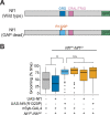
Fig 3. Functional Nf1 GAP-related domain (GRD) is required to maintain normal grooming frequency.
(A) Diagram of the wild type (top) and mutated (bottom) Nf1 rescue constructs. Major protein domains, the R1320P mutation, and GFP C-terminal fusions are depicted. (B) Grooming durations for (left to right): (1) nSyb-Gal4 genetic control, and Nf1P1/Nf1E1 mutants expressing (2) wild-type UAS-Nf1 rescue, (3) UAS-Nf1R1320P rescue, (4) nSyb-Gal4 alone, (5) UAS-Nf1 alone, and (6) UAS-Nf1R1320P alone. n = 40; p < 0.001, Kruskal-Wallis; **p < 0.01, n.s.: not significant (Dunn/Sidak).
Fig 4
Fig 4. Excessive grooming results from the loss of Nf1 during a critical developmental window.
Gal80ts was used to restrict knockdown (KD) of Nf1 with RNAi to different time windows during development and adulthood. (A-G) In each panel, elav-Gal4 > UAS-Nf1 RNAi, tub-Gal80ts (knockdown [KD]) is compared to UAS-Nf1 RNAi (UAS) and elav-Gal4 and tub-Gal80ts (Gal4) controls. n = 20. *p < 0.05, **p < 0.01, ***p < 0.001 (Dunn-Sidak). (A) Knockdown during embryo through larval L2 stage. p = 0.37 (Kruskal-Wallis). (B) Larval L3 and pupal stages. p < 0.001 (Kruskal-Wallis). (C) Larval L3 stage. p < 0.001 (Kruskal-Wallis). (D) Pupal stage. p < 0.001 (Kruskal-Wallis). (E) Early pupal stage. p < 0.001 (Kruskal-Wallis). (F) Late pupal stage. p = 0.05 (Kruskal-Wallis). (G) Knockdown in adulthood, following eclosion. p = 0.1 (Kruskal-Wallis).
Fig 5
Fig 5. Octopamine-tyramine receptor+ cells are abundant throughout development.
(A) Maximum-intensity projection of oct-tyrR+ neurons (green) and anti-neuroglian (Nrg) (magenta) immunostaining in the 0-hr pupal nervous system. (B) 48-hr pupal brain. (C) 48-hr pupal VNS. (D) 96-hr pupal brain. (E) 96-hr pupal VNS. APF: after puparium formation.
Fig 6
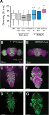
Fig 6. Loss of Nf1 across combinations of distributed circuits drives elevated grooming.
(A) Grooming duration in flies with Nf1 knockdown in one or both sets of neurons labeled by the R13F10 (R13) and R11B06 (R11) Gal4 drivers. n = 20; Kruskal-Wallis; *p < 0.05 compared to UAS and Gal4 controls, n.s. not significant compared to UAS or Gal4 controls (Dunn/Sidak). (B) Maximum-intensity projection showing the expression of R13F10-Gal4 (green) and anti-neuroglian immunostaining (magenta) in a 48-hr pupal brain. (C) 48-hr pupal VNS, as in panel B. (D) GFP channel of VNS shown in panel C. (E) Maximum-intensity projection showing the expression of R11B06-Gal4 (green) and anti-neuroglian immunostaining (magenta) in a 48-hr pupal brain. (F) 48-hr pupal VNS, as in panel B. (G) GFP channel of the VNS shown in panel F.

References

    1. Hyman SL, Shores A, North KN. (2005) The nature and frequency of cognitive deficits in children with neurofibromatosis type 1. Neurology 65: 1037–44. 10.1212/01.wnl.0000179303.72345.ce - DOI - PubMed
    1. Diggs-Andrews KA, Gutmann DH. (2013) Modeling cognitive dysfunction in neurofibromatosis-1. Trends Neurosci 36: 237–47. 10.1016/j.tins.2012.12.002 - DOI - PMC - PubMed
    1. Constantino JN, Zhang Y, Holzhauer K, Sant S, Long K, Vallorani A, Malik L, Gutmann DH. (2015) Distribution and Within-Family Specificity of Quantitative Autistic Traits in Patients with Neurofibromatosis Type I. J Pediatr 167: 621–6 e1. 10.1016/j.jpeds.2015.04.075 - DOI - PMC - PubMed
    1. Eijk S, Mous SE, Dieleman GC, Dierckx B, Rietman AB, de Nijs PFA, Ten Hoopen LW, van Minkelen R, Elgersma Y, Catsman-Berrevoets CE, Oostenbrink R, Legerstee JS. (2018) Autism Spectrum Disorder in an Unselected Cohort of Children with Neurofibromatosis Type 1 (NF1). J Autism Dev Disord, 10.1007/s10803-018-3478-0 - DOI - PMC - PubMed
    1. Garg S, Green J, Leadbitter K, Emsley R, Lehtonen A, Evans DG, Huson SM. (2013) Neurofibromatosis type 1 and autism spectrum disorder. Pediatrics 132: e1642–8. 10.1542/peds.2013-1868 - DOI - PubMed

Publication types

LinkOut - more resources