. 2020 Sep 15;465(2):119-129.
doi: 10.1016/j.ydbio.2020.06.012. Epub 2020 Jul 19.

Periocular neural crest cell differentiation into corneal endothelium is influenced by signals in the nascent corneal environment

Affiliations

Periocular neural crest cell differentiation into corneal endothelium is influenced by signals in the nascent corneal environment

Anna Babushkina et al. Dev Biol. .

Abstract

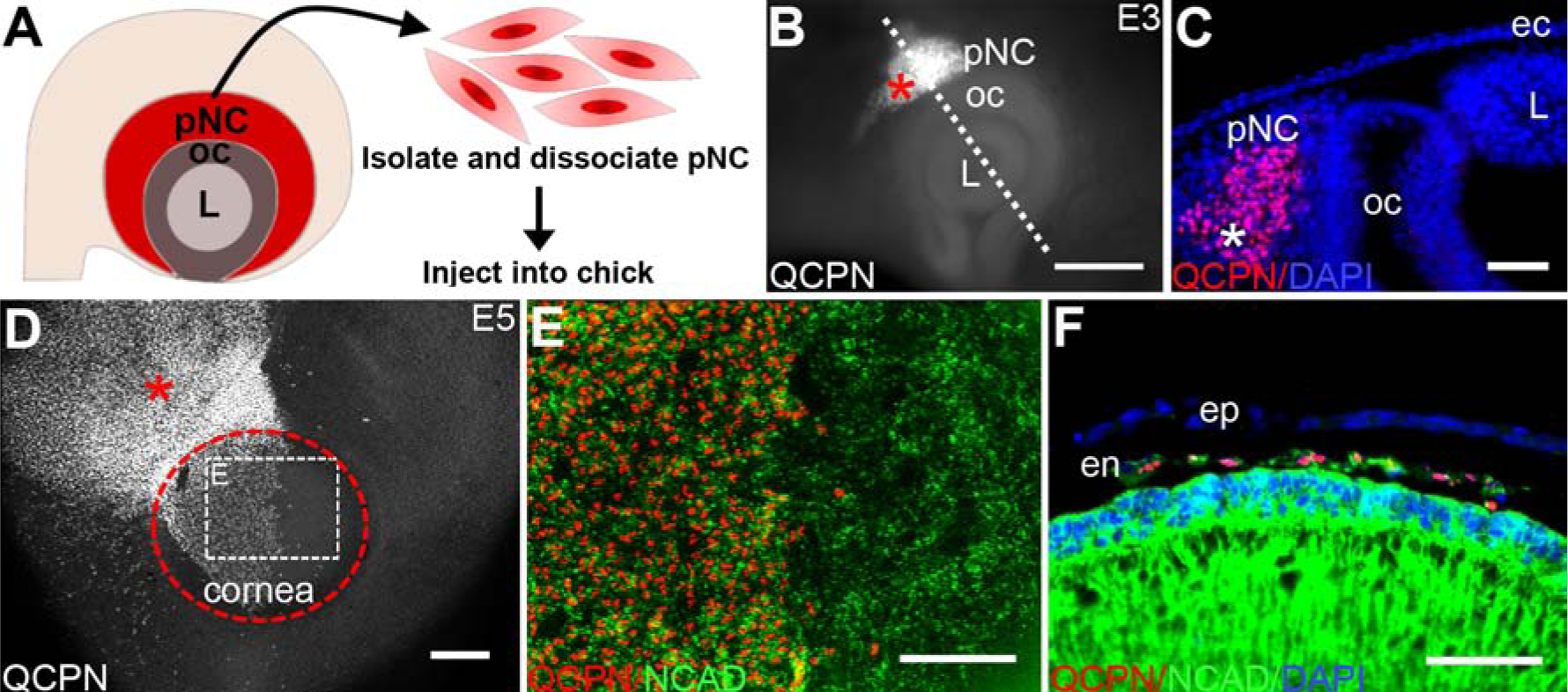
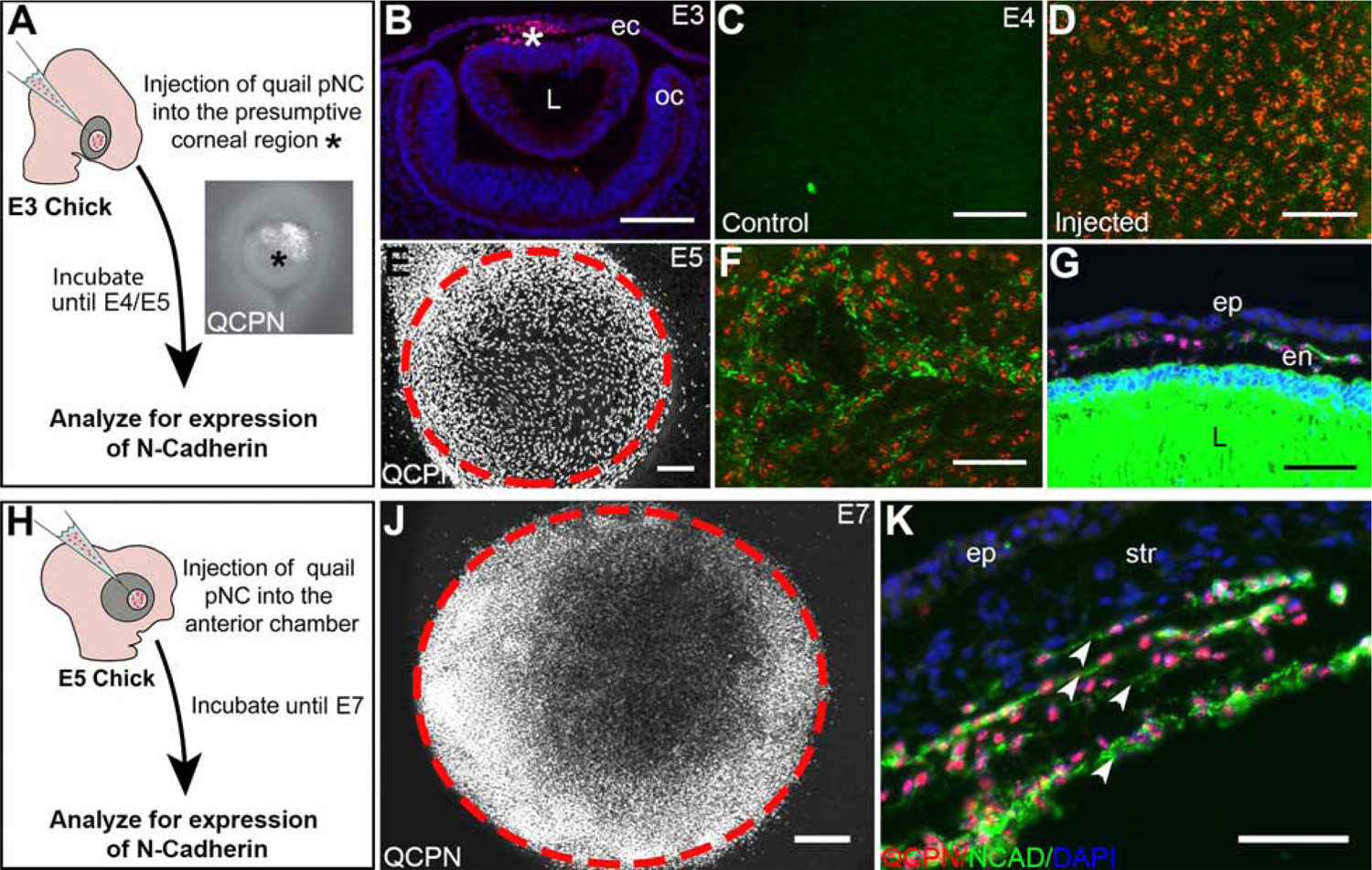
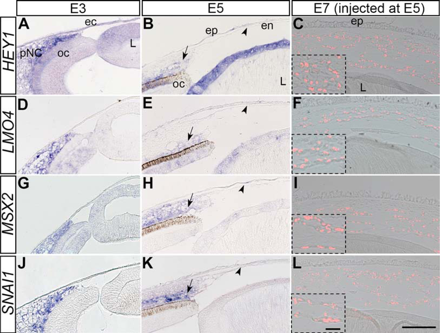
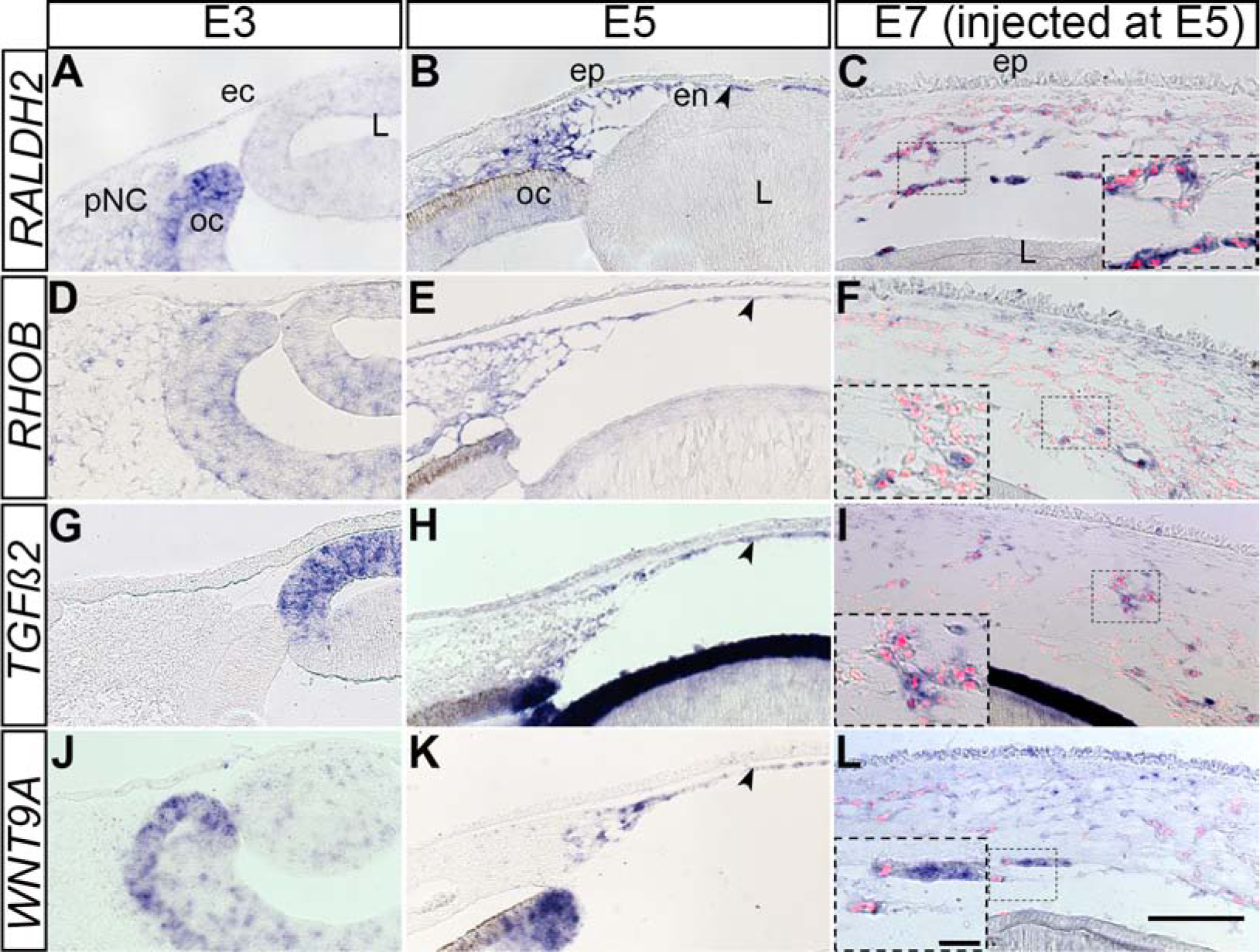
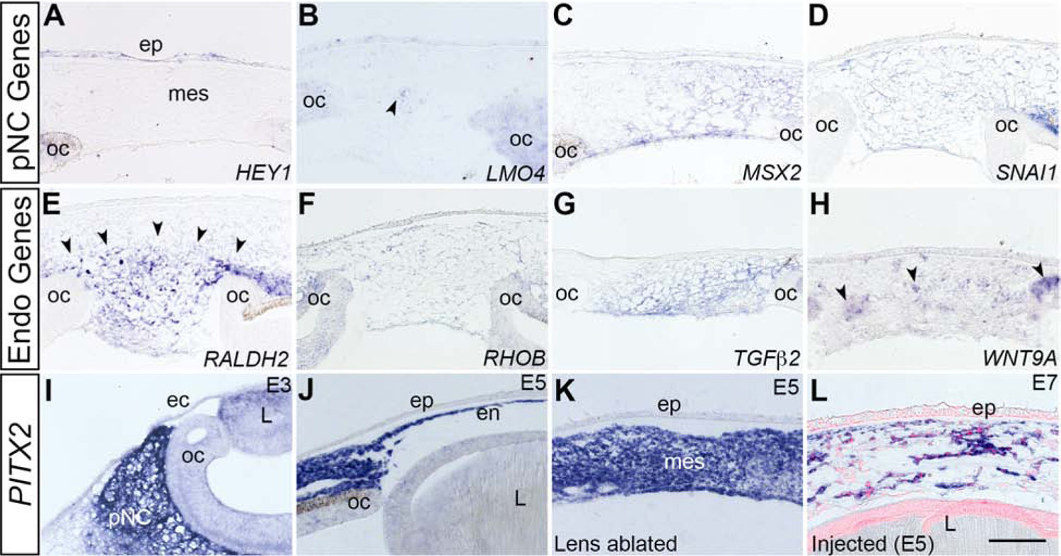
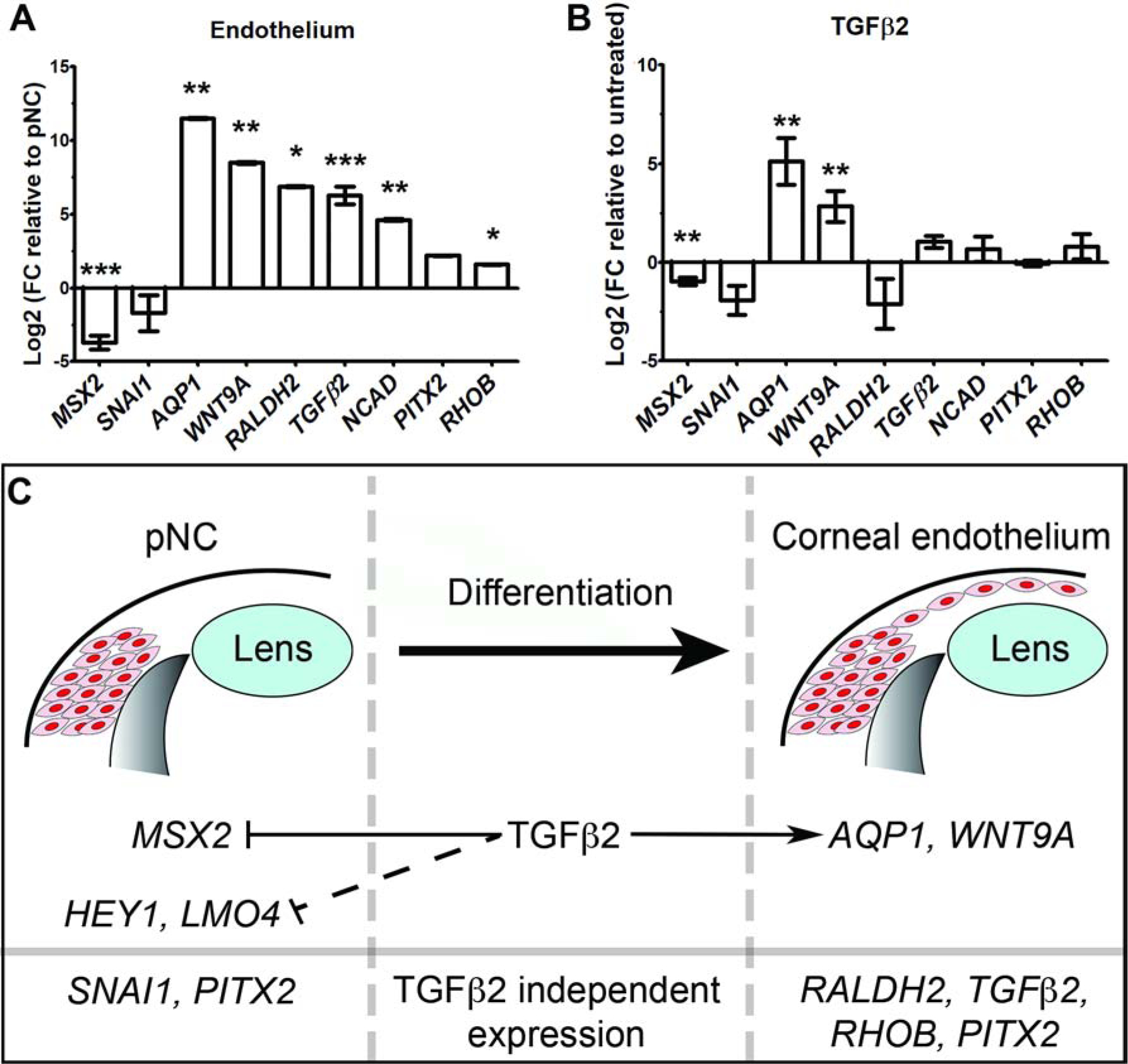
During ocular development, periocular neural crest cells (pNC) migrate into the region between the lens and presumptive corneal epithelium to form the corneal endothelium and stromal keratocytes. Although defects in neural crest cell development are associated with ocular dysgenesis, very little is known about the molecular mechanisms involved in this process. This study focuses on the corneal endothelium, a monolayer of specialized cells that are essential for maintaining normal hydration and transparency of the cornea. In avians, corneal endothelial cells are first to be specified from the pNC during their migration into the presumptive corneal region. To investigate the signals required for formation of the corneal endothelium, we utilized orthotopic and heterotopic injections of dissociated quail pNC into chick ocular regions. We find that pNC are multipotent and that the nascent cornea is competent to induce differentiation of ectopically injected pNC into corneal endothelium. Injected pNC downregulate expression of multipotency transcription factors and upregulate genes that are consistent with ontogenesis of the chick corneal endothelium. Importantly, we showed that TGFβ2 is expressed by the nascent lens and the corneal endothelium, and that TGFβ signaling plays a critical role in changing the molecular signature of pNC in vitro. Collectively, our results demonstrate the significance of the ocular environmental cues towards pNC differentiation, and have potential implications for clinical application of stem cells in the anterior segment.

Keywords: Corneal development; Corneal endothelium; Periocular neural crest; TGFβ2.


Conflict of interest statement

Declaration of competing interest No competing interests to declare.

Figures

Figure 1.
Figure 1.. Differentiation of E2.5 quail pNC following orthotopic graft into chick.
(A) Schematic overview of the isolation and dissociation procedures for obtaining quail pNC. (B) Wholemount immunostaining of an E3 chick eye showing QCPN-positive injected quail pNC (asterisk) in the periocular region. Dotted line represents the plane of cross-section in (C) showing localization of quail pNC (asterisk) within the periocular mesenchyme. (D) Wholemount immunostaining of an E5 chick eye showing the extent of migration QCPN-positive injected pNC (asterisk) in the periocular region and cornea (delineated by the dotted circle). (E) High magnification of selected region in (D) showing expression of NCAD by QCPN-positive cells and endogenous chick corneal endothelium. (F) Cross-section though the anterior eye of E5 injected embryo showing a monolayer of QCPN-positive cells co-expressing NCAD. ec, ectoderm; en, corneal endothelium; ep, corneal epithelium; L, lens; oc, optic cup. Scale bars represent 100 μm (B, D, E, F); 50 μm (C).
Figure 2.
Figure 2.. pNC injected directly into the presumptive corneal region at different timepoints of development differentiate into corneal endothelial cells.
(A) Schematic overview of the injection of pNC into E3 presumptive corneal region (asterisk) and further experimental procedures. (B) cross-section through E3 eye, showing QCPN-positive pNC (asterisk) in the presumptive corneal region between the ectoderm and lens. (C) Wholemount immunostaining of an E4 chick eye showing no NCAD expression in the presumptive corneal region prior to the pNC migration comparing to (D) Wholemount immunostaining of an E4 chick eye showing the NCAD expression surrounding some of the QCPN-positive cells 1 day post-injection at E3. (E) Wholemount immunostaining of an E5 chick eye showing the localization QCPN-positive pNC in the cornea (delineated by the dotted circle) and surrounding region 2 days post injection at E3. (F) Injected pNC stain positive for NCAD at E5. (G) Cross-section of an E5 eye showing that the injected pNC form an NCAD-positive monolayered corneal endothelium. (H) Schematic overview of the injection of the pNC into E5 presumptive cornea and further experimental procedures. (J) Wholemount immunostaining of an E7 chick eye showing the localization QCPN-positive pNC in the cornea (delineated by the cells and dotted circle) following 2 days post injection at E5. (K) Cross-section through E7 eye, showing that injected cells form multiple layers of QCPN- and NCAD-positive cells (arrowheads). ec, ectoderm; en, corneal endothelium; ep, corneal epithelium; L, lens; oc, optic cup; str, corneal stroma. Scale bars represent 100 μm (B,E); 50 μm (C, D, F, G, J, K).
Figure 3.
Figure 3.. Expression of pNC-related genes during normal development of the corneal endothelium and by the injected pNC.
Section in situ hybridization was performed on E3, E5, and E7 injected eyes. (A, D, G, J) E3 chick eyes showing the localization of HEY1, LMO4, MSX2 and SNAI1 in the periocular region. (B, E, H, K) E5 chick eyes showing no detection of HEY1, LMO4, MSX2 and SNAI1 expression by the corneal endothelium (arrowheads), despite their expression in other ocular regions. (C, F, I, L) E7 chick eyes showing no detection of HEY1, LMO4, MSX2 and SNAI1 expression by QCPN-positive cells 2 days post injection. Inserts are higher magnifications of selected regions. ec, ectoderm; en, corneal endothelium; ep, corneal epithelium; L, lens; oc, optic cup; str, corneal stroma. Scale bars represent 100 μm (A-L) and the scale bars for the inserts represent 20 μm (C, F, I, L).
Figure 4.
Figure 4.. Expression of corneal endothelium genes during normal ocular development and by the injected pNC.
Section in situ hybridization was performed on E3, E5, and E7 injected eyes. (A, D, G, J) E3 chick eyes showing little or no expression of RALDH2, RHOB, TGFβ2 and WNT9A in the periocular region. (B, E, H, K) E5 chick eyes robust expression of RALDH2, RHOB, TGFβ2 and WNT9A by the corneal endothelium (arrowheads). (C, F, I, L) E7 chick eyes showing strong expression of expression of RALDH2, RHOB, TGFβ2 and WNT9A by QCPN-positive cells 2 days post injection. Inserts are higher magnifications of selected regions. ec, ectoderm; en, corneal endothelium; ep, corneal epithelium; L, lens; oc, optic cup; str, corneal stroma. Scale bars represent 100 μm (A-L) and the scale bars for the inserts represent 20 μm (C, F, I, L).
Figure 5.
Figure 5.. Expression patterns of pNC- and corneal endothelium-related genes in the mesenchyme of malformed corneas following lens-ablation.
Lens ablations were performed at E3 and section in situ hybridization was performed at E5. (A-D) pNC-related genes show different patterns of expression in mesenchyme: (A) HEY1 is not detected. (B) LMO4 is expressed by a few cells (arrowheads). (C-D) MSX2 and SNAI1 are robustly expressed. (E-H) Corneal endothelium-related genes (RALDH2, RHOB, TGFβ2 and WNT9A) are all expressed in mesenchyme (RALDH2, arrowheads), (WNT9A, arrowheads), (I-L) PITX2 shows expression in the pNC, the endothelium, the ablated mesenchyme and in the injected cells in stages E3–E7. ec, surface ectoderm; ep, corneal epithelium, L, lens; mes, mesenchyme; oc, optic cup; pNC, periocular neural crest cells; str, stroma. Scale bar represents 100 μm (A-H).
Figure 6.
Figure 6.. Molecular mechanisms of pNC differentiation into the corneal endothelium.
(A-B) Comparative qPCR analysis of pNC and endothelial mRNA expression under different conditions. GAPDH was used as reference gene. Independent sample replicates n=3. *P<0.05, ** P<0.01, *** P<0.001 (Welch`s two-tailed t-test). Data represented as mean ± s.d. (A) Fold change (FC) of corneal endothelial genes compared to E2.5 pNC (baseline). (B) Fold change in gene expression of TGFβ2-treated pNC compared to untreated control culture (baseline). (C) Schematic representation of the regulation of pNC and corneal endothelial genes by TGFβ2 signaling.

References

    1. Adini I, Rabinovitz I, Sun JF, Prendergast GC, and Benjamin LE, 2003. RhoB controls Akt trafficking and stage-specific survival of endothelial cells during vascular development. Genes Dev. 17, 2721–2732. - PMC - PubMed
    1. Alexander NR, Tran NL, Rekapally H, Summers CE, Glackin C, and Heimark RL, 2006. N-cadherin Gene Expression in Prostate Carcinoma Is Modulated by Integrin-Dependent Nuclear Translocation of Twist1. Cancer Res. 66, 3365–3369. - PubMed
    1. Al-Yousuf N, Mavrikakis I, Mavrikakis E, and Daya SM, 2004. Penetrating keratoplasty: indications over a 10 year period. Br. J. Ophthalmol 88, 998–1001. - PMC - PubMed
    1. Anderson DJ, 1997. Cellular and Molecular Biology of Neural Crest Lineage Determination. Trends Genet. 13, 276–280. - PubMed
    1. Bard JBL, Hay ED, and Meller SM, 1975. Formation of the Endothelium of the Avian Cornea: A Study of Cell Movement in Vivo. Dev. Biol 42, 334–361. - PubMed

Publication types

Substances

LinkOut - more resources