. 2020 Jul 21;32(3):107931.
doi: 10.1016/j.celrep.2020.107931.

RSK2 Maintains Adult Estrogen Homeostasis by Inhibiting ERK1/2-Mediated Degradation of Estrogen Receptor Alpha

Affiliations

RSK2 Maintains Adult Estrogen Homeostasis by Inhibiting ERK1/2-Mediated Degradation of Estrogen Receptor Alpha

Katarzyna A Ludwik et al. Cell Rep. .

Abstract

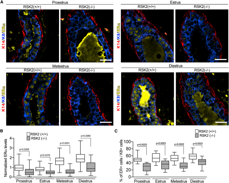
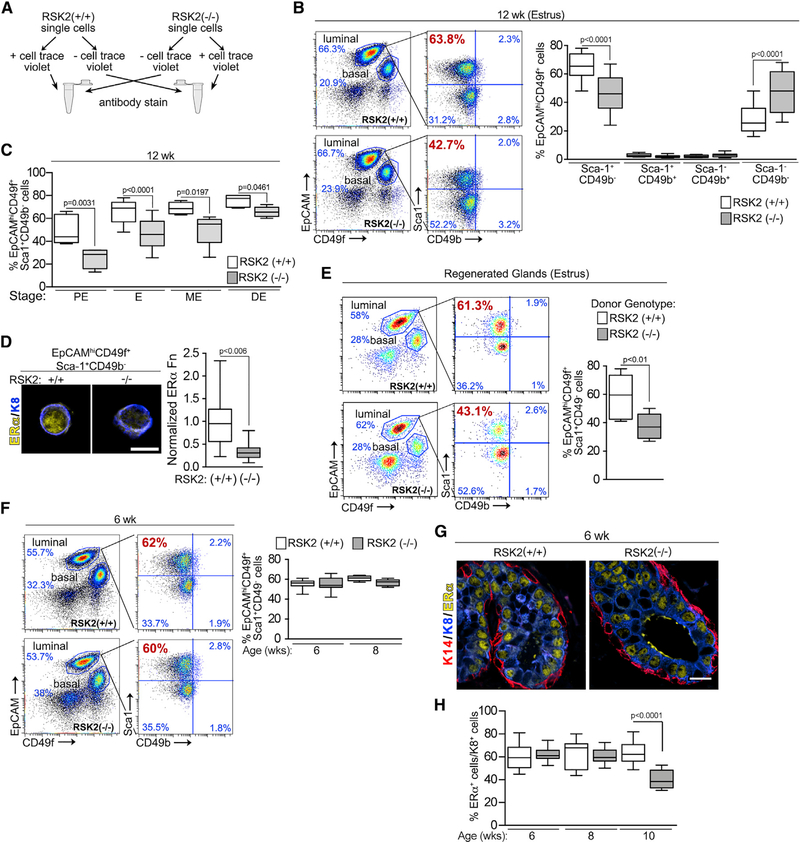
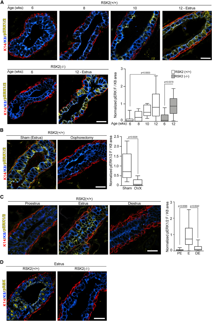
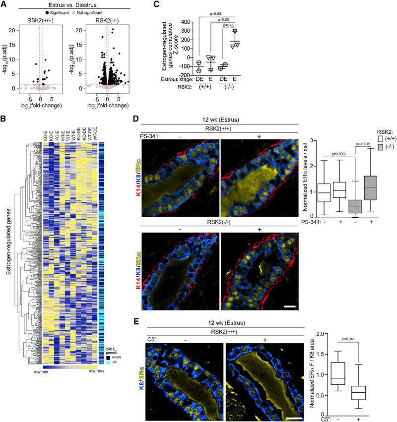
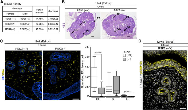
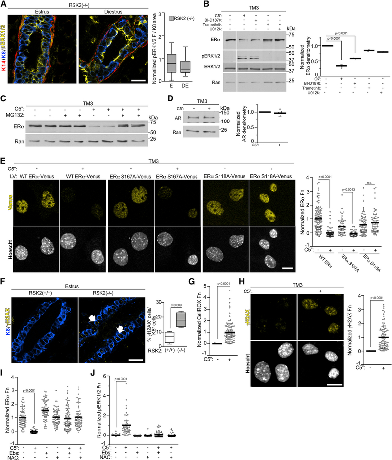
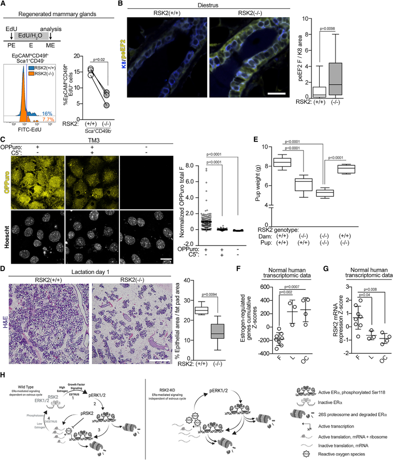
In response to estrogens, estrogen receptor alpha (ERα), a critical regulator of homeostasis, is degraded through the 26S proteasome. However, despite the continued presence of estrogen before menopause, ERα protein levels are maintained. We discovered that ERK1/2-RSK2 activity oscillates during the estrous cycle. In response to high estrogen levels, ERK1/2 is activated and phosphorylates ERα to drive ERα degradation and estrogen-responsive gene expression. Reduction of estrogen levels results in ERK1/2 deactivation. RSK2 maintains redox homeostasis, which prevents sustained ERK1/2 activation. In juveniles, ERK1/2-RSK2 activity is not required. Mammary gland regeneration demonstrates that ERK1/2-RSK2 regulation of ERα is intrinsic to the epithelium. Reduced RSK2 and enrichment in an estrogen-regulated gene signature occur in individuals taking oral contraceptives. RSK2 loss enhances DNA damage, which may account for the elevated breast cancer risk with the use of exogenous estrogens. These findings implicate RSK2 as a critical component for the preservation of estrogen homeostasis.

Keywords: ERK1/2; ERα; MAPK; RSK2; estrogen; growth factors; mammary gland; p90 ribosomal S6 kinase; p90RSK; transgenic mice.


Conflict of interest statement

Declaration of Interests The authors D.A.L. and G.A.O. have a patent related to this work.

Figures

Figure 1.
Figure 1.. RSK2 Regulates ERα Protein Levels in the Adult Mammary Gland throughout the Estrous Cycle
(A) ERα protein expression in the adult mammary gland of WT and RSK2-KO mice during the estrous cycle. Scale bar: 20 μm. (B) ERα protein levels are lower in the RSK2-KO mice at all stages of the estrous cycle in adult mammary glands as determined by IF. ERα protein levels normalized to the average level observed in the WT mice at proestrus (median ± quartile, n ≥ 3 mice/genotype and stage, one-way ANOVA with Holm-Sidak’s correction for multiple comparisons). (C) Loss of RSK2 results in a decrease in the number of ERα cells relative to K8+ cells at all stages of the estrous cycle in adult mammary glands (median ± quartile, n ≥ 4 mice/genotype, ≥150 cells/mouse, one-way ANOVA with Holm-Sidak’s correction for multiple comparisons). See Figure S1 and Table S1.
Figure 2.
Figure 2.. RSK2 Maintains the EpCAMhiCD49f+Sca1+CD49b (NCL) Population within the Adult Mammary Gland throughout the Estrous Cycle
(A) Schematic of FACS protocol. (B) FACS analysis of adult mammary glands isolated from females during estrus. Gating strategy of luminal cells by further subdivision using Sca-1 and CD49b. The percentage of NCL cells within the luminal population at estrus decreases in adult RSK2-KO mice (median ± quartile, n≥6mice/genotype, one-way ANOVA with Holm-Sidak’s correction for multiple comparisons). (C) Loss of RSK2 results in a reduction in the percentage of NCL cells at all stages of the estrous cycle in adult mammary glands (median ± quartile, n≥3 mice/genotype and stage, one-way ANOVA with Holm-Sidak’s correction for multiple comparisons). PE, proestrus; E, estrus; ME, metestrus; DE, diestrus. (D) ERα protein levels are decreased in cells isolated from the NCL population of RSK2-KO mice (median ± quartile, n = 3 mice/genotype, >20 cells/mouse, Student’s t test). Scale bar: 10 μm. Fn, fluorescence. (E) RSK2 regulation of the NCL population is intrinsic to the epithelium (median ± quartile, n = 3 mice/genotype, Student’s t test). (F) The percentage of NCL cells within the luminal population is similar between WT and RSK2-KO juvenile female mice (median ± quartile, n≥3 mice/genotype and age group, one-way ANOVA with Holm-Sidak’s correction for multiple comparisons). (G and H) The levels of ERα protein expression (G) and the number of ERα cells (H) relative to K8+ cells are similar in WT and RSK2-KO juvenile female mice (median ± quartile, n = 3 mice/genotype, ≥5 fields/mouse, one-way ANOVA with Holm-Sidak’s correction for multiple comparisons). Scale bar: 20 μm. See Figures S1 and S2 and Table S1.
Figure 3.
Figure 3.. ERK1/2-RSK2 Signaling Is Activated Only in the Adult Mammary Gland
(A) ERK1/2 activity is increased in the adult compared with juvenile animals (median ± quartile, n≥2 mice/genotype and age, ≥3 fields/mouse, one-way ANOVA with Holm-Sidak’s correction for multiple comparisons). Scale bar: 20 μm. (B) ERK1/2 activity in the mammary gland depends on estrogen (median ± quartile, n ≥ 2 mice/genotype and procedure, ≥3 fields/mouse, Student’s t test). Scale bar: 20 μm. (C) ERK1/2 activity varies during the estrous cycle in the WT mice adult mammary gland (median ± quartile, n ≥ 2 mice/genotype, ≥3 fields/mouse, one-way ANOVA with Tukey’s correction for multiple comparisons). Scale bar: 20 μm. (D) Active nuclear RSK2 is the predominant RSK in adult mammary glands. Scale bar: 20 μm. See Figures S3 and Table S1.
Figure 4.
Figure 4.. RSK2 Is a Negative Regulator of ERα-Mediated Signaling
(A) RSK2-KO mice show greater numbers of DEGs between estrus and diestrus (right panel) than do WT mice (left panel). Genes with a fold-change ≥ |1.5| (log2[fold-change]≥|0.5|) and a false discovery rate (FDR)-adjusted p < 0.05 are shown as black dots, and genes with a fold-change < |1.5| (log2[fold-change] < |0.5|) and an FDR-adjusted p value > 0.05 are shown as gray dots. The dashed line indicates the cutoff values. (B) Heatmap illustrating that the gene expression of NCL cells isolated from RSK2-KO mice in estrus correlates with a 24-h estrogen-regulated gene signature identified from MCF-7 cells (Dutertre et al., 2010). (C) Quantitative assessment of enrichment for estrogen-regulated genes. Cumulative Z scores were generated for each mouse by summing individual Z scores of genes upregulated in estrogen-regulated signature and subtracting individual Z scores of genes downregulated (mean ± SD, each point represents a mouse; one-way ANOVA with Holm-Sidak’s correction for multiple comparisons). (D) Loss of RSK2 increases ERα turnover. Adult mice staged at estrus were treated with vehicle or PS-341 (5 mg/kg) intraperitoneally (i.p.) for 4 h before euthanasia and isolation of the mammary gland. ERα protein levels were normalized to those observed in the WT mice at estrus (median ± quartile, n = 3 mice/genotype and condition, ≥200 cells/mouse, one-way ANOVA with Holm-Sidak’s correction for multiple comparisons). Scale bar: 20 μm. (E) RSK2 kinase activity is necessary to maintain ERα protein levels. Adult mice staged at estrus were treated with vehicle or C5′′-n-propyl cyclitol SL0101 (C5′′) (40 mg/kg) IP twice every 7 h before euthanasia and isolation of the mammary gland (median ± quartile, n≥3 mice/genotype, ≥3 fields/mouse, Student’s t test). See Figures S4 and S6 and Tables S1, S2, and S3.
Figure 5.
Figure 5.. RSK2 Maintains ERα Protein Levels in the Uterine Epithelium
(A) RSK2-KO mice show a fertility defect (n ≥ 15 dams/genotype, Chi-square test p = 0.0299). (B) The hypothalamic-pituitary-ovarian axis is not disrupted in RSK2-KO female mice. H&E sections of ovaries isolated from adult mice in estrus. Scale bar: 1 mm, PF, primary follicle; SF, secondary follicle; TF, tertiary follicle; CL, corpus luteum. (C) RSK2-KO mice have reduced ERα protein levels in the glandular and luminal epithelium of the uterus (median ± quartile, n = 3 mice/genotype, >120 cells/mouse, Student’s t test). Scale bar: 40 μm. GE, glandular epithelium; S, stroma; LE, luminal epithelium. (D) Active ERK1/2 is confined to the epithelium of the uterus. Scale bar: 40 μm. See Figures S1 and S5.
Figure 6.
Figure 6.. ERK1/2 Drives ERα Degradation through Phosphorylation of Ser-118
(A) ERK1/2 activity remains elevated during diestrus in the adult mammary gland (median ± quartile, n = 3 mice, ≥3 fields/mice, Student’s t test). (B) RSK2 is a negative regulator of ERK1/2 activity. Serum-starved TM3 was treated for 6 h with vehicle, C5′′-n-propyl cyclitol SL0101 (C5′′) (20 μM), BI-D1870 (10 μM), trametinib (1 μM), or U0126 (10 μM). The white vertical line indicates that conditions not relevant to the manuscript were removed. ERα levels were normalized to Ran and then to the vehicle (mean, n = 3, one-way ANOVA with Dunnett’s correction for multiple comparisons). (C) RSK1/2 inhibition stimulates ERα degradation through the 26S proteasome pathway. Serum-starved TM3 was treated for 6 h with vehicle, C5′′ (20 μM) with or without a 1 h of pre-treatment with MG132 (10 μM). (D) RSK2 does not regulate androgen receptor (AR) degradation. Serum-starved TM3 was treated for 6 h with vehicle or C5′′ (20 μM). AR levels were normalized to Ran and then to the vehicle (mean, n = 3 in duplicate, Student’s t test). (E) Phosphorylation of Ser-118A is required for ERα degradation. Cells transduced with WT or mutant ERα-VENUS were treated with vehicle or C5′′ (20 μM) as in (B). The range was normalized to WT ERα (mean, n = 3, >150 cells/condition/experiment, one-way ANOVA with Holm-Sidak’s correction for multiple comparisons). Scale bar: 10 μm. (F) Loss of RSK2 increases double-stranded DNA breaks (γ-H2AX foci) in the mammary gland (median ± quartile, n ≥ 4 mice/genotype, ≥3 fields/mouse, Student’s t test). (G) RSK1/2 inhibition increases ROS. Serum-starved TM3 was treated as in (B). The data were normalized to the range with and without C5′′ (20 μM) (mean, n = 3, >100 cells/condition/experiment, Student’s t test). (H) RSK1/2 inhibition increases DNA damage in vitro. Cells treated for 72 h with vehicle or C5′′ (20 μM). The data were normalized to the range with and without C5′′ (mean, n = 3, >80 cells/condition/experiment, Student’s t test). (I) Inhibition of ROS rescues ERα levels. Serum-starved TM3 was treated for 6 h with vehicle or C5′′ (20 μM) with or without ebselen (Ebs) (50 μM) or N-acetyl cysteine (NAC) (15 mM) for the final 2 h. The range was normalized to ERα levels in the absence of anti-oxidants (mean, n = 3, >50 cells/condition/experiment, one-way ANOVA with Dunnett’s correction for multiple comparisons). (J) Inhibition of ROS inhibits ERK1/2 activation. Cells treated and analyzed as in (I). See Figures S3 and S6 and Table S1.
Figure 7.
Figure 7.. RSK2 Is Necessary for Alveolar Expansion
(A) RSK2-KO NCL cells show a decrease in proliferation as compared with the WT cells. RSK2-KO or WT MECs were used to regenerate the mammary gland in a WT mouse. These mice were staged in proestrus and administered 5-ethynyl-2′-deoxyuridine (EdU) throughout one estrus cycle. The mammary glands were isolated and analyzed by FACS (n = 3 glands/genotype; paired Student’s t test). (B) RSK2 regulates eEF2K activity in vivo (median ± quartile, n ≥ 3 mice/genotype, ≥5 fields/mouse, Student’s t test). (C) Inhibition of RSK1/2 decreases translation in vitro. Serum-starved TM3 was treated for 6 h with vehicle or C5′′ (20 μM). The range was normalized to the o-propargyl-puromycin (OPP) in the absence and presence of C5′′ (mean, n ≥ 3, >150 cells/condition/experiment, one-way ANOVA with Holm-Sidak’s correction for multiple comparisons). (D) Alveolar expansion is reduced in RSK2-KO dams as shown by H&E stains of mammary glands isolated from dams 1 d after birth (median ± quartile, n ≥ 3 mice/genotype, ≥3 fields/mouse, Student’s t test). Scale bar: 1 mm. (E) Pups nursed by RSK2-KO dams are smaller than those nursed by WT dams. Weanling weight at 21 d nursed by a dam with the indicated genotype (median ± quartile, n = 3 litters matched for size/dam genotype, one-way ANOVA with Holm-Sidak’s correction for multiple comparisons). (F) The estrogen-regulated signature is enriched in the luteal phase or with oral contraceptive use. Cumulative patient Z scores were generated for each individual by summing individual Z scores of genes upregulated in estrogen-regulated signature and subtracting individual Z scores of genes downregulated (mean ± SD, n = 8; F, follicular, 3 L, luteal; 4 OC, oral contraceptive; one-way ANOVA with Tukey’s correction for multiple comparisons). (G) RSK2 mRNA levels are decreased in response to the luteal phase or oral contraceptives based on Z score analysis as in (F). (H) Schematic illustrating maintenance of estrogen homeostasis by RSK2. See Discussion for further explanation. See Figure S7 and Table S1.

References

    1. Barros RP, and Gustafsson JA (2011). Estrogen receptors and the metabolic network. Cell Metab. 14, 289–299. - PubMed
    1. Blair RM, Fang H, Branham WS, Hass BS, Dial SL, Moland CL, Tong W, Shi L, Perkins R, and Sheehan DM (2000). The estrogen receptor relative binding affinities of 188 natural and xenochemicals: structural diversity of ligands. Toxicol. Sci 54, 138–153. - PubMed
    1. Brill B, Boecher N, Groner B, and Shemanko CS (2008). A sparing procedure to clear the mouse mammary fat pad of epithelial components for transplantation analysis. Lab. Anim 42, 104–110. - PubMed
    1. Brisken C, and Ataca D (2015). Endocrine hormones and local signals during the development of the mouse mammary gland. Wiley Interdiscip. Rev. Dev. Biol 4, 181–195. - PubMed
    1. Bunone G, Briand PA, Miksicek RJ, and Picard D (1996). Activation of the unliganded estrogen receptor by EGF involves the MAP kinase pathway and direct phosphorylation. EMBO J. 15, 2174–2183. - PMC - PubMed

Publication types

MeSH terms

Substances