. 2020 Jul 20;12(7):1972.
doi: 10.3390/cancers12071972.

Inhibition of Methyltransferase DOT1L Sensitizes to Sorafenib Treatment AML Cells Irrespective of MLL-Rearrangements: A Novel Therapeutic Strategy for Pediatric AML

Affiliations

Inhibition of Methyltransferase DOT1L Sensitizes to Sorafenib Treatment AML Cells Irrespective of MLL-Rearrangements: A Novel Therapeutic Strategy for Pediatric AML

Annalisa Lonetti et al. Cancers (Basel). .

Abstract

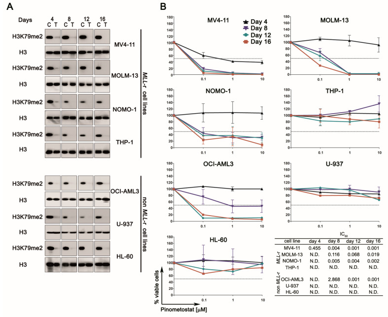
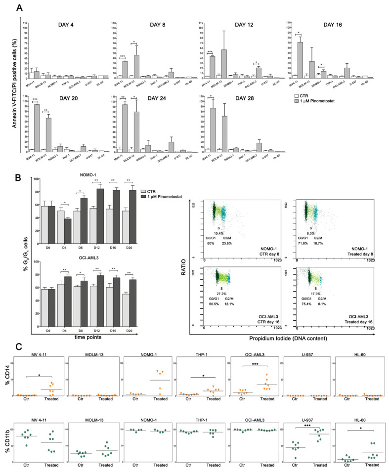
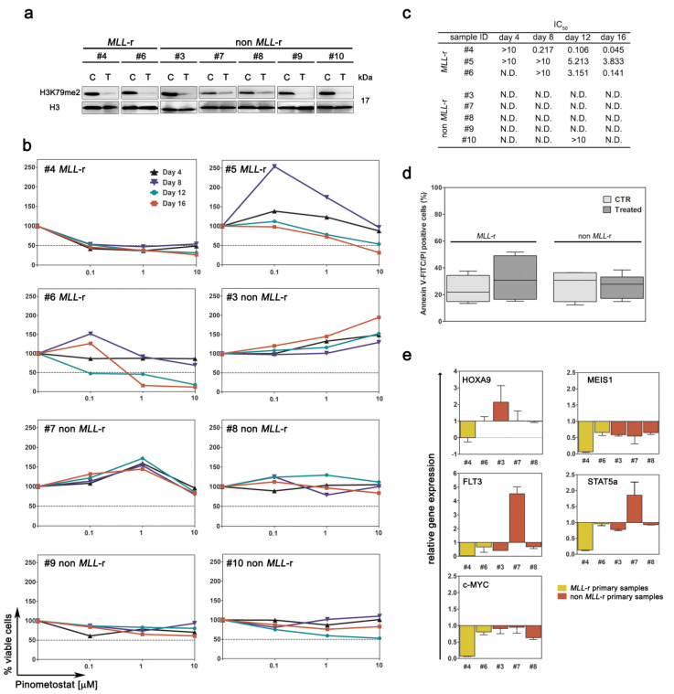
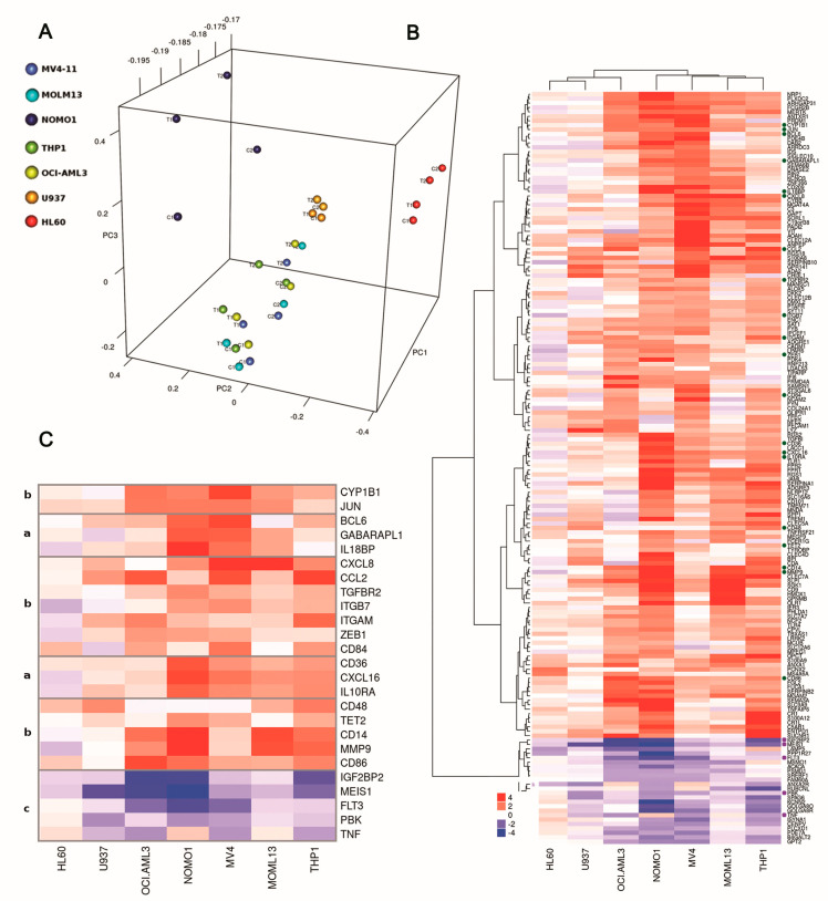
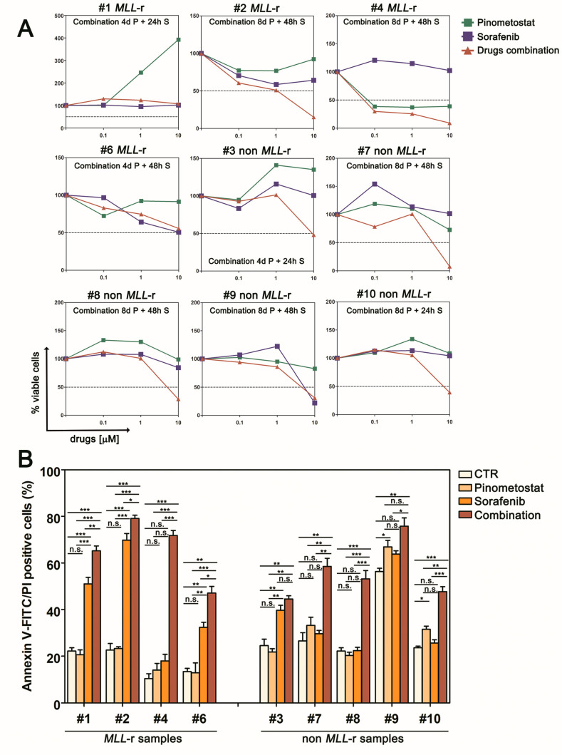
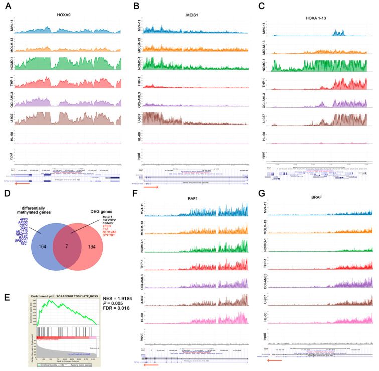
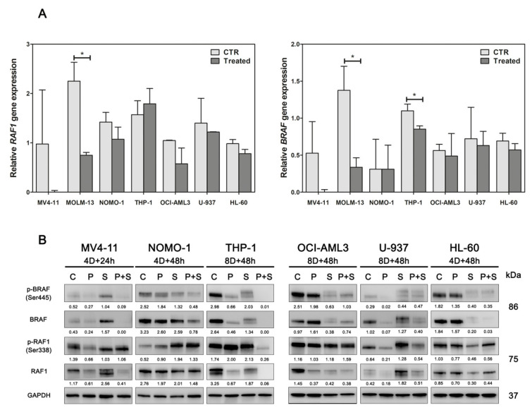
Pediatric acute myeloid leukemia (AML) is an aggressive malignancy with poor prognosis for which there are few effective targeted approaches, despite the numerous genetic alterations, including MLL gene rearrangements (MLL-r). The histone methyltransferase DOT1L is involved in supporting the proliferation of MLL-r cells, for which a target inhibitor, Pinometostat, has been evaluated in a clinical trial recruiting pediatric MLL-r leukemic patients. However, modest clinical effects have been observed. Recent studies have reported that additional leukemia subtypes lacking MLL-r are sensitive to DOT1L inhibition. Here, we report that targeting DOT1L with Pinometostat sensitizes pediatric AML cells to further treatment with the multi-kinase inhibitor Sorafenib, irrespectively of MLL-r. DOT1L pharmacologic inhibition induces AML cell differentiation and modulates the expression of genes with relevant roles in cancer development. Such modifications in the transcriptional program increase the apoptosis and growth suppression of both AML cell lines and primary pediatric AML cells with diverse genotypes. Through ChIP-seq analysis, we identified the genes regulated by DOT1L irrespective of MLL-r, including the Sorafenib target BRAF, providing mechanistic insights into the drug combination activity. Our results highlight a novel therapeutic strategy for pediatric AML patients.

Keywords: BRAF; ChIP-seq; DOT1L; Pinometostat; Sorafenib; pediatric acute myeloid leukemia; targeted therapy.


Conflict of interest statement

The authors declare no potential conflicts of interest.

Figures

Figure 1
Figure 1
Pharmacological targeting of DOT1L in human acute myeloid leukemia (AML) cell lines with or without mixed lineage leukemia rearranged (MLL-r). (A) Western blotting analysis of histones isolated from MLL-r and non-MLL-r AML cell lines treated with 1 µM of Pinometostat or DMSO 0.01%. H3K79me2 levels were analyzed every 4 days for a period of 16 days. Molecular weights are indicated on the right. C: control cells; T: treated cells. (B) Growth curves of MLL-r and non-MLL-r AML cell lines treated with increasing concentrations of Pinometostat or DMSO 0.01% as vehicle control for up to 16 days and summary of IC50 concentrations. Results of three independent replicates are presented as means ± SD.
Figure 2
Figure 2
Impact of Pinometostat treatment on apoptosis, cell cycle, and differentiation. MLL-r and non-MLL-r human AML cell lines were treated with 1 µM of Pinometostat or DMSO 0.01% for up to 28 days and analyzed every 4 days. (A) Flow cytometry analysis of apoptosis measured by Annexin V-FITC/PI staining. Results of three independent replicates are presented as means ± SD. (B) Increase in G0/G1 phase cells in Pinometostat-sensitive NOMO-1 and OCI-AML3 cell lines, as measured by flow cytometry (left panels), and representative dot plots showing G0/G1 cell cycle phase increase at two time points (day 8 and 16, right panels). Cell cycle analyses were performed every 4 days for a period of 20 days (D0–D20). Results of two independent replicates are presented as means ± SD. (C) Modulation of CD14 (upper panels) and CD11b (lower panels) myeloid surface markers in AML cell lines. CD14 and CD11b were analyzed by flow cytometry every 4 days for a period of 28 days, and markers’ expression in Pinometostat-treated AML cell lines was compared to that of control cells. Ctr: control cells (DMSO 0.01%). Asterisks indicate levels of significance (* p ≤ 0.05, ** p ≤ 0.01, *** p ≤ 0.001).
Figure 3
Figure 3
Pharmacological targeting of DOT1L in primary AML cells derived from pediatric patients with or without MLL-r. Primary AML cells were treated with Pinometostat for up to 16 days. (A) Western blot analysis of H3K79me2 levels following 8 days of treatment with 1 µM of Pinometostat or DMSO 0.01%. Molecular weights are indicated on right. C: control cells; T: treated cells. (B) Growth curves of primary AML cells treated with increasing concentrations of Pinometostat for up to 16 days. (C) Summary of IC50 concentrations. (D) Average percentage of apoptotic cells measured by flow cytometry in MLL-r and non-MLL-r primary AML cells treated for 16 days with 1 µM of Pinometostat or DMSO 0.01%. Ctr: control cells. Data are presented as means ± SD. (E) Modulation of gene expression induced by 8 days of treatment with 1 µM of Pinometostat and quantified by RT-PCR. Relative mRNA expression in treated vs. control cells was calculated using the 2−ΔΔCt methods. Results of three independent replicates are presented as means ± SD.
Figure 4
Figure 4
Global gene expression profiling in MLL-r and non-MLL-r human AML cell lines treated with Pinometostat. All AML cell lines were treated with 1 µM Pinometostat or DMSO 0.01% for 8 days, because in previous analyses, this drug exposure time significantly impacted proliferation, differentiation, and transcription processes. (A) Principal component analysis (PCA) plot of gene expression data. Two replicates for each cell line and condition are shown. C1: control cells, replicate 1; T1: treated cells, replicate 1; C2: control cells, replicate 2; T2: treated cells, replicate 2. (B) Heatmap and hierarchical clustering of fold changes in gene expression in response to Pinometostat for the 171 genes concordantly up- or down-regulated in at least three out of seven AML cell lines. The dendrogram clusters genes based on MLL-r. Genes involved in sustaining cancer cells or in hematopoietic cell differentiation are indicated with dots and are detailed in (C) (group a: up-regulated genes only in MLL-r cells; group b: up-regulated genes shared between MLL-r and non-MLL-r cells; group c: down-regulated genes shared between MLL-r and non-MLL-r cells). The color scale reflects the fold change in gene expression in response to Pinometostat, ranging from down-regulated (blue) to up-regulated (red).
Figure 5
Figure 5
Pinometostat sensitizes primary cells from pediatric AML patients to Sorafenib treatment. (A) Growth curves of primary AML cells pre-treated with Pinometostat before Sorafenib addition (Pinometostat/Sorafenib ratio 1:1). The pre-treatment model consists of 4 or 8 days of treatment with Pinometostat followed by 24 or 48 h treatment with Sorafenib. Combination times are indicated in each panel. (B) Flow cytometry analysis of apoptosis measured by Annexin V-FITC/PI staining in primary AML cells treated with 10 µM of Pinometostat (8 days), Sorafenib (48 h), or combined drugs (8 days + 48 h). Ctr: control cells (DMSO 0.01%). Results of three independent replicates are presented as means ± SD.
Figure 6
Figure 6
H3K79 methylation in MLL-r and non-MLL-r human AML cell lines. (AC) are H3K79me2 profiles of MLL target genes. (D) Venn diagram showing the overlap between the 172 genes differentially methylated in MLL-r compared to non-MLL-r human AML cell lines and the 171 genes differentially expressed upon Pinometostat treatment and shared between at least three AML cell lines. Overlapping genes are reported (black: down-regulated genes; red: up-regulated genes). Additional genes with important roles in regulating signaling cascades, transcription, or cell development, and differentially H3K79me2 enriched in MLL-r compared to non-MLL-r, are indicated in blue. DEG: differentially expressed genes. (E) Gene set enrichment analysis (GSEA) of commonly H3K79me2 enriched genes in both MLL-r and non-MLL-r AML cells showing the enrichment in Sorafenib’s drug signature. The normalized enrichment score (NES) and the statistically significant False Discovery Rate (FDR) and p values are indicated. (F) and (G) are H3K79me2 profiles of RAF1 and BRAF, which encode for Sorafenib targets.
Figure 7
Figure 7
Impact of Pinometostat and Sorafenib on RAF1 and BRAF expression. (A) Assessment of BRAF and RAF1 gene expression in AML cell lines treated with the DOT1L inhibitor Pinometostat for 16 days. CTR: control cells (DMSO 0.01%); Treated: cells treated with 1 µM Pinometostat. Results of three independent replicates are presented. Data are expressed as 2ΔΔCt ± SD, and the universal human reference RNA was used as calibrator. (B) Western blot analysis of BRAF and RAF1 total and phosphorylated proteins in AML cell lines treated with 1 µM of Pinometostat and Sorafenib as single agents or in combination. Combination times are indicated for each cell line (4/8D: 4/8 days). Antibody to GAPDH served as a loading control. Molecular weights are indicated on right. C: control cells (DMSO 0.01%); P: Pinometostat treatment; S: Sorafenib treatment; P+S: combined treatment (Pinometostat and Sorafenib). Densitometric data were normalized on GAPDH.

References

    1. Pui C.H., Carroll W.L., Meshinchi S., Arceci R.J. Biology, risk stratification, and therapy of pediatric acute leukemias: An update. J. Clin. Oncol. 2011;29:551–565. doi: 10.1200/JCO.2010.30.7405. - DOI - PMC - PubMed
    1. Zwaan C.M., Kolb E.A., Reinhardt D., Abrahamsson J., Adachi S., Aplenc R., de Bont E.S.J.M., de Moerloose B., Dworzak M., Gibson B., et al. Collaborative Efforts Driving Progress in Pediatric Acute Myeloid Leukemia. J. Clin. Oncol. 2015;33:2949–2962. doi: 10.1200/JCO.2015.62.8289. - DOI - PMC - PubMed
    1. Steger D.J., Lefterova M.I., Ying L., Stonestrom A.J., Schupp M., Zhuo D., Vakoc A.L., Kim J.E., Chen J., Lazar M.A., et al. DOT1L/KMT4 recruitment and H3K79 methylation are ubiquitously coupled with gene transcription in mammalian cells. Mol. Cell. Biol. 2008;28:2825–2839. doi: 10.1128/MCB.02076-07. - DOI - PMC - PubMed
    1. Nguyen A.T., Zhang Y. The diverse functions of Dot1 and H3K79 methylation. Genes Dev. 2011;25:1345–1358. doi: 10.1101/gad.2057811. - DOI - PMC - PubMed
    1. Bernt K.M., Zhu N., Sinha A.U., Vempati S., Faber J., Krivtsov A.V., Feng Z., Punt N., Daigle A., Bullinger L., et al. MLL-rearranged leukemia is dependent on aberrant H3K79 methylation by DOT1L. Cancer Cell. 2011;20:66–78. doi: 10.1016/j.ccr.2011.06.010. - DOI - PMC - PubMed