. 2020 Jul 20;8(3):49.
doi: 10.3390/toxics8030049.

Mechanism of Vascular Toxicity in Rats Subjected to Treatment with a Tyrosine Kinase Inhibitor

Affiliations

Mechanism of Vascular Toxicity in Rats Subjected to Treatment with a Tyrosine Kinase Inhibitor

Claudia Reyes-Goya et al. Toxics. .

Abstract

Sunitinib (Su) is a tyrosine kinase inhibitor with antiangiogenic and antineoplastic effects that is recommended therapy for renal cell carcinoma, gastrointestinal stromal tumors, and pancreatic neuroendocrine tumors. Arterial hypertension is one of the adverse effects observed in the treatment with Su. The aim of this work was to deepen our understanding of the underlying mechanisms involved in the development of this side effect. Studies on endothelial function, vascular remodeling and nicotinamide adenine dinucleotide phosphate oxidase (NADPH oxidase) system were carried out in thoracic aortas from rats treated with Su for three weeks. Animals subjected to Su treatment presented with increased blood pressure and reduced endothelium-dependent vasodilation, the latter being reverted by NADPH oxidase blockade. Furthermore, vascular remodeling and stronger Masson trichrome staining, together with enhanced immunofluorescence signal for collagen 1 alpha 1 (Col1α1), were observed in aortas from treated animals. These results were accompanied by a significant elevation in superoxide anion production and the activity/protein/gene expression of NADPH oxidase isoforms (NOX1, NOX2, and NOX4), which was also prevented by NOX inhibition. Furthermore, a decrease in nitric oxide (NO) levels and endothelial nitric oxide synthase (eNOS) activation was observed in aortas from Su-treated animals. All these results indicate that endothelial dysfunction secondary to changes in vascular remodeling and oxidative stress might be responsible for the typical arterial hypertension that develops following treatment with Su.

Keywords: arterial hypertension; endothelial dysfunction; tyrosine kinase inhibitor; vascular remodeling.


Conflict of interest statement

The authors declare no conflicts of interest.

Figures

Figure 1
Figure 1
(A) Weight gain, (B) food intake, (C) water intake, (D) systolic blood pressure, (E) diastolic blood pressure, and (F) heart rate in control and sunitinib (Su)-treated (25 mg/kg/day) animals. Values are expressed as mean ± S.E.M. of 15 animals per group. ** p < 0.01, *** p < 0.001 vs. control group.
Figure 2
Figure 2
Dose-response curve of (A) vasoconstriction mediated by phenylephrine (Phe) (10−9–3 × 10⁻⁵ mol/L); (B) ACh-mediated (10−9–3 × 10⁻⁵ mol/L) vasodilation, including preincubation with ML171 (10−4 mol/L); (C) ACh-mediated vasodilation in the presence of L-NAME (Nω- nitro-L-arginine methyl ester (10−4 mol/L); and (D) vasorelaxation response to sodium nitroprusside (SNP) (10−10–3 × 10⁻⁶ mol/L) in phenylephrine (Phe)-precontracted vessels from control and sunitinib-treated (25 mg/kg/day) animals. The results correspond to mean ± S.E.M. of at least six experiments (n = 6–12). Results are expressed as relative percentages of the maximum contraction induced by 60 mmol/L KCl (A), or to the contraction induced by a submaximal dose of Phe (BD). ** p < 0.01; *** p < 0.001 vs. control group, # p < 0.05, ## p < 0.01, ### p < 0.001 vs. Su group.
Figure 3
Figure 3
Aorta thickness (A and B; delimited by arrowheads), cross-sectional area (CSA) of tunica media (C), lumen area (D) and media/lumen ratio (E) in control and Su-treated (25 mg/kg/day) animals. Magnification: 10×. Values are expressed as mean ± S.E.M. of six animals per group. * p < 0.05; ** p < 0.01; *** p < 0.001 vs. control group.
Figure 4
Figure 4
Masson’s trichrome staining for collagen fibers in blue (A,B, marked with arrowheads), double immunostaining of collagen 1 alpha 1 (Col1α1) (red) and α-smooth muscle actin (α-SMA) from vascular smooth muscle cells (VSMCs) (green) (CF), and Col1𝛼1 (red) and CD31 (green) (G,H), and protein/mRNA expression of Col1 (I) and TGF-β1 (J,K), in aortas from control and sunitinib-treated (25 mg/kg/day) animals. Magnification 10× for Masson’s trichrome staining. Magnifications: 10× (A,B), 20× (C,E) and 40× (D,FH). Values are expressed as mean ± S.E.M. of at least 6 animals per group (n = 6–8). * p < 0.05; ** p < 0.01; *** p < 0.001 vs. control group.
Figure 5
Figure 5
Superoxide anion production in thoracic aortas. (A) Representative dihydroethidium (DHE) staining, (B) superoxide anion quantification, (C) NADPH oxidase activity in aorta homogenates, and (D) 3-nitrotyrosine protein expression, in aortas from control and sunitinib-treated (25 mg/kg/day) animals. Magnification: 10×. Values are expressed as mean ± S.E.M. of at least four animals per group (n = 4–8). * p < 0.05, *** p < 0.001 vs. control group; ## p < 0.01, ### p < 0.001 vs. Su group; ++ p < 0.01, +++ p < 0.001 vs. Su + ML171 group; ººº p < 0.001 vs. Su + GKT136901 group; yyy p < 0.001 vs. Su + VAS2870 group.
Figure 6
Figure 6
Double immunostaining of NOX1 (red) and α-SMA (VSMCs) (green) (AD), and NOX1 (red) and CD31 (green) (EH), in aortas from control and sunitinib-treated (25 mg/kg/day) animals. Magnifications: 40× (A,C,E,G) and 100× (B,D,F,H). (IK) Protein and mRNA expression of NOX1 (I), NOX2 (J), and NOX4 (K). Values are expressed as mean ± S.E.M. of eight animals per group. * p < 0.05; *** p < 0.001 vs. control group.
Figure 6
Figure 6
Double immunostaining of NOX1 (red) and α-SMA (VSMCs) (green) (AD), and NOX1 (red) and CD31 (green) (EH), in aortas from control and sunitinib-treated (25 mg/kg/day) animals. Magnifications: 40× (A,C,E,G) and 100× (B,D,F,H). (IK) Protein and mRNA expression of NOX1 (I), NOX2 (J), and NOX4 (K). Values are expressed as mean ± S.E.M. of eight animals per group. * p < 0.05; *** p < 0.001 vs. control group.
Figure 7
Figure 7
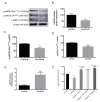
Protein expression of (A) p-endothelial nitric oxide synthase (eNOS) (Ser1177) and p-eNOS (Thr495); (A,B) total eNOS (t-eNOS); (C) ratio p-eNOS (Ser1177)/total eNOS; (D) ratio p-eNOS (Thr495)/total eNOS; (E) t-eNOS mRNA expression; and (F) NO levels in absence or presence of NOX inhibitors, in aortas from control and sunitinib-treated (25 mg/kg/day) animals. Values are expressed as mean ± S.E.M. of eight animals per group. * p < 0.05; ** p < 0.01; *** p < 0.001 vs. control group.; ### p < 0.001 vs. Su group; + p < 0.05 vs. Su + ML171 group; ºº p < 0.01 vs. Su + GKT136901 group.

References

    1. Le Tourneau C., Faivre S., Raymond E. New developments in multitargeted therapy for patients with solid tumours. Cancer Treat. Rev. 2008;34:37–48. doi: 10.1016/j.ctrv.2007.09.003. - DOI - PubMed
    1. Jiao Q., Bi L., Ren Y., Song S., Wang Q., Wang Y.S. Advances in studies of tyrosine kinase inhibitors and their acquired resistance. Mol. Cancer. 2018;17:1–12. doi: 10.1186/s12943-018-0801-5. - DOI - PMC - PubMed
    1. O’Farrell A.M., Abrams T.J., Yuen H.A., Ngai T.J., Louie S.G., Yee K.W.H., Wong L.M., Hong W., Lee L.B., Town A., et al. SU11248 is a novel FLT3 tyrosine kinase inhibitor with potent activity in vitro and in vivo. Blood. 2003 doi: 10.1182/blood-2002-07-2307. - DOI - PubMed
    1. Manir K.S., Banerjee D., Bhowmick R., Roy C. Sunitinib-induced acute severe hypothyroidism in a case of metastatic gastrointestinal stromal tumor: A case report. J. Cancer Res. Ther. 2018 doi: 10.4103/0973-1482.199443. - DOI - PubMed
    1. Faivre S., Niccoli P., Castellano D., Valle J.W., Hammel P., Raoul J.L., Vinik A., Van Cutsem E., Bang Y.J., Lee S.H., et al. Sunitinib in pancreatic neuroendocrine tumors: Updated progression-free survival and final overall survival from a phase III randomized study. Ann. Oncol. 2017 doi: 10.1093/annonc/mdw561. - DOI - PubMed